Review
. 2019 Jun 10:303:130-150.
doi: 10.1016/j.jconrel.2019.04.025. Epub 2019 May 3.

Liposomes used as a vaccine adjuvant-delivery system: From basics to clinical immunization

Affiliations
Review

Liposomes used as a vaccine adjuvant-delivery system: From basics to clinical immunization

Ning Wang et al. J Control Release. .

Abstract

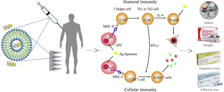
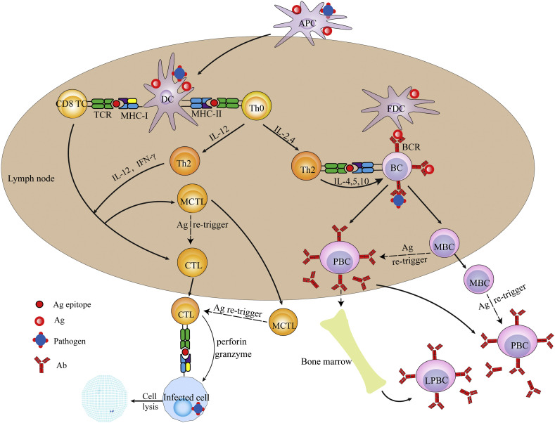
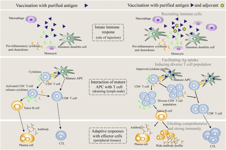
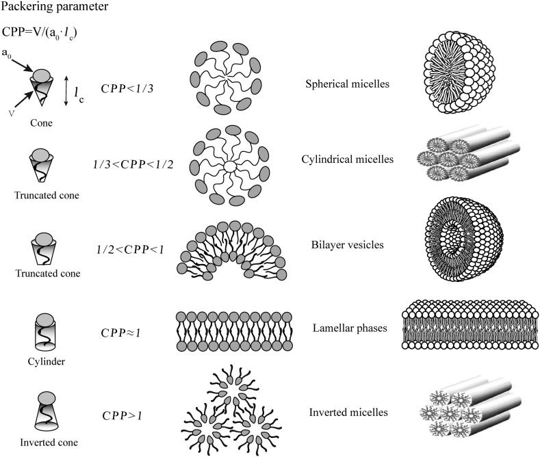
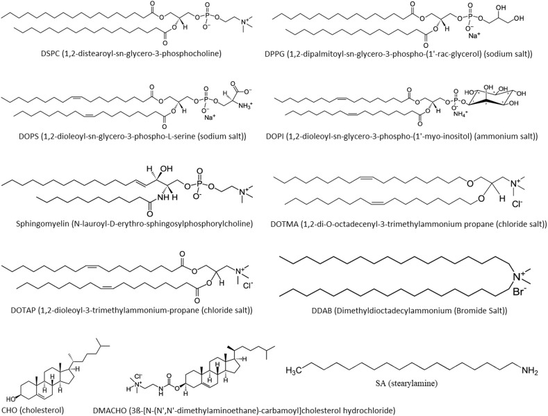
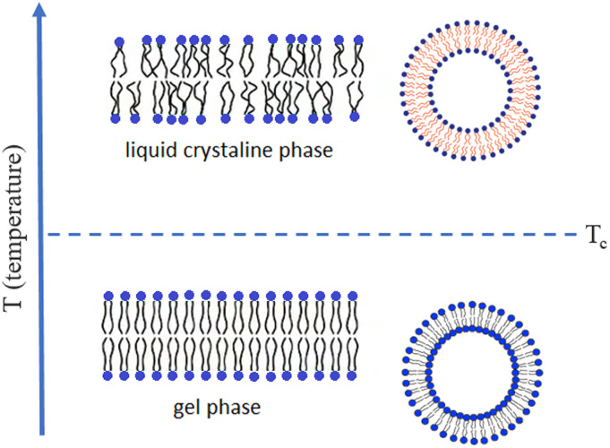
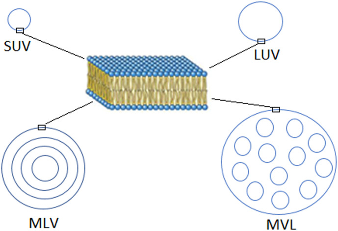
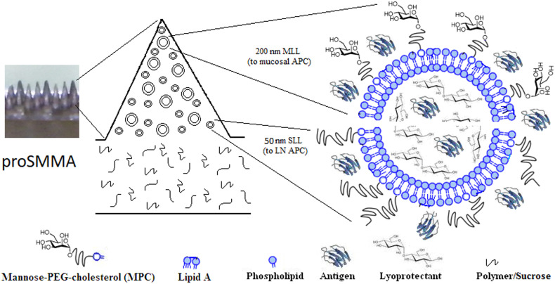
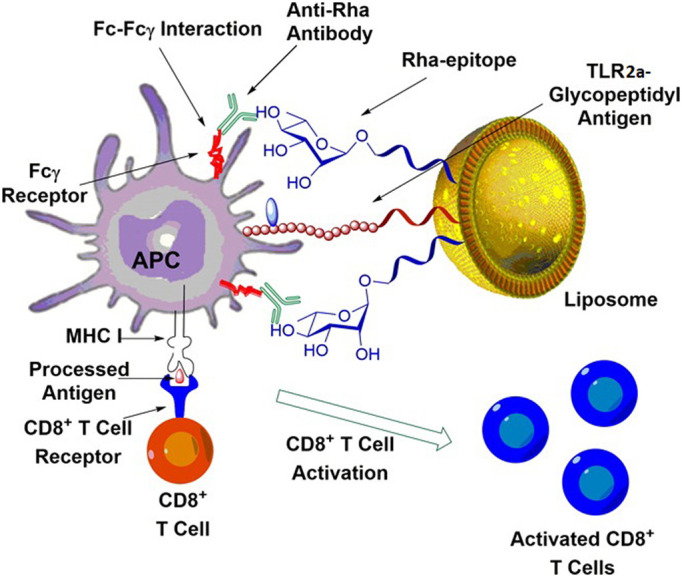
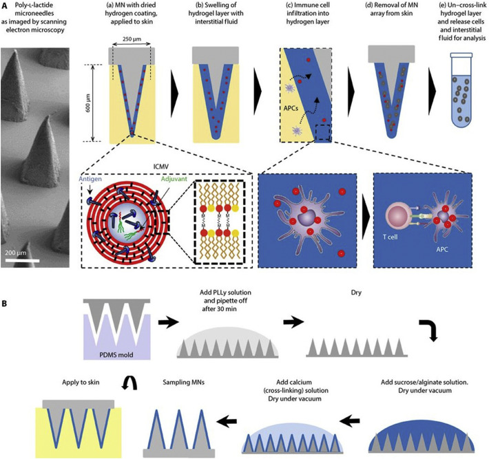
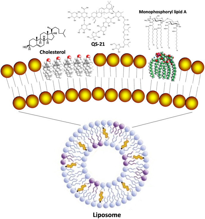
Liposomes are widely utilized as a carrier to improve therapeutic efficacy of agents thanks to their merits of high loading capacity, targeting delivery, reliable protection of agents, good biocompatibility, versatile structure modification and adjustable characteristics, such as size, surface charge, membrane flexibility and the agent loading mode. In particular, in recent years, through modification with immunopotentiators and targeting molecules, and in combination with innovative immunization devices, liposomes are rapidly developed as a multifunctional vaccine adjuvant-delivery system (VADS) that has a high capability in inducing desired immunoresponses, as they can target immune cells and even cellular organelles, engender lysosome escape, and promote Ag cross-presentation, thus enormously enhancing vaccination efficacy. Moreover, after decades of development, several products developed on liposome VADS have already been authorized for clinical immunization and are showing great advantages over conventional vaccines. This article describes in depth some critical issues relevant to the development of liposomes as a VADS, including principles underlying immunization, physicochemical properties of liposomes as the immunity-influencing factors, functional material modification to enhance immunostimulatory functions, the state-of-the-art liposome VADSs, as well as the marketed vaccines based on a liposome VADS. Therefore, this article provides a comprehensive reference to the development of novel liposome vaccines.

Keywords: Antigen cross-presentation; Cellular immunity; Immunoresponse; Mucosal vaccination; Pathogen-associated molecular pattern; Vaccine adjuvant-delivery system.


Figures

Unlabelled Image
Graphical abstract
Fig. 1
Fig. 1
Schematic description of the process for establishing the humoral and cellular immunity triggered by a pathogen (or vaccine). Abbreviations: APC, antigen presentation cell; DC, dendritic cell; FDC, follicular dendritic cell; CTL, cytotoxic T lymphocyte; TCR, T cell receptor; BCR, B cell receptor; Th0, naïve CD4+ T cell; Th1, type 1 helper T cell; Th2, type 2 helper T cell; PBC, plasma B cell; LPBC, long-lived plasma B cell.
Fig. 2
Fig. 2
The immunoresponses induced by immunization with vaccines in the presence or absence of an adjuvant.
Fig. 3
Fig. 3
Molecular shape and critical packing parameter (CPP) of amphiphilic molecules, and the self-assembly entities formed of different amphiphiles (v, the lipophilic chain volume; ao, the cross-section area of hydrophilic head group, lc, the length of lipophilic chain).
Fig. 4
Fig. 4
Molecular structure of some of the lipids that are frequently used for constructing liposomes.
Fig. 5
Fig. 5
Phospholipid bilayer membrane fluidity of liposomes. When the ambient temperature (T) < Tc of phospholipids, liposome membrane is in the gel phase; when T > Tc, liposome membrane in the liquid crystalline phase; while T increases from below Tc to above Tc, liposome membrane changes from the gel to the liquid crystalline phase.
Fig. 6
Fig. 6
Structural features of different types of liposomes.
Fig. 7
Fig. 7
Schematic of proSMMA designed as a robust mucosal VADS consisting of a mixture of 50 nm stealth MPLA-liposomes and mannosylated MPLA-liposomes fabricated into a microneedle array.
Fig. 8
Fig. 8
Schematic structure the VADS constructed with the Rha epitope-anchored liposomes loaded with Pam3Cys-linked MUC1 glycopeptide Ags, and the principle underlying the immunostimulatory action of the VADS. Reprinted with permission from Reference [142].
Fig. 9
Fig. 9
Schematic of description of preparation of SSMNs (stimulatory sampling microneedles) for inducing and monitoring immunoresponses. (A) Schematic of structure of a SSMN and its action mechanism. (B) Microneedle engineering process. PLLy, poly-l-lysine; PDMS, polydimethylsiloxane. Partially reprinted with permission from Reference [145].
Fig. 10
Fig. 10
Structure of VZV virus and anatomy of sensory dorsal root ganglia, where VZV (varicella zoster virus) reside silently after primary infection. Reprinted with permission from ViralZone (Source: ViralZone: www.expasy.org/viralzone, SIB Swiss Institute of Bioinformatics).
Unlabelled Image
Fig. 11
Schematic description of AS01b which is a liposome-based VADS. It is used for boosting the efficacy of a subunit vaccine, Shingrix®, against VZV (varicella zoster virus), which contains the Ag of gE. Reprinted with a little adaption with permission from Reference [157].

References

    1. De Gregorio E., Rappuoli R. From empiricism to rational design: a personal perspective of the evolution of vaccine development. Nat. Rev. Immunol. 2014;14(7):505–514. - PMC - PubMed
    1. Plotkin S.A. Vaccines: the fourth century. Clin. Vaccine Immunol. 2009;16(12):1709–1719. - PMC - PubMed
    1. Gao Y., McKay P.F., Mann J.F.S. Advances in HIV-1 vaccine development. Viruses. 2018;10(4) - PMC - PubMed
    1. Koff W.C., Burton D.R., Johnson P.R., Walker B.D., King C.R., Nabel G.J., Ahmed R., Bhan M.K., Plotkin S.A. Accelerating next-generation vaccine development for global disease prevention. Science. 2013;340(6136) - PMC - PubMed
    1. Del Giudice G., Rappuoli R., Didierlaurent A.M. Correlates of adjuvanticity: A review on adjuvants in licensed vaccines. Semin. Immunol. 2018;39:14–21. - PubMed

Publication types