Review
. 2019 Jul:209:10-24.
doi: 10.1016/j.biomaterials.2019.04.009. Epub 2019 Apr 14.

The cell in the ink: Improving biofabrication by printing stem cells for skeletal regenerative medicine

Affiliations
Review

The cell in the ink: Improving biofabrication by printing stem cells for skeletal regenerative medicine

G Cidonio et al. Biomaterials. 2019 Jul.

Abstract

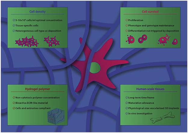
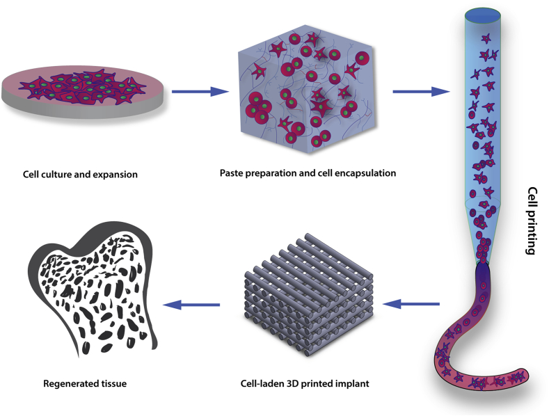
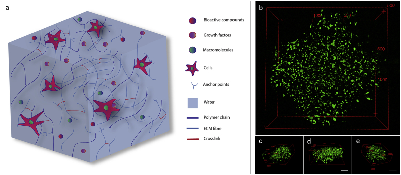
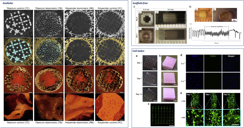
Recent advances in regenerative medicine have confirmed the potential to manufacture viable and effective tissue engineering 3D constructs comprising living cells for tissue repair and augmentation. Cell printing has shown promising potential in cell patterning in a number of studies enabling stem cells to be precisely deposited as a blueprint for tissue regeneration guidance. Such manufacturing techniques, however, face a number of challenges including; (i) post-printing cell damage, (ii) proliferation impairment and, (iii) poor or excessive final cell density deposition. The use of hydrogels offers one approach to address these issues given the ability to tune these biomaterials and subsequent application as vectors capable of delivering cell populations and as extrusion pastes. While stem cell-laden hydrogel 3D constructs have been widely established in vitro, clinical relevance, evidenced by in vivo long-term efficacy and clinical application, remains to be demonstrated. This review explores the central features of cell printing, cell-hydrogel properties and cell-biomaterial interactions together with the current advances and challenges in stem cell printing. A key focus is the translational hurdles to clinical application and how in vivo research can reshape and inform cell printing applications for an ageing population.

Keywords: 3D printing; Additive manufacturing; Biofabrication; Bioink; Bioprinting; Cell printing; Hydrogels.


Figures

Image 1
Graphical abstract
Fig. 1
Fig. 1
The cell printing paradigm. Cell printing involves cell incorporation or encapsulation within biomaterials possessing viscoelastic properties that allow their use as a “bioink”. Upon printing, cell-laden 3D printed structures are fabricated with the aim, ultimately, of implantation into the patient to regenerate the specific tissue of interest.
Fig. 2
Fig. 2
Hydrogels resembling extracellular matrix. (a) Polymeric cell carrier matrices need to mimic the physiological environment that cells experience and sense within the human body. Cells can use filopodia extensions to attach to several anchor-like proteins within the gel matrix or interact with the polymeric chains. Bioactive compounds, growth factors and macromolecules can be included or preserved from natural tissue to stimulate cell activity or differentiation toward a specific lineage. (b) Skeletal stem cells (SSCs) encapsulated in GelMA hydrogels and printed into a 3D lattice structure. 3D reconstruction of a SSCs-laden cross-section illustrating cells stretching and spreading in three dimensions. Right (c), front (d) and left (e) 3D reconstructions of SSCs encapsulated in printed GelMA demonstrating uniform and consistent cell attachment throughout the cross-section. Scale bars: (b–e) 500 μm.
Fig. 3
Fig. 3
Current biofabrication approaches. Acellular. Reconstruction of bone ingrowth and segmentation of micro CT images of acellular 3D implants in sheep distal femur defect. First and second row: 2D slices of the 3D implant. Bone is labelled in yellow and metal in blue. Third and fourth row: bone ingrowth and close up high resolution detail of nascent bone. Pixel size 0.5 μm, panels' scale bars are 40 μm. Adapted with permission from Ref. [39] CC BY license. Scaffold-free. i) Scaffold-free tubular cardiac constructs on a needle array. Representative images of tubular constructs just after printing (A,B) and after culture for 7 days (C, D). ii) Needle-free tubular constructs (A) cultured for 7 days (B) and further electrically stimulated and effects analysed (C). Adapted with permission from Ref. [46]. Cell-laden. Representative images of the whole constructs prepared with hdECM (a) and adECM (b) at progressing days of cell culture. Confocal image of the whole construct prepared with adECM at day 14 of culture (c) showing live cells (green) and dead cells (red) (scale bar, 2 mm). An image of the whole construct reconstructed from B32 images taken at different positions. Representative images of apoptosis through TUNEL (d) and Live/Dead (e) assays. TUNEL assay displays minimal apoptotic cells, indicating the generated stress at 2 s−1 shear rate at the nozzle wall did not produce a deleterious effect on the encapsulated cells with comparable apoptosis to the non-printed gel (0 s−1). Cell viability was >95% at day 1 and > 90% at both day 7 and 14. Scale bars: (a–c) 2 mm, (d,e) 100 mm. Adapted from Ref. [50] with permission from publisher.
Fig. 4
Fig. 4
Cell seeding density influence on 3D cell-laden biomaterial strands and nozzle extrusion. (a) Extruded cell-laden filaments can retain a number of cells proportional to the cell seeding density. Polymeric chains (dark blue) concentration and distribution directly influence cell proliferation capability. A lower cell seeding density results in poor cell distribution within the printed strand showing low cell-to-cell interaction and a limited proliferation rate. Increasing the cell seeding density results in a printed strand filled with cells with a high degree of cell-to-cell interaction and cell death. Proliferation and viability is limited by physical stresses imposed by neighbouring cells. Cell density can be tuned to achieve an even distribution of cell encapsulated within the printed filament in such a way that cells can maintain the required interaction with other cells to remain mitotically active and proliferate. (b) Maintaining constant the number of cells loaded in a printing syringe but changing the nozzle aperture will affect cell printability. Large nozzles (>800 μm) allow limited cell-nozzle walls and cell-to-cell interactions resulting in a widespread distribution of cells in suspension within the bioink. These settings can ensure high cell survival upon extrusion but result in low resolution of the overall construct. In contrast, narrower nozzles (<250 μm) offering smaller surface area for the same number of cells force biopaste encapsulated cells to interact with one another resulting in high density at the nozzle aperture. A narrow orifice can produce high resolution, as well as high cell death upon printing. Medium size conical nozzles (250 μm-800μm) ensure an optimal cells distribution within the nozzle and an increase in print resolution without influencing, extensively, cell survival.
Fig. 5
Fig. 5
Three-dimensional diagram showing the correlation between cell viability and pressure provided by the 3D printing system and bioink polymer content. Values listed in percentages. The highest cell viability upon printing is observed when a low polymer content hydrogel is used in combination with low pressure. Increasing polymer content and lowering extrusion pressure results in the absence of bioink deposition and reduced cell viability upon printing. High pressure and the use of a low polymer content paste for cell encapsulation produces structurally weak scaffolds. Elevated print pressure and polymer content lead to a significant decrease and ultimately, a total loss of viable printed cells. Indicative 3D model plotted with Matlab (MathWorks, R2018a).
Fig. 6
Fig. 6
Viability of 3D constructs fabricated by cell printing depends on initial cell density. SSCs were encapsulated at (a–c) low density ( ≤1 × 106 cell ml−1) and (d–f) high density (>5 × 106 cell ml−1) GelMA bioinks. (a) Viability of 3D printed GelMA is adapted from Ref. [85] (Copyright 2016 American Chemical Society). Living cells are marked green, dead cells in red. (b) Low cell density seeded on 3D printed GelMA scaffolds. (c) Quantification of viability of 3D printed hMSCs in GelMA is adapted from Ref. [85] (Copyright 2016 American Chemical Society). (d) High cell density is encapsulated and printed in GelMA bioink. Living cells are in green, pre-labelled cells to visualise distribution in blue – from previously employed protocol [31]. (e) Equal density was seeded on top of the 3D printed scaffolds to show visual comparison between different cell seeding numbers. (f) Viability was determined by confocal microscopy (Leica SPS5) cell counting in a ROI 10 × . Pre-encapsulation staining of all cells in blue with lypophilic dye (Vybrant DiD, ThermoFisher) and metabolically active cells in green (Calcein AM, ThermoFisher) was done using previously employed methodology [31]. Statistical analysis was carried out using two-way ANOVA (*p < 0.05, ****p < 0.0001). Scale bars: (a,d) 200 μm, (b,e) 100 μm. (For interpretation of the references to colour in this figure legend, the reader is referred to the Web version of this article.)
Fig. 7
Fig. 7
Scaling up cell printing approaches. (i–a) Illustration of a computer aided design (CAD) example of an assembled hemispherical construct for osteochondral joint resurfacing. (i–b) A biphasic hemispherical construct with stained GelMA hydrogel micro-spheres representing chondrogenic (red) and osteogenic (blue) phase of an osteochondral construct fabricated by applying the bottom-up automated tissue bioassembly strategy. Scale bars: 2 mm. Fluorescence microscopy images of (ii - a) a manually assembled construct and (ii - b) a construct assembled using the bioassembly system stained with Calcein AM (live cells, green) and Propidium Iodide (dead cells, red). (iii - a-f) Histological analysis of assembled micro-tissues and associated tissue fusion in adjacent culture over 28 days. Histological sections stained with (iii - a-c) Safranin-O/Haematoxylin/fast green or (iii - e, f) Collagen II antibodies. Bioassembled HAC-laden 9.5%GelMA-0.5%HepMA micro-spheres (iii - g, h) stained with Calcein AM (live cells, green) and Propidium Iodide (dead cells, red) (iii - g) or DAPI(blue) and Aggrecan (purple) antibodies (iii - h) after 35 days culture in chondrogenic differentiation media. Adapted with permission [100] CC BY license. (iv - a) Outline of experimental groups (solid and micro channelled) and control groups (empty and BMP-2); pre-implantation chondrogenic culture conditions; and implantation of primed hydrogel (channelled) into a 5 mm femoral defect. (iv - b) Biochemical analysis (Total DNA/construct (n = 3) and sGAG/DNA (n = 5) of both groups after 4 weeks of in vitro culture. (iv - c) Immunohistochemical staining for collagen II pre-implantation, 4 × scale-bar 1 mm. Adapted with permission [77] CC BY license.

Similar articles

Cited by

References

    1. Kang H.-W., Lee S.J., Ko I.K., Kengla C., Yoo J.J., Atala A. A 3D bioprinting system to produce human-scale tissue constructs with structural integrity. Nat. Biotechnol. 2016;34:312–319. - PubMed
    1. Klebe R.J. Cytoscribing: a method for micropositioning cells and the construction of two- and three-dimensional synthetic tissues. Exp. Cell Res. 1988;179:362–373. - PubMed
    1. V Murphy S., Atala A. 3D bioprinting of tissues and organs. Nat. Biotechnol. 2014;32:773–785. - PubMed
    1. Biggs M.J.P., Richards R.G., Gadegaard N., Wilkinson C.D.W., Oreffo R.O.C., Dalby M.J. The use of nanoscale topography to modulate the dynamics of adhesion formation in primary osteoblasts and ERK/MAPK signalling in STRO-1+ enriched skeletal stem cells. Biomaterials. 2009;30:5094–5103. - PubMed
    1. Hong S., Sycks D., Chan H.F., Lin S., Lopez G.P., Guilak F., Leong K.W., Zhao X. 3D printing: 3D printing of highly stretchable and tough hydrogels into complex, cellularized structures (Adv. Mater. 27/2015. Adv. Mater. 2015;27 4034–4034. - PMC - PubMed

Publication types