

DNA methylation, transcriptome and genetic copy number signatures of diffuse cerebral WHO grade II/III gliomas resolve cancer heterogeneity and development
- PMID: 31023364
- PMCID: PMC6482573
- DOI: 10.1186/s40478-019-0704-8
DNA methylation, transcriptome and genetic copy number signatures of diffuse cerebral WHO grade II/III gliomas resolve cancer heterogeneity and development
Abstract
Background: Diffuse lower WHO grade II and III gliomas (LGG) are slowly progressing brain tumors, many of which eventually transform into a more aggressive type. LGG is characterized by widespread genetic and transcriptional heterogeneity, yet little is known about the heterogeneity of the DNA methylome, its function in tumor biology, coupling with the transcriptome and tumor microenvironment and its possible impact for tumor development.
Methods: We here present novel DNA methylation data of an LGG-cohort collected in the German Glioma Network containing about 85% isocitrate dehydrogenase (IDH) mutated tumors and performed a combined bioinformatics analysis using patient-matched genome and transcriptome data.
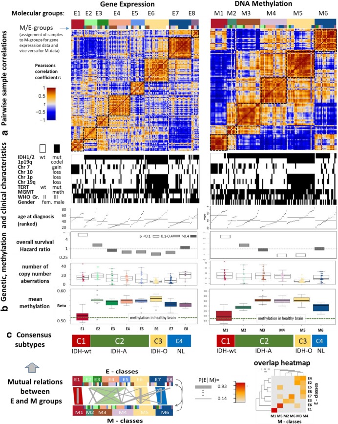
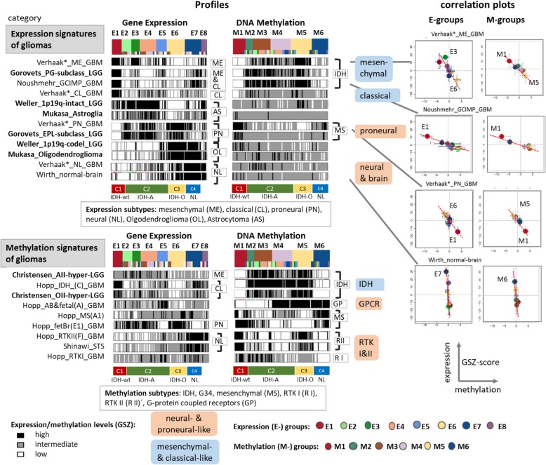
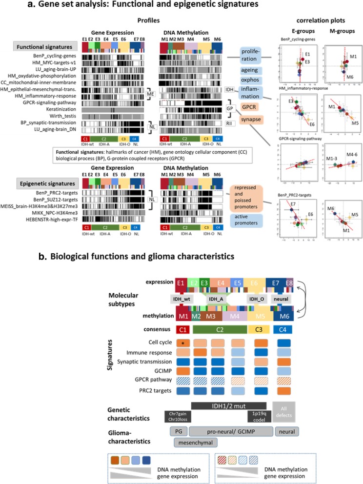
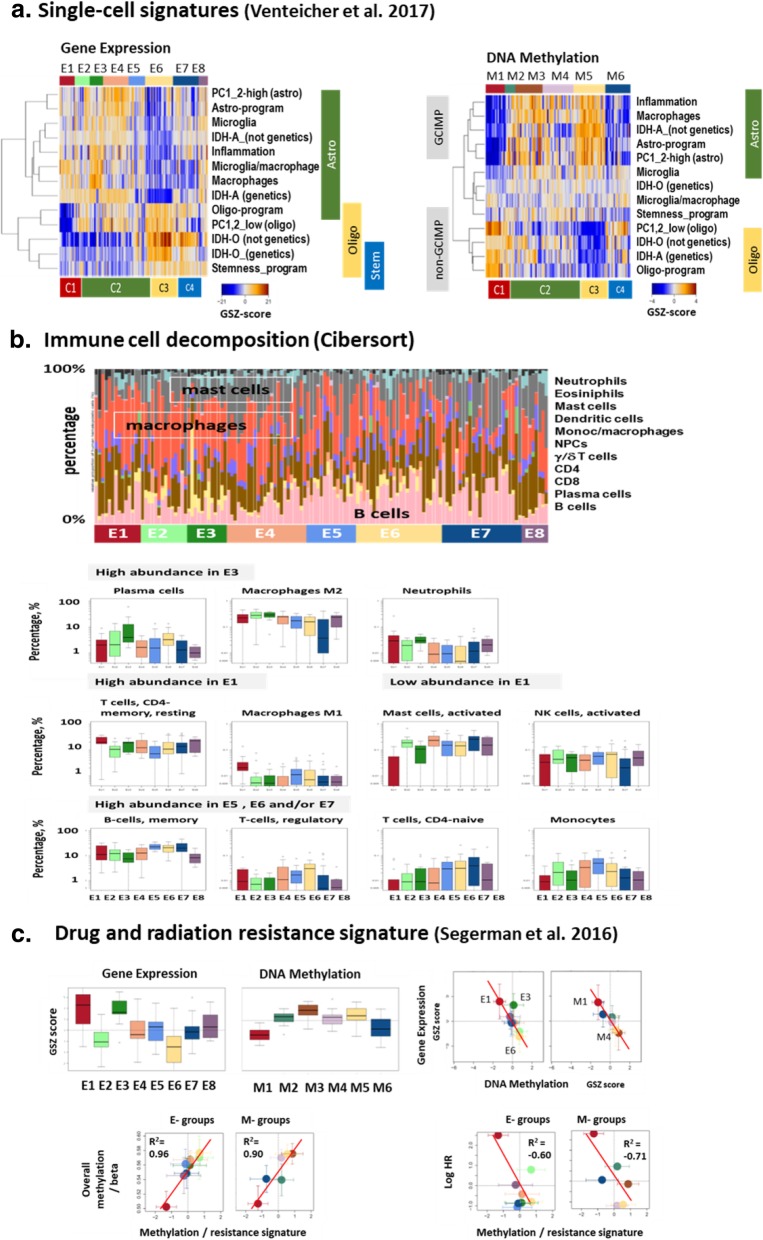
Results: Stratification of LGG based on gene expression and DNA-methylation provided four consensus subtypes. We characterized them in terms of genetic alterations, functional context, cellular composition, tumor microenvironment and their possible impact for treatment resistance and prognosis. Glioma with astrocytoma-resembling phenotypes constitute the largest fraction of nearly 60%. They revealed largest diversity and were divided into four expression and three methylation groups which only partly match each other thus reflecting largely decoupled expression and methylation patterns. We identified a novel G-protein coupled receptor and a cancer-related 'keratinization' methylation signature in in addition to the glioma-CpG island methylator phenotype (G-CIMP) signature. These different signatures overlap and combine in various ways giving rise to diverse methylation and expression patterns that shape the glioma phenotypes. The decrease of global methylation in astrocytoma-like LGG associates with higher WHO grade, age at diagnosis and inferior prognosis. We found analogies between astrocytoma-like LGG with grade IV IDH-wild type tumors regarding possible worsening of treatment resistance along a proneural-to-mesenchymal axis. Using gene signature-based inference we elucidated the impact of cellular composition of the tumors including immune cell bystanders such as macrophages.
Conclusions: Genomic, epigenomic and transcriptomic factors act in concert but partly also in a decoupled fashion what underpins the need for integrative, multidimensional stratification of LGG by combining these data on gene and cellular levels to delineate mechanisms of gene (de-)regulation and to enable better patient stratification and individualization of treatment.
Keywords: Astrocytoma; Cellular composition; DNA methylation; Epigenetics; Glioma; Molecular subtypes; Prognosis; Tumor microenvironment.
Conflict of interest statement
Ethics approval and consent to participate
All patients gave written informed consent for participation in the GGN and its translational research projects.
Consent for publication
Not applicable.
Competing interests
MW has received research grants from Abbvie, Acceleron, Actelion, Bayer, Merck, Sharp & Dohme (MSD), Merck (EMD), Novocure, OGD2, Piqur, Roche and Tragara, and honoraria for lectures or advisory board participation or consulting from Abbvie, BMS, Celgene, Celldex, Merck, Sharp & Dohme (MSD), Merck (EMD), Novocure, Orbus, Pfizer, Progenics, Roche, Teva and Tocagen. US has received honoraria for lectures or advisory board participation from medac, GSK, mundipharma, Novartis, Novocure, Roche. DK has received research grants from Novocure, Northwest biotherapeutics, Kyowa, and honoraria for lectures or advisory board participation or consulting from Baxter and Kyowa. WW received study drug for clinical research from Apogenix, Roche and Pfizer. JCT has received research grants BrainLab and honoraria for lectures from BrainLab, Siemens, Merck, Roche and medac.
All other authors declare that they have no competing interests.
Publisher’s Note
Springer Nature remains neutral with regard to jurisdictional claims in published maps and institutional affiliations.
Figures

Similar articles
-
DNA methylation signatures for 2016 WHO classification subtypes of diffuse gliomas.Clin Epigenetics. 2017 Apr 4;9:32. doi: 10.1186/s13148-017-0331-9. eCollection 2017. Clin Epigenetics. 2017. PMID: 28392842 Free PMC article.
-
SHOX2 is a Potent Independent Biomarker to Predict Survival of WHO Grade II-III Diffuse Gliomas.EBioMedicine. 2016 Nov;13:80-89. doi: 10.1016/j.ebiom.2016.10.040. Epub 2016 Oct 28. EBioMedicine. 2016. PMID: 27840009 Free PMC article.
-
Molecular subtyping reveals immune alterations in IDH wild-type lower-grade diffuse glioma.J Pathol. 2020 Jul;251(3):272-283. doi: 10.1002/path.5468. Epub 2020 Jun 15. J Pathol. 2020. PMID: 32418210
-
DNA methylation in adult diffuse gliomas.Brief Funct Genomics. 2016 Nov;15(6):491-500. doi: 10.1093/bfgp/elw019. Epub 2016 Jun 10. Brief Funct Genomics. 2016. PMID: 27288434 Review.
-
Molecular features assisting in diagnosis, surgery, and treatment decision making in low-grade gliomas.Neurosurg Focus. 2015 Mar;38(3):E2. doi: 10.3171/2015.1.FOCUS14745. Neurosurg Focus. 2015. PMID: 25727224 Review.
Cited by
-
Metabolic Signature-Based Subtypes May Pave Novel Ways for Low-Grade Glioma Prognosis and Therapy.Front Cell Dev Biol. 2021 Nov 23;9:755776. doi: 10.3389/fcell.2021.755776. eCollection 2021. Front Cell Dev Biol. 2021. PMID: 34888308 Free PMC article.
-
Identification of Prognostic Signatures of Alternative Splicing in Glioma.J Mol Neurosci. 2020 Oct;70(10):1484-1492. doi: 10.1007/s12031-020-01581-0. Epub 2020 Jun 29. J Mol Neurosci. 2020. PMID: 32602029
-
The Transcriptome and Methylome of the Developing and Aging Brain and Their Relations to Gliomas and Psychological Disorders.Cells. 2022 Jan 21;11(3):362. doi: 10.3390/cells11030362. Cells. 2022. PMID: 35159171 Free PMC article.
-
Deciphering Glioblastoma: Fundamental and Novel Insights into the Biology and Therapeutic Strategies of Gliomas.Curr Issues Mol Biol. 2024 Mar 13;46(3):2402-2443. doi: 10.3390/cimb46030153. Curr Issues Mol Biol. 2024. PMID: 38534769 Free PMC article. Review.
-
Pediatric diffuse intrinsic pontine glioma radiotherapy response prediction: MRI morphology and T2 intensity-based quantitative analyses.Eur Radiol. 2024 Dec;34(12):7962-7972. doi: 10.1007/s00330-024-10855-9. Epub 2024 Jun 21. Eur Radiol. 2024. PMID: 38907098 Free PMC article.
References
-
- Brat DJ, Aldape K, Colman H, Holland EC, Louis DN, Jenkins RB, Kleinschmidt-DeMasters BK, Perry A, Reifenberger G, Stupp R, et al. cIMPACT-NOW update 3: recommended diagnostic criteria for “diffuse astrocytic glioma, IDH-wildtype, with molecular features of glioblastoma, WHO grade IV”. Acta Neuropathol. 2018;136:805–810. doi: 10.1007/s00401-018-1913-0. - DOI - PMC - PubMed
Publication types
MeSH terms
LinkOut - more resources
Full Text Sources
Medical
Molecular Biology Databases
