. 2019 Jun 5;27(6):1074-1086.
doi: 10.1016/j.ymthe.2019.04.003. Epub 2019 Apr 9.

Aberrant Clonal Hematopoiesis following Lentiviral Vector Transduction of HSPCs in a Rhesus Macaque

Affiliations

Aberrant Clonal Hematopoiesis following Lentiviral Vector Transduction of HSPCs in a Rhesus Macaque

Diego A Espinoza et al. Mol Ther. .

Abstract

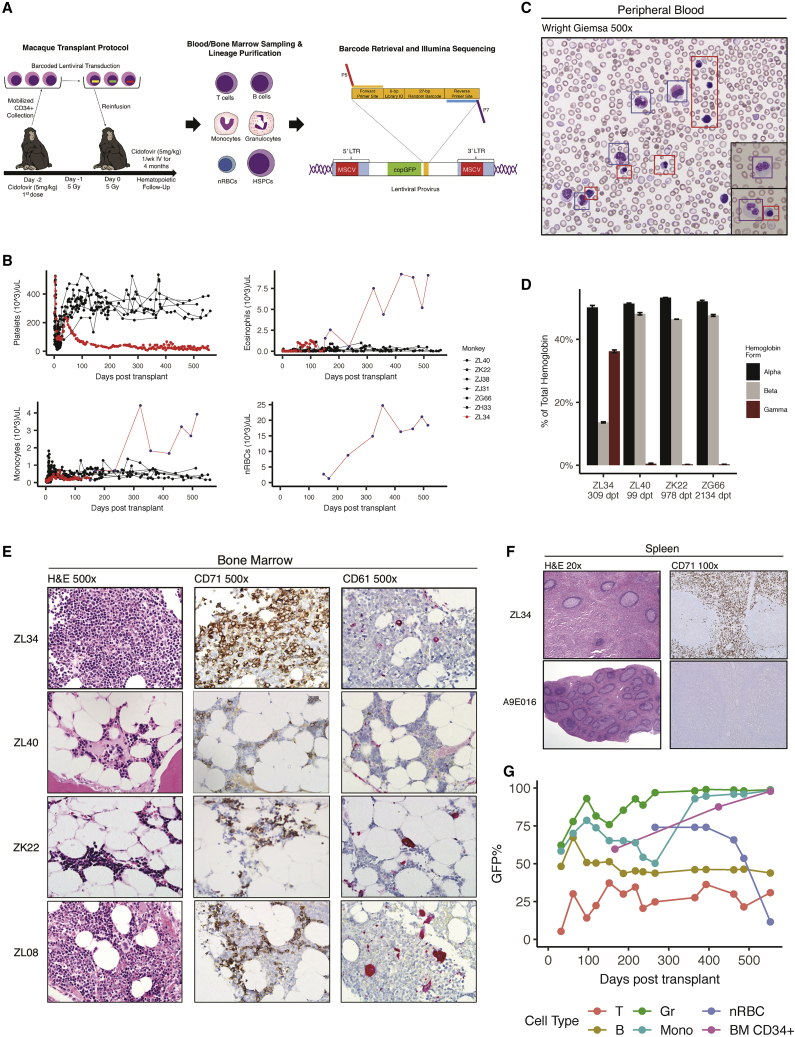
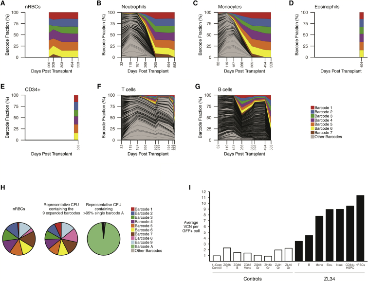
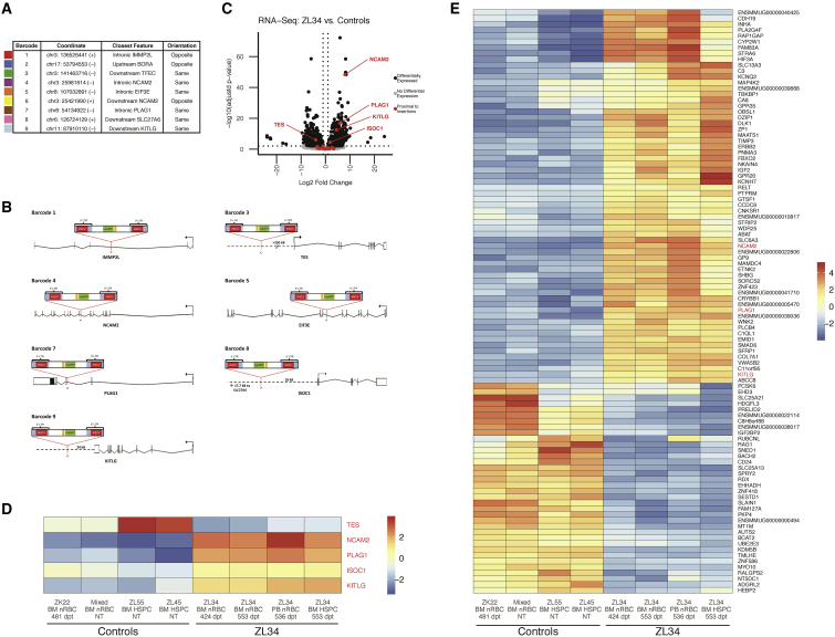
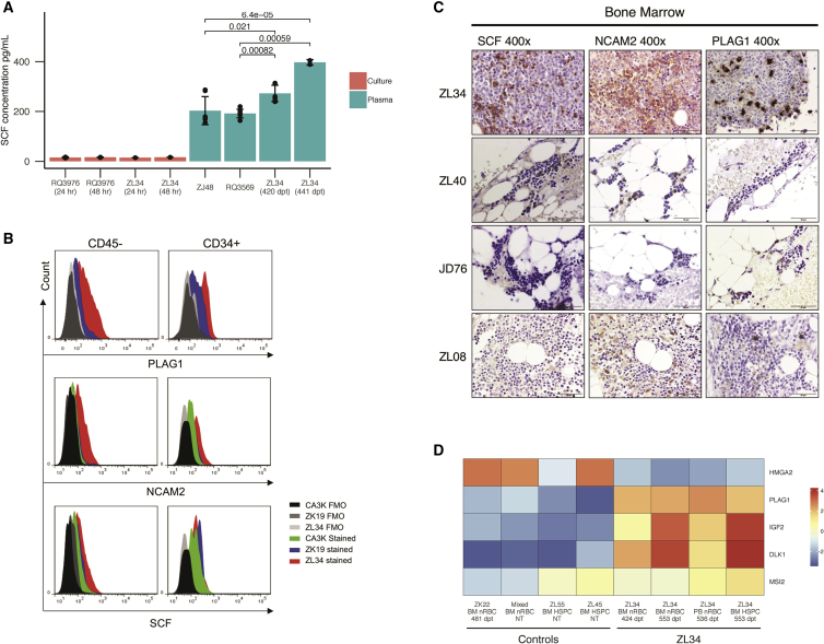
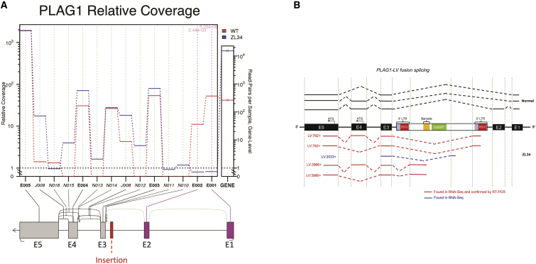
Lentiviral vectors (LVs) are used for delivery of genes into hematopoietic stem and progenitor cells (HSPCs) in clinical trials worldwide. LVs, in contrast to retroviral vectors, are not associated with insertion site-associated malignant clonal expansions and, thus, are considered safer. Here, however, we present a case of markedly abnormal dysplastic clonal hematopoiesis affecting the erythroid, myeloid, and megakaryocytic lineages in a rhesus macaque transplanted with HSPCs that were transduced with a LV containing a strong retroviral murine stem cell virus (MSCV) constitutive promoter-enhancer in the LTR. Nine insertions were mapped in the abnormal clone, resulting in overexpression and aberrant splicing of several genes of interest, including the cytokine stem cell factor and the transcription factor PLAG1. This case represents the first clear link between lentiviral insertion-induced clonal expansion and a clinically abnormal transformed phenotype following transduction of normal primate or human HSPCs, which is concerning, and suggests that strong constitutive promoters should not be included in LVs.

Keywords: gene therapy; genotoxicity; lentiviral vector; non-human primate.


Figures

Figure 1
Figure 1
Rhesus Macaque Autologous Transplantation Model and Development of Aberrant Hematopoiesis in Rhesus Macaque ZL34 (A) Transplantation timeline for ZL34. HSPCs were mobilized with G-CSF and AMD3100, collected via apheresis, enriched for CD34+ HSPCs, and transduced with a barcoded lentiviral vector. Following total body irradiation, transduced autologous CD34+ HSPCs were reinfused, and cidofovir was administered. The integrated form of the provirus is shown, detailing the position of the MSCV promoter and/or enhancer(s), copGFP marker gene, and high-diversity barcode library consisting of a 6-bp library ID and 27-bp random nucleotides, flanked by sequencing primer binding sites. (B) Platelet, eosinophil, monocyte, and nRBC concentrations in the blood over time in seven macaques receiving cells transduced with the barcoded LV containing the MSCV promoter and/or enhancer, including ZL34. Counts were determined by Coulter counter (red or black circles) or by automated PB smear scanning with Cellavision DM (blue dots, ZL34). (C) Wright’s-stained ZL34 PB smear from day 424 post-transplantation, showing frequent eosinophils (blue squares), nucleated erythroid cells (red squares), which are not found in normal rhesus macaque blood, and hypogranular neutrophils with dysplastic nuclei (black insets, purple squares). (D) HPLC assay of hemoglobin chain composition in the blood partitioned into α-, β-, and γ-globin molecules for ZL34 309 days post-transplantation (dpt) and three barcoded macaques without clonal expansions (ZL40, ZK22, and ZG66 99–2134 days post-transplantation). ZL34 values correspond to a predicted fetal hemoglobin of 70%. Error bars denote SD for three technical replicates. (E) BM biopsies from ZL34 (day 424), two healthy transplanted macaques (ZK22 and ZL40), and one healthy non-transplanted macaque (ZL08). CD71, immunohistochemistry for the erythroid precursor-specific transferrin receptor (CD71); CD61, immunohistochemistry for the megakaryocyte-specific glycoprotein IIIa (CD61). Note that ZL34’s megakaryocytes are small and uninuclear. (F) Spleen from ZL34 and a control-transplanted macaque (A9E016). (G) GFP%+ cells by flow cytometry over time in ZL34 PB T cells, B cells, granulocytes, monocytes, and nRBCs and BM CD34+ cells.
Figure 2
Figure 2
Clonal Tracking via Barcode Retrieval in ZL34 (A–G) Longitudinal clonal tracking of barcodes retrieved from nRBCs (A), neutrophils (B), monocytes (C), eosinophils (D), BM CD34+ cells (E), T cells (F), and B cells (G). Each graph shows the fractional contributions from individual barcodes over time, with each colored ribbon representing the expanded barcodes detected via standard barcode recovery and all other barcodes shaded in gray. (H) Modified barcode recovery using the alternative forward primer on ZL34 nRBCs (393 days post-transplant), showing retrieval of 9 expanded barcodes. Modified barcode recovery using the same primers was performed on single-cell-derived GFP+ CFUs from ZL34 BM CD34+ cells (266 days post-transplant) plated at low density. Representative barcode analysis on a myeloid CFU containing all 9 barcodes is shown, matching those found in the circulating nRBC, confirming integration of the 9 barcodes in one original HSPC. For comparison, barcode analysis on a myeloid CFU from the same plate found to contain a single barcode, barcode A, is shown, confirming lack of cellular contamination across the plate that would complicate analysis. The small sector shown in gray represents multiple other barcodes with very low read counts, likely resulting from residual single cells not forming CFUs. (I) Vector copy number per GFP+ cell quantified via qPCR on Gr (granulocytes), Mono (monocytes), T cells, B cells, Eos (eosinophils), nRBCs, and CD34+ BM HSPCs from ZL34 (black bars, days 494–553 post-transplantation) and additional barcoded transplanted macaques (white bars, days 278–1,085 post-transplantation).
Figure 3
Figure 3
Identification of Insertion Sites and Effect on Gene Expression in Hematopoietic Cells (A) Localization in the rhesus genome, closest gene, and proviral orientation relative to the closest gene for the 9 insertion sites corresponding to the 9 expanded barcodes. (B) Orientation and location of the provirus relative to the 5 proximally differentially expressed genes and 2 genes with intronic disruptions. (C) Volcano plot of RNA-seq data from normal control versus ZL34 nRBCs and CD34+ cells showing all differentially expressed genes (black) as defined by adj. p < 0.01 and log2(fold change) > 1 or log2(fold change) < −1. Non-differentially expressed genes are shown in gray. Differentially expressed genes within 1 Mb upstream to 1 Mb downstream of any of the 9 insertion sites are colored in red and labeled. Two control BM nRBC samples (one from transplanted macaque ZK22 and one pooled from two non-transplanted macaques) and two control BM CD34+ samples (from two different non-transplanted macaques) were analyzed along with two independent BM nRBC samples, one PB nRBC sample, and one CD34+ sample from ZL34. (D) Mean-centered gene expression levels of NCAM2, KITLG, PLAG1, ISOC1, and TES. Expression levels are regularized log transformation (rlog) values from DESeq2. NT, non-transplanted. (E) Mean-centered gene expression levels of the top 100 differentially expressed genes (by adj. p value) in ZL34 compared with control nRBC and CD34+ HSPC samples. Expression levels are rlog values from DESeq2. A complete list of differentially expressed genes is given in Table S3.
Figure 4
Figure 4
Analysis of Dysregulated Expression of KITLG, NCAM2, and PLAG1 (A) Stem cell factor (SCF) concentrations in serum and in culture media from high-density cultured PB MNCs for ZL34 and controls, as determined by ELISA. Error bars are shown for technical replicates. p values are shown and were determined by Welch’s two-sample t test. (B) Expression of PLAG1, NCAM2, and SCF, assessed by flow cytometry in ZL34 and control non-transplanted normal macaque (ZK19) and a macaque recovering from malarial anemia (CA3K). Gating of BM CD45 cells was used as an alternative marker for nRBCs in these studies because of destruction of the CD71 epitope during permeabilization for intracellular staining. More than 85% of CD45 cells are nRBCs (Figure S9A). ZK19 is a non-transplanted normal monkey. (C) ZL34 and control BM samples (ZL08, non-transplanted; JD46 and ZL40, barcoded animals) stained via immunohistochemistry for PLAG1, SCF, and NCAM2. (D) Mean-centered gene expression levels for PLAG1, its putative upstream regulator HMGA2, and the PLAG1 downstream targets IGF2, DLK1, and MSI2 in ZL34 and control samples. Expression levels are rlog values from DESeq2.
Figure 5
Figure 5
Abnormal Splicing Related to Lentiviral Insertions (A) Differential expression of exons within ZL34 relative to control samples, as determined from RNA-seq data, showing significant downregulation of exons 1 and 2 within ZL34 PLAG1 transcripts relative to control macaque nRBCs and HSPCs. (B) Hybrid vector-PLAG1 mRNA species detected via RNA-seq fusion transcript analysis and/or confirmation by PCR and Sanger sequencing in ZL34 and control nRBCs. Lines denote transcripts detected in ZL34 (red) and normal controls (black). The results show the absence of exons 1 and 2 in ZL34 PLAG1 transcripts along with hybrid vector-PLAG1 transcripts.

Comment in

References

    1. Naldini L., Trono D., Verma I.M. Lentiviral vectors, two decades later. Science. 2016;353:1101–1102. - PubMed
    1. Aiuti A., Biasco L., Scaramuzza S., Ferrua F., Cicalese M.P., Baricordi C., Dionisio F., Calabria A., Giannelli S., Castiello M.C. Lentiviral hematopoietic stem cell gene therapy in patients with Wiskott-Aldrich syndrome. Science. 2013;341:1233151. - PMC - PubMed
    1. Biffi A., Montini E., Lorioli L., Cesani M., Fumagalli F., Plati T., Baldoli C., Martino S., Calabria A., Canale S. Lentiviral hematopoietic stem cell gene therapy benefits metachromatic leukodystrophy. Science. 2013;341:1233158. - PubMed
    1. Cartier N., Hacein-Bey-Abina S., Bartholomae C.C., Veres G., Schmidt M., Kutschera I., Vidaud M., Abel U., Dal-Cortivo L., Caccavelli L. Hematopoietic stem cell gene therapy with a lentiviral vector in X-linked adrenoleukodystrophy. Science. 2009;326:818–823. - PubMed
    1. Eichler F., Duncan C., Musolino P.L., Orchard P.J., De Oliveira S., Thrasher A.J., Armant M., Dansereau C., Lund T.C., Miller W.P. Hematopoietic Stem-Cell Gene Therapy for Cerebral Adrenoleukodystrophy. N. Engl. J. Med. 2017;377:1630–1638. - PMC - PubMed

Publication types

MeSH terms