. 2019 Jun 7;294(23):9161-9171.
doi: 10.1074/jbc.RA119.008552. Epub 2019 Apr 25.

Interaction of the late endo-lysosomal lipid PI(3,5)P2 with the Vph1 isoform of yeast V-ATPase increases its activity and cellular stress tolerance

Affiliations

Interaction of the late endo-lysosomal lipid PI(3,5)P2 with the Vph1 isoform of yeast V-ATPase increases its activity and cellular stress tolerance

Subhrajit Banerjee et al. J Biol Chem. .

Abstract

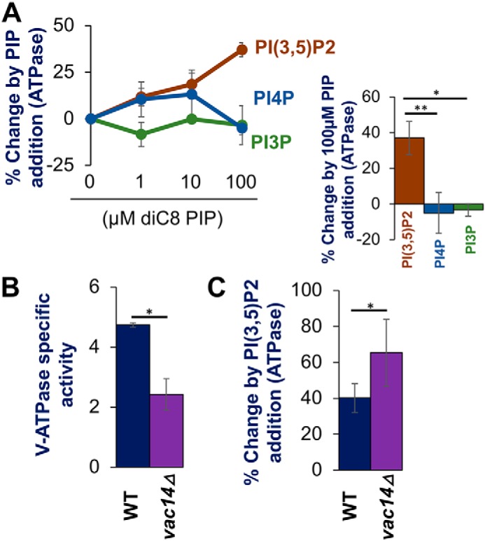
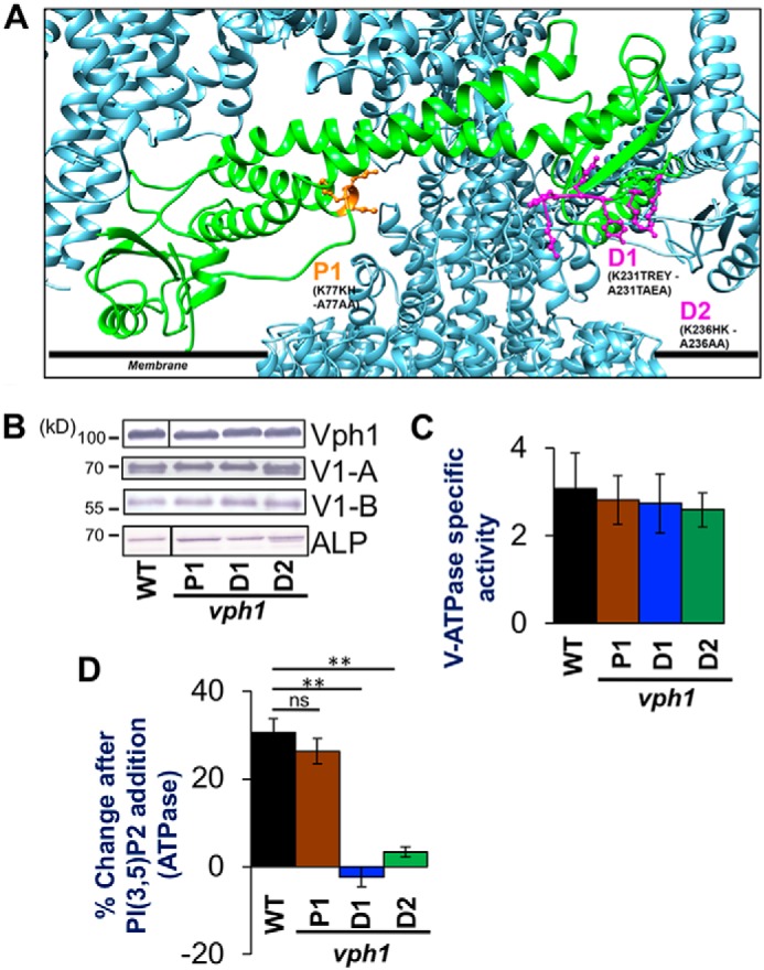
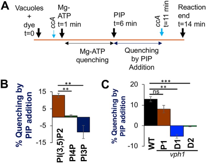
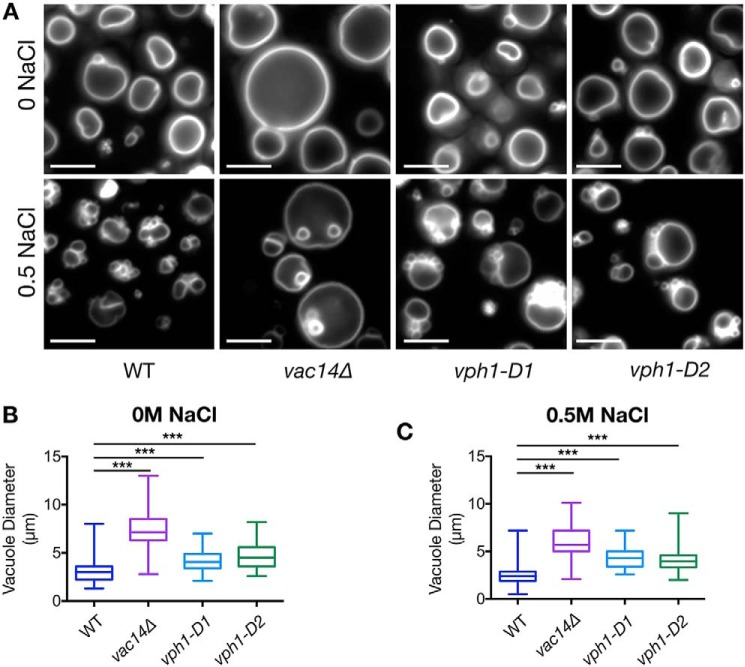
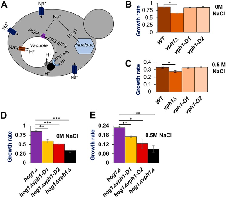
The low-level endo-lysosomal signaling lipid, phosphatidylinositol 3,5-bisphosphate (PI(3,5)P2), is required for full assembly and activity of vacuolar H+-ATPases (V-ATPases) containing the vacuolar a-subunit isoform Vph1 in yeast. The cytosolic N-terminal domain of Vph1 is also recruited to membranes in vivo in a PI(3,5)P2-dependent manner, but it is not known if its interaction with PI(3,5)P2 is direct. Here, using biochemical characterization of isolated yeast vacuolar vesicles, we demonstrate that addition of exogenous short-chain PI(3,5)P2 to Vph1-containing vacuolar vesicles activates V-ATPase activity and proton pumping. Modeling of the cytosolic N-terminal domain of Vph1 identified two membrane-oriented sequences that contain clustered basic amino acids. Substitutions in one of these sequences (231KTREYKHK) abolished the PI(3,5)P2-dependent activation of V-ATPase without affecting basal V-ATPase activity. We also observed that vph1 mutants lacking PI(3,5)P2 activation have enlarged vacuoles relative to those in WT cells. These mutants exhibit a significant synthetic growth defect when combined with deletion of Hog1, a kinase important for signaling the transcriptional response to osmotic stress. The results suggest that PI(3,5)P2 interacts directly with Vph1, and that this interaction both activates V-ATPase activity and protects cells from stress.

Keywords: Saccharomyces cerevisiae; acidification; lysosome; osmoregulation; osmotic stress; phosphatidylinositol 3,5-bisphosphate; phosphatidylinositol signaling; proton pump; vacuolar ATPase; vacuole.


Conflict of interest statement

The authors declare that they have no conflicts of interest with the contents of this article

Figures

Figure 1.
Figure 1.
Exogenous PI(3,5)P2 enhances activity of V-ATPase on vacuolar vesicles isolated from WT and vac14Δ mutant cells. A: left, dose-response curves showing changes in ATPase activity with addition of the indicated concentrations PIP lipids to vacuolar vesicles from WT cells. Right: comparison of change from exogenous addition of 100 μm diC8 PIP lipids. B, V-ATPase–specific activity of WT (black bar) and vac14Δ (purple bar) (in μmol min−1 mg−1) from isolated vacuoles. C, percentage change in V-ATPase activity by exogenous addition of 100 μm diC8 PI(3,5)P2 to vacuolar vesicles isolated from WT (black bar) and vac14Δ (purple bar) yeast. In each case, values represent a mean value from three or more independent experiments. Error bars indicates mean ± S.E. A paired t test was run to analyze significance in differences of means. * indicates p < 0.05, ** indicates p < 0.005, and *** indicates p < 0.0005.
Figure 2.
Figure 2.
PI(3,5)P2 activation mutations in Vph1 eliminates PI(3,5)P2-dependent V-ATPase activation. A, structural model of PHYRE2.0 generated Vph1-NT (green chain) replacing partial Vph1NT in the available cryo-electron microscope generated structure of the yeast V-ATPase (PDB 3J9T) using Matchmaker (21, 39). Polypeptide chains at the V1-Vo interface are colored in metallic blue. The sites on Vph1-NT that are mutated are indicated in ball and stick as P1 (orange), D1 and D2 (both in purple). The residues and their corresponding mutations are written in black letters. Numbers indicate the position of the amino acid in the polypeptide chain. The black line indicates the approximate position of the cytosolic leaflet of the organelle membrane. B, immunoblots of vacuolar protein samples from WT, vph1-P1, vph1-D1, and vph1-D2 yeast strains separated by SDS-PAGE. Immunoblots were probed for the vacuolar proteins indicated on the right. Lines indicate nonadjacent lanes from the same gel. Alkaline phosphatase is used as loading control. C, V-ATPase-specific activity (μmol min−1 mg−1) of WT (black bar), vph1-P1 (brown bar), vph1-D1 (blue bar), and vph1-D2 (green bar) vacuoles isolated from a corresponding strain. D, percentage change in V-ATPase activity by exogenous addition of 100 μm diC8 PI(3,5)P2 to vacuoles isolated from WT (black bar), vph1-P1 (brown bar), vph1-D1 (blue bar), and vph1-D2 (green bar) yeast. In each case, values represent a mean value from three or more independent experiments. Error bars indicate mean ± S.E. A paired t test was run to analyze the significance in differences of mean, where indicated. ns indicates p > 0.05 and ** indicates p < 0.005.
Figure 3.
Figure 3.
PI(3,5)P2 promotes ATP-dependent proton pumping in isolated vacuolar vesicles. A, schematic of acridine orange quenching assay: vacuoles were loaded with the dye for 1 min followed by addition of 0.5 mm ATP and 1 mm MgSO4 (MgATP) (t = 1 min). After allowing an initial fluorescence quenching for 5 min, 10 μm diC8 PIP lipid was added to the reaction (t = 6 min) and fluorescent reading was continued. After 5 min (t = 11 min), the V-ATPase inhibitor ConA (ccA) (thick blue arrow) was added to observe de-quenching of fluorescence and the reaction was ended 3 min later (t = 14 min). ConA (thin blue arrow) was added to test if quenching is V-ATPase dependent before Mg-ATP addition. B, percentage fluorescence quenching on 10 μm diC8 PIP addition to vacuole vesicles, isolated from WT yeast, normalized to Mg-ATP dependent fluorescence quenching: PI(3,5)P2 (brown bar), PI(4)P (green bar), and PI(3)P (dark blue bar). C, percentage of fluorescence quenching on addition of 10 μm diC8 PI(3,5)P2 to vacuoles normalized to MgATP-dependent fluorescence quenching: vacuole vesicles are isolated from WT (black bar), vph1-P1 (brown bar), vph1-D1 (blue bar), and vph1-D2 (green bar) yeast. In each case, values represent a mean value from three independent experiments. Error bars indicate mean ± S.E. A paired t test was run to analyze significance in differences of mean. ns indicates p > 0.05, * indicates p < 0.05, ** indicates p < 0.005, and *** indicates p < 0.0005.
Figure 4.
Figure 4.
Loss of V-ATPase activation by PI(3,5)P2 compromises vacuolar morphology and fragmentation under hypertonic stress. A, fluorescent micrographs of WT, vac14Δ, vph1-D1, and vph1-D2 strains, stained with the vacuolar dye FM4-64. Images of stained cells, either unexposed to NaCl (upper panel) or, briefly exposed to 0.5 m NaCl (lower panel) are presented. White bar on the lower left corner of individual images is a scale bar representing 5 μm dimension. Images are representative of two independent experiments performed using ∼200 cells for each strain on each occasion. B and C, box and whisker plots representing diameters (in μm) of the largest vacuole of yeast cells in the absence (B) and presence of 0.5 m salt (C). The line within the box represent a median value of vacuolar diameter. A paired t test was run to analyze significance in differences of mean. *** indicates p < 0.0005.
Figure 5.
Figure 5.
Synthetic growth defects between vph1-PI(3,5)P2 activation mutations with hog1Δ under hyperosmotic stress. A, cartoon depicts two pathways involved in physiological response of yeast to hyperosmotic stress by Na+ ions: (i) the initial protection pathway that relies on PI(3,5)P2 synthesis and increased assembly and activity of V-ATPase, generating H+ gradient to be utilized for Na+ import to the vacuole via Na+/H+ exchangers. (ii) HOG pathway that relies on activation and nuclear translocation of the mitogen-activated protein kinase Hog1, which generates osmo-protective gene regulation. B and C, growth rate (doublings per hour) of wildtype (WT), vph1Δ, vph1-D1, and vph1-D2 yeast strains in medium containing 0 m NaCl (B) and 0.5 m NaCl (C). D and E, growth rate (doublings per hour) of the indicated strains (labeled below corresponding bars on the x axis) with 0 m NaCl (D) and 0.5 NaCl (E) in the growth media. Mean growth rates are obtained from three independent experiments. Error bars indicate the mean ± S.E. ns indicates p > 0.05, * indicates p < 0.05, ** indicates p < 0.005, and *** indicates p < 0.0005.

References

    1. Kane P. M. (2006) The where, when, and how of organelle acidification by the yeast vacuolar H+-ATPase. Microbiol. Mol. Biol. Rev. 70, 177–191 10.1128/MMBR.70.1.177-191.2006 - DOI - PMC - PubMed
    1. Forgac M. (2007) Vacuolar ATPases: rotary proton pumps in physiology and pathophysiology. Nat. Rev. Mol. Cell Biol. 8, 917–929 10.1038/nrm2272 - DOI - PubMed
    1. Casey J. R., Grinstein S., and Orlowski J. (2010) Sensors and regulators of intracellular pH. Nat. Rev. Mol. Cell Biol. 11, 50–61 10.1038/nrm2820 - DOI - PubMed
    1. Brett C. L., Kallay L., Hua Z., Green R., Chyou A., Zhang Y., Graham T. R., Donowitz M., and Rao R. (2011) Genome-wide analysis reveals the vacuolar pH-stat of Saccharomyces cerevisiae. PLoS ONE 6, e17619 10.1371/journal.pone.0017619 - DOI - PMC - PubMed
    1. Oot R. A., Couoh-Cardel S., Sharma S., Stam N. J., and Wilkens S. (2017) Breaking up and making up: the secret life of the vacuolar H+-ATPase. Protein Sci. 26, 896–909 10.1002/pro.3147 - DOI - PMC - PubMed

Publication types

MeSH terms

Substances

Associated data