Review
. 2019 Apr 5:13:141.
doi: 10.3389/fncel.2019.00141. eCollection 2019.

Circuit Investigations With Open-Source Miniaturized Microscopes: Past, Present and Future

Affiliations
Review

Circuit Investigations With Open-Source Miniaturized Microscopes: Past, Present and Future

Daniel Aharoni et al. Front Cell Neurosci. .

Abstract

The ability to simultaneously image the spatiotemporal activity signatures from many neurons during unrestrained vertebrate behaviors has become possible through the development of miniaturized fluorescence microscopes, or miniscopes, sufficiently light to be carried by small animals such as bats, birds and rodents. Miniscopes have permitted the study of circuits underlying song vocalization, action sequencing, head-direction tuning, spatial memory encoding and sleep to name a few. The foundation for these microscopes has been laid over the last two decades through academic research with some of this work resulting in commercialization. More recently, open-source initiatives have led to an even broader adoption of miniscopes in the neuroscience community. Open-source designs allow for rapid modification and extension of their function, which has resulted in a new generation of miniscopes that now permit wire-free or wireless recording, concurrent electrophysiology and imaging, two-color fluorescence detection, simultaneous optical actuation and read-out as well as wide-field and volumetric light-field imaging. These novel miniscopes will further expand the toolset of those seeking affordable methods to probe neural circuit function during naturalistic behaviors. Here, we will discuss the early development, present use and future potential of miniscopes.

Keywords: 3D printing; behavior; freely moving animals; miniaturization; miniscope; open-source; systems neurobiology.


Figures

Figure 1
Figure 1
Miniaturized one-photon excitation microscope design. (A) A typical design for a miniaturized one-photon excitation microscope used in combination with gradient refractive index (GRIN) lenses. It is comprised of an excitation LED light source, half-ball lens light collimator, excitation filter (Exc. filter) and dichroic mirror for reflecting excitation light down toward the specimen and transmitting emitted fluorescence up to the imaging detector. Emitted light is focused onto a CMOS imaging sensor using an achromatic lens after passing through an emission filter (Em. filter). The use of GRIN lenses permits imaging from both superficial (left) or deep-lying (right) brain structures. Adjustment of the focal plane in the specimen is achieved by moving the image sensor towards or away from the achromatic lens. For superficial imaging, the objective GRIN lens is placed directly on the brain surface, for deep brain imaging the objective GRIN lens is mounted inside the scope and combined with a thinner relay GRIN lens that is implanted into the brain to image from cells transduced with a fluorescent activity reporter (green dots). (B) An open-source first-generation UCLA Miniscope, which is mounted, via a baseplate, on a mouse for the duration of the recording session. Mice carry these 3-g miniscopes without any overt effects on overall behavior, although cabled versions may affect social interactions with other mice. Scale bar ~10 mm.
Figure 2
Figure 2
Open-source miniscopes released in the public domain. (A) FinchScope (https://github.com/gardner-lab/FinchScope), image credit: W.A. Liberti III. (B) miniScope (https://github.com/giovannibarbera/miniscope_v1.0). (C) UCLA Miniscope (http://www.miniscope.org). (D) CHEndoscope (https://github.com/jf-lab/chendoscope), image credit: A. Jacob, Josselyn lab.
Figure 3
Figure 3
Electrowetting Lenses (EWLs). (A) Image of an object brought into focus with an EWL. (B) Commercially available miniature EWLs weigh as little as 0.2 g with an outer diameter of ~6 mm. (C) Example use of an EWL (Varioptic Arctic 25H0) to focus onto a 1951 USAF test target with a ±200 μm focal length shift.
Figure 4
Figure 4
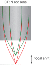
Focal shift caused by chromatic aberrations. Schematic showing focal shift in a GRIN rod lens when imaging two colors.
Figure 5
Figure 5
cScope macroscope developed at Princeton University. Schematic showing the optics used to increase the field-of-view for a head-mounted microscope. Schematic courtesy S.Y. Thiberge, Princeton University.

References

    1. Adams J. K., Boominathan V., Avants B. W., Vercosa D. G., Ye F., Baraniuk R. G., et al. . (2017). Single-frame 3D fluorescence microscopy with ultraminiature lensless FlatScope. Sci. Adv. 3:e1701548. 10.1126/sciadv.1701548 - DOI - PMC - PubMed
    1. Aharoni D., Khakh B. S., Silva A. J., Golshani P. (2019). All the light that we can see: a new era in miniaturized microscopy. Nat. Methods 16, 11–13. 10.1038/s41592-018-0266-x - DOI - PMC - PubMed
    1. Ahrens M. B., Li J. M., Orger M. B., Robson D. N., Schier A. F., Engert F., et al. . (2012). Brain-wide neuronal dynamics during motor adaptation in zebrafish. Nature 485, 471–477. 10.1038/nature11057 - DOI - PMC - PubMed
    1. Antipa N., Kuo G., Heckel R., Mildenhall B., Bostan E., Ng R., et al. (2017). DiffuserCam: lensless single-exposure 3D imaging. arXiv [Preprint] 1710.02134 [cs.CV]. Available online at: http://arxiv.org/abs/1710.02134
    1. Bando Y., Sakamoto M., Kim S., Ayzenshtat I., Yuste R. (2019). Comparative evaluation of genetically encoded voltage indicators. Cell Rep. 26, 802.e4–813.e4. 10.1016/j.celrep.2018.12.088 - DOI - PMC - PubMed