. 2019 Apr 10:10:350.
doi: 10.3389/fphar.2019.00350. eCollection 2019.

Cannabinoid CB1 and CB2 Receptor-Mediated Arrestin Translocation: Species, Subtype, and Agonist-Dependence

Affiliations

Cannabinoid CB1 and CB2 Receptor-Mediated Arrestin Translocation: Species, Subtype, and Agonist-Dependence

Mikkel Søes Ibsen et al. Front Pharmacol. .

Abstract

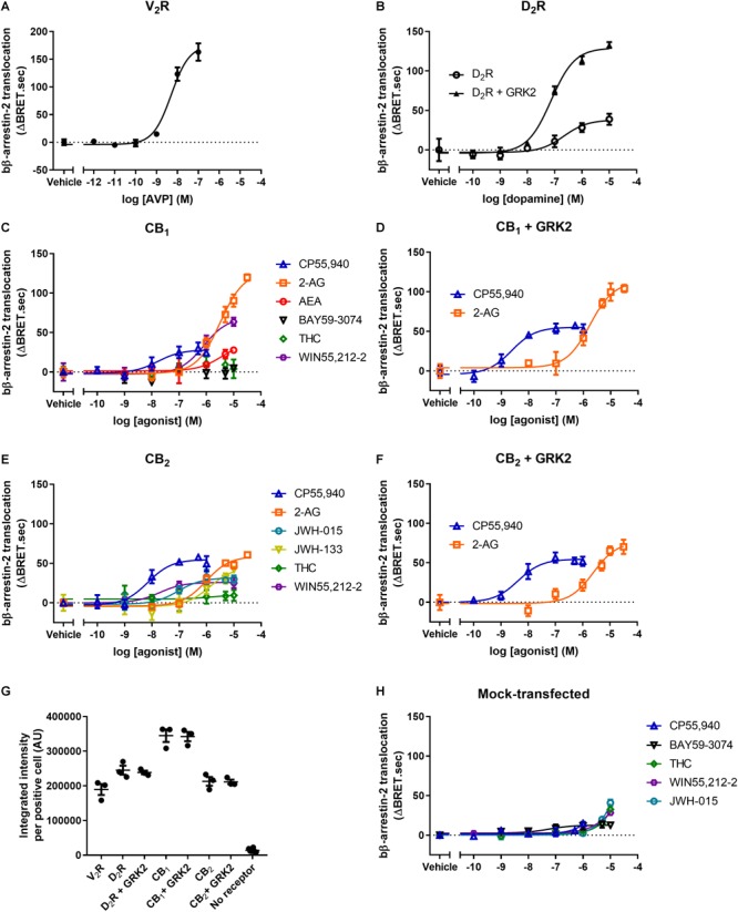
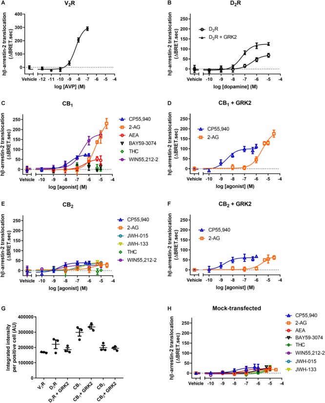
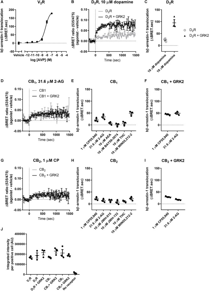
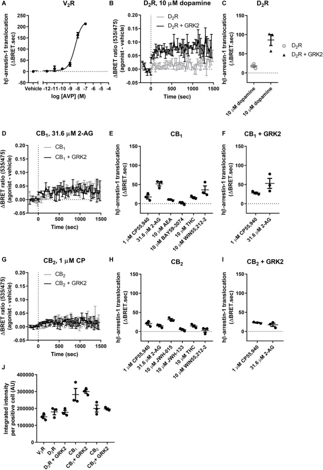
Arrestin translocation and signaling have come to the fore of the G protein-coupled receptor molecular pharmacology field. Some receptor-arrestin interactions are relatively well understood and considered responsible for specific therapeutic or adverse outcomes. Coupling of arrestins with cannabinoid receptors 1 (CB1) and 2 (CB2) has been reported, though the majority of studies have not systematically characterized the differential ligand dependence of this activity. In addition, many prior studies have utilized bovine (rather than human) arrestins, and the most widely applied assays require reporter-tagged receptors, which prevent meaningful comparison between receptor types. We have employed a bioluminescence resonance energy transfer (BRET) method that does not require the use of tagged receptors and thereby allows comparisons of arrestin translocation between receptor types, as well as with cells lacking the receptor of interest - an important control. The ability of a selection of CB1 and CB2 agonists to stimulate cell surface translocation of human and bovine β-arrestin-1 and -2 was assessed. We find that some CB1 ligands induce moderate β-arrestin-2 translocation in comparison with vasopressin V2 receptor (a robust arrestin recruiter); however, CB1 coupling with β-arrestin-1 and CB2 with either arrestin elicited low relative efficacies. A range of efficacies between ligands was evident for both receptors and arrestins. Endocannabinoid 2-arachidonoylglycerol stood out as a high efficacy ligand for translocation of β-arrestin-2 via CB1. Δ9-tetrahydrocannabinol was generally unable to elicit translocation of either arrestin subtype via CB1 or CB2; however, control experiments revealed translocation in cells not expressing CB1/CB2, which may assist in explaining some discrepancy with the literature. Overexpression of GRK2 had modest influence on CB1/CB2-induced arrestin translocation. Results with bovine and human arrestins were largely analogous, but a few instances of inconsistent rank order potencies/efficacies between bovine and human arrestins raise the possibility that subtle differences in receptor conformation stabilized by these ligands manifest in disparate affinities for the two arrestin species, with important potential consequences for interpretation in ligand bias studies. As well as contributing important information regarding CB1/CB2 ligand-dependent arrestin coupling, our study raises a number of points for consideration in the design and interpretation of arrestin recruitment assays.

Keywords: G protein-coupled receptor (GPCR); arrestin; cannabinoid; cannabinoid receptor 1 (CB1); cannabinoid receptor 2 (CB2); signaling; signaling bias; vasopressin.


Figures

FIGURE 1
FIGURE 1
bβ-arrestin-2 translocation to V2R, D2R, CB1, and CB2. bβ-Arrestin-2 translocation to plasma membrane in cells expressing (A) V2R stimulated with AVP, (B) D2R with or without co-expressed GRK2 stimulated with dopamine, (C) CB1 stimulated with CP55,940, 2-AG, AEA, BAY, THC, and WIN55,212-2, (D) CB1 with co-expressed GRK2 stimulated with CP55,940 and 2-AG, (E) CB2 stimulated with CP55,940, 2-AG, JWH-015, JWH-133, THC, and WIN55,212-2, and (F) CB2 with co-expressed GRK2 stimulated with CP55,940 and 2-AG. (G) Receptor expression of V2R, D2R, D2R+GRK2, CB1, CB1+GRK2, CB2, CB2+GRK2, and no-receptor transfected controls in cells used in experiments (A–G). Receptor expression per cell was quantified in cells positive for expression by ICC. (H) bβ-Arrestin-2 translocation to plasma membrane in mock-transfected cells. Error bars are ±standard deviation of representative data with three technical replicates (A–F), or standard error of the means from three independent biological replicates (G,H).
FIGURE 2
FIGURE 2
hβ-arrestin-2 translocation to V2R, D2R, CB1, and CB2. hβ-Arrestin-2 translocation to plasma membrane in cells expressing (A) V2R stimulated with AVP, (B) D2R with or without co-expressed GRK2 stimulated with dopamine, (C) CB1 stimulated with CP55,940, 2-AG, AEA, BAY, THC, and WIN55,212-2, (D) CB1 with co-expressed GRK2 stimulated with CP55,940 and 2-AG, (E) CB2 stimulated with CP55,940, 2-AG, JWH-015, JWH-133, THC, and WIN55,212-2, and (F) CB2 with co-expressed GRK2 stimulated with CP55,940 and 2-AG. (G) Receptor expression of V2R, D2R, D2R+GRK2, CB1, CB1+GRK2, CB2, and CB2+GRK2 in cells used in experiments (A–G). Receptor expression was quantified in cells positive for expression by ICC. (H) hβ-Arrestin-2 translocation to plasma membrane in mock-transfected cells. Error bars are ±standard deviation of representative data with three technical replicates (A–F), or standard error of the means from three independent biological replicates (G,H).
FIGURE 3
FIGURE 3
bβ-Arrestin-1 translocation to V2R, D2R, CB1, and CB2. (A) bβ-Arrestin-1 translocation to V2R stimulated with AVP. (B) ΔBRET ratio curves of bβ-arrestin-1 translocation to D2R with or without co-expressed GRK2 stimulated with 10 μM dopamine. (C) AUC quantification of translocation to D2R with or without co-expressed GRK2 stimulated with 10 μM dopamine as shown in (B). (D) ΔBRET ratio curves of bβ-arrestin-1 translocation to CB1 with or without co-expressed GRK2 stimulated with 31.6 μM 2-AG. (E) AUC of bβ-arrestin-1 translocation to CB1 stimulated with 1 μM CP55,940, 31.6 μM 2-AG, 10 μM AEA, 10 μM BAY, 10 μM THC, and 10 μM WIN55,212-2. (F) AUC of bβ-arrestin-1 translocation to CB1 with co-expressed GRK2 stimulated with 1 μM CP55,940 and 31.6 μM 2-AG. (G) ΔBRET ratio curves of bβ-arrestin-1 translocation to CB2 with or without co-expressed GRK2 stimulated with 1 μM CP55,940. (H) AUC of bβ-arrestin-1 translocation to CB2 stimulated with 1 μM CP55,940, 31.6 μM 2-AG, 10 μM JWH-015, 10 μM JWH-133, 10 μM THC, and 10 μM WIN55,212-2. (I) AUC of bβ-arrestin-1 translocation to CB2 with co-expressed GRK2 stimulated with 1 μM CP55,940 and 31.6 μM 2-AG. (J) Receptor expression of V2R, D2R, D2R+GRK2, CB1, CB1+GRK2, CB2, and CB2+GRK2 in cells used in experiments (A–G). Receptor expression was quantified in cells positive for expression by ICC. Error bars represent ±standard deviation of representative data with three technical replicates (A, B, D, G) or standard error of the means from three independent biological replicates (C, E, F, H, I, J).
FIGURE 4
FIGURE 4
hβ-Arrestin-1 translocation to V2R, D2R, CB1, and CB2. (A) hβ-Arrestin-1 translocation to V2R stimulated with AVP. (B) ΔBRET ratio curves of hβ-arrestin-1 translocation to D2R with or without co-expressed GRK2 stimulated with 10 μM dopamine. (C) AUC quantification of translocation to D2R with or without co-expressed GRK2 stimulated with 10 μM dopamine as shown in B. (D) ΔBRET ratio curves of hβ-arrestin-1 translocation to CB1 with or without co-expressed GRK2 stimulated with 31.6 μM 2-AG. (E) AUC of hβ-arrestin-1 translocation to CB1 stimulated with 1 μM CP55,940, 31.6 μM 2-AG, 10 μM AEA, 10 μM BAY, 10 μM THC, and 10 μM WIN55,212-2. (F) AUC of hβ-arrestin-1 translocation to CB1 with co-expressed GRK2 stimulated with 1 μM CP55,940 and 31.6 μM 2-AG. (G) ΔBRET ratio curves of hβ-arrestin-1 translocation to CB2 with or without co-expressed GRK2 stimulated with 1 μM CP. (H) AUC of hβ-arrestin-1 translocation to CB2 stimulated with 1 μM CP55,940, 31.6 μM 2-AG, 10 μM JWH-015, 10 μM JWH-133, 10 μM THC, and 10 μM WIN55,212-2. (I) AUC of hβ-arrestin-1 translocation to CB2 with co-expressed GRK2 stimulated with 1 μM CP55,940 and 31.6 μM 2-AG. (J) Receptor expression of V2R, D2R, D2R+GRK2, CB1, CB1+GRK2, CB2, and CB2+GRK2 in cells used in experiments (A–G). Receptor expression was quantified in cells positive for expression by ICC. Error bars represent ±standard deviation of representative data with three technical replicates (A, B, D, G) or standard error of the means from three independent biological replicates (C, E, F, H, I, J).

References

    1. Armstrong S. P., Seeber R. M., Ayoub M. A., Feldman B. J., Pfleger K. D. G. (2013). Characterization of three vasopressin receptor 2 variants: an apparent polymorphism (V266A) and Two Loss-of-Function Mutations (R181C and M311V). PLoS One 8:e65885. 10.1371/journal.pone.0065885 - DOI - PMC - PubMed
    1. Atwood B. K., Lopez J., Wager-Miller J., Mackie K., Straiker A. (2011). Expression of G protein-coupled receptors and related proteins in HEK293, AtT20, BV2, and N18 cell lines as revealed by microarray analysis. BMC Genomics 12:14. 10.1186/1471-2164-12-14 - DOI - PMC - PubMed
    1. Atwood B. K., Wager-Miller J., Haskins C., Straiker A., Mackie K. (2012). Functional selectivity in CB(2) cannabinoid receptor signaling and regulation: implications for the therapeutic potential of CB(2) ligands. Mol. Pharmacol. 81 250–263. 10.1124/mol.111.074013 - DOI - PMC - PubMed
    1. Bakshi K., Mercier R. W., Pavlopoulos S. (2007). Interaction of a fragment of the cannabinoid CB1 receptor C-terminus with arrestin-2. Febs Lett. 581 5009–5016. 10.1016/j.febsiet.2007.09.030 - DOI - PMC - PubMed
    1. Bash R., Rubovitch V., Gafni M., Sarne Y. (2003). The stimulatory effect of cannabinoids on calcium uptake is mediated by Gs GTP-binding proteins and cAMP formation. Neurosignals 12 39–44. 10.1159/000068915 - DOI - PubMed