. 2019 Apr 9:10:365.
doi: 10.3389/fphar.2019.00365. eCollection 2019.

Duloxetine, a Balanced Serotonin-Norepinephrine Reuptake Inhibitor, Improves Painful Chemotherapy-Induced Peripheral Neuropathy by Inhibiting Activation of p38 MAPK and NF-κB

Affiliations

Duloxetine, a Balanced Serotonin-Norepinephrine Reuptake Inhibitor, Improves Painful Chemotherapy-Induced Peripheral Neuropathy by Inhibiting Activation of p38 MAPK and NF-κB

Jing Meng et al. Front Pharmacol. .

Abstract

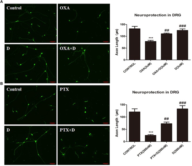
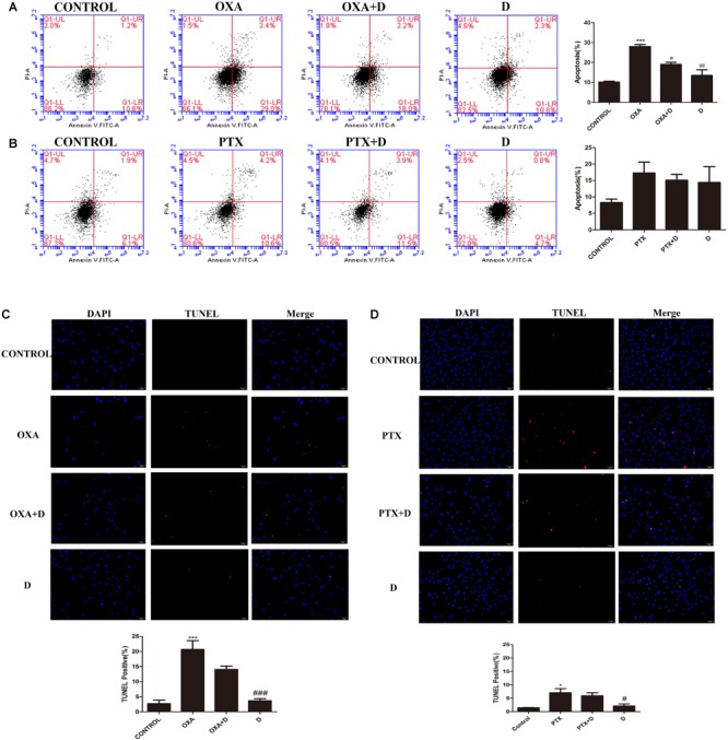
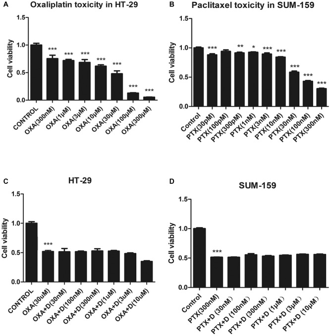
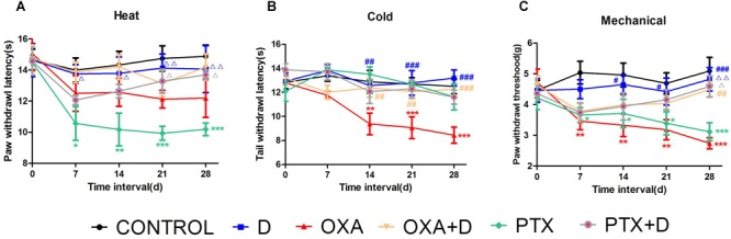
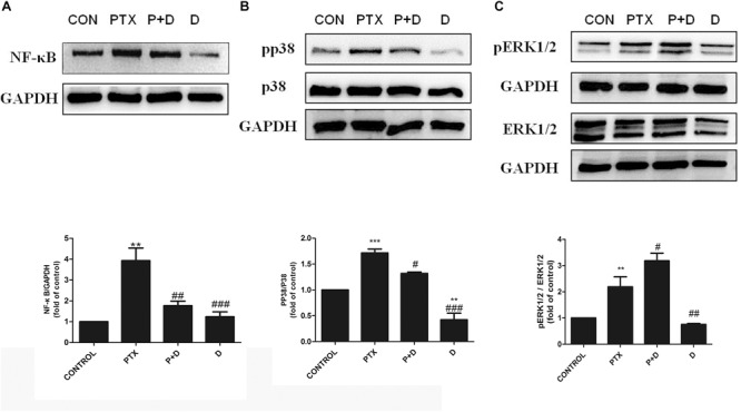
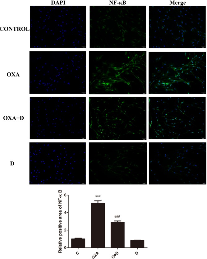
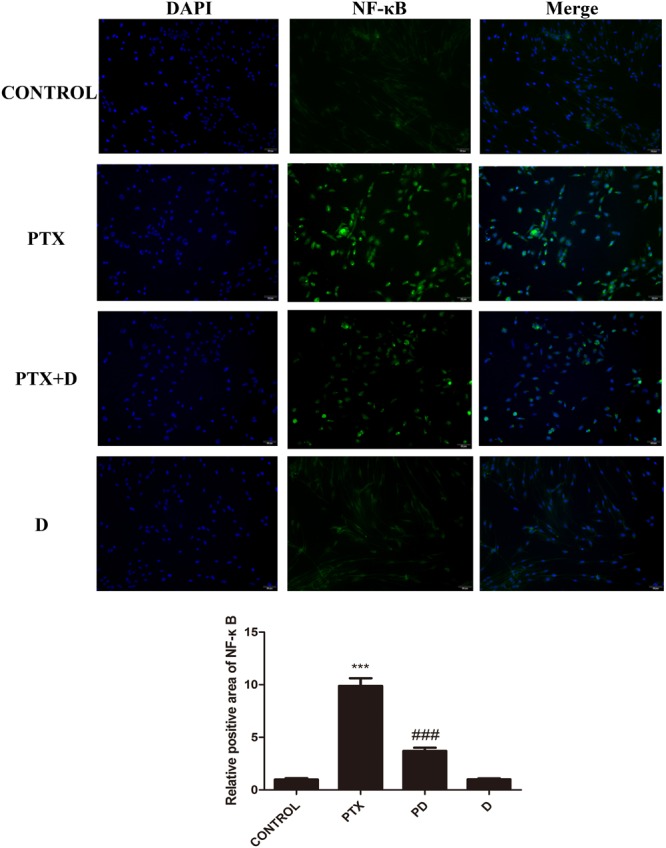
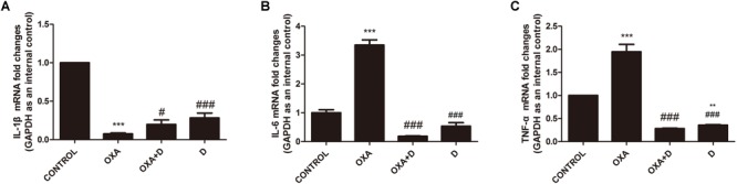
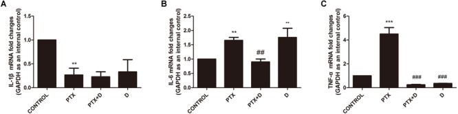
Chemotherapy-induced peripheral neuropathy (CIPN) is a severe, toxic side effect that frequently occurs in anticancer treatment and may result in discontinuation of treatment as well as a serious reduction in life quality. The CIPN incidence rate is as high as 85-90%. Unfortunately, there is currently no standard evidence-based CIPN treatment. In several clinical trials, it has been reported that duloxetine can improve CIPN pain induced by oxaliplatin (OXA) and paclitaxel (PTX); thus, The American Society of Clinical Oncology (ASCO) recommends duloxetine as the only potential treatment for CIPN. However, this guidance lacks the support of sufficient evidence. Our study shows that duloxetine markedly reduces neuropathic pain evoked by OXA or PTX. Duloxetine acts by inhibiting the activation of p38 phosphorylation, thus preventing the activation and nuclear translocation of the NF-κB transcription factor, reducing the inflammatory response and inhibiting nerve injury by regulating nerve growth factor (NGF). Furthermore, in this study, it is shown that duloxetine does not affect the antitumor activity of OXA or PTX. This study not only provides biological evidence to support the use of duloxetine as the first standard CIPN drug but will also lead to potential new targets for CIPN drug development.

Keywords: chemotherapy-induced peripheral neewopathy (CIPN); dorsal root ganglia (DRG); duloxetine; eripheral neuropathic pain; oxaliplatin (OXA); paclitaxel (PTX).


Figures

FIGURE 1
FIGURE 1
Effect of duloxetine on axonal injury caused by oxaliplatin or paclitaxel (A) Primary rat DRG neurons were grown and axons were extended for 24 h. Neurons were then treated with OXA (3 μM) or duloxetine (3 μM) for 48 h. Cells were then subjected to immunofluorescence staining, and axon length measurements were performed (∗∗∗p < 0.001 vs. control; ##p < 0.01; ###p < 0.001 vs. oxaliplatin alone). (B) Primary rat DRG neuronal cells were grown and axons were extended for 24 h. Neurons were then treated with PTX (300 nM) or duloxetine (300 nM) for 24 h. Cells were then subjected to immunofluorescence staining, and axon length measurements were performed (∗∗∗p < 0.001 vs. control; ##p < 0.01; ###p < 0.001 vs. paclitaxel alone). The results are expressed as the mean ± SEM (n = 10).
FIGURE 2
FIGURE 2
Effect of duloxetine on DRG neuronal apoptosis through flow cytometry and TUNEL assay. After growing in culture for 24 h, DRG neuronal cells were exposed to OXA (3 μM) with (or without) duloxetine (3 μM) for another 48 h (A,C) or exposed to PTX (300 nM) with (or without) duloxetine (300 nM) for another 24 h (B,D). (A,B):Cells were then double-stained with annexin V-FITC/PI. Annexin V-FITC fluorescence was measured with the FL1 channel, and PI fluorescence was measured with the FL3 channel. Representative pictures are from one of three independent experiments with similar results. (C,D) The cell death of DRG was examined by TUNEL assay. (p < 0.05, ∗∗∗p < 0.001 compared with control; #p < 0.05, ##p < 0.01, ###p < 0.001 compared with model). The data are presented as mean ± SEM. Representative pictures are from one of three independent experiments with similar results (×200).
FIGURE 3
FIGURE 3
Effect of duloxetine on the anticancer activity of OXA or PTX in vitro. (A,B) When HT-29 or SUM-159 cancer cells were grown in medium, OXA or PTX reduced their cell viability by 20–80%. (C,D) Various concentrations of duloxetine together with OXA or PTX showed no significant changes in cell viability when compared to treatments with OXA or PTX alone. (p < 0.05, ∗∗p < 0.01,∗∗∗p < 0.001 vs. control). All the data are the mean ± SEM for each experiment (n = 4).
FIGURE 4
FIGURE 4
Partial prevention by duloxetine against OXA or PTX-induced peripheral neuropathy in ICR mice. (A) The effect of duloxetine on the heat withdrawal latency in mice treated with vehicle or chemotherapeutic drugs (OXA and PTX) was evaluated. (B) The effect of duloxetine on the cold threshold in mice treated with vehicle or chemotherapeutic drugs (OXA and PTX) was determined. (C) The effect of duloxetine on the mechanical withdrawal threshold in mice treated with vehicle or chemotherapeutic drugs (OXA and PTX) was assessed. (p < 0.05, ∗∗p < 0.01,∗∗∗p < 0.001 vs. control; #p < 0.05,##p < 0.01, ###p < 0.001 vs. OXA; Δp < 0.05, ΔΔp < 0.01 vs. PTX). The data are presented as the mean ± SEM (n = 5–8).
FIGURE 5
FIGURE 5
Effect of duloxetine on IENF retraction induced by OXA or PTX. Paw biopsies were obtained from the hind paws of mice when behavior tests were finished. Tissues were fixed and stained with antibodies (PGP9.5) for IENFs. (A) Representative images from six groups are shown. (B) IENF density = number of nerve fibers crossing the basement membrane/length of the basement membrane (mm). (Magnification × 400. p < 0.05,∗∗p < 0.01,∗∗∗p < 0.001). The data are presented as the mean ± SEM (n = 5).
FIGURE 6
FIGURE 6
Effect of duloxetine on phosphorylation of p38 MAPK and ERK1/2 expression in the neuropathic mouse DRG following oxaliplatin treatment. (A) Duloxetine significantly decreased the expression of NF-κB protein in the DRG of OXA-treated mice. (B) Duloxetine significantly decreased the expression of pp38 protein in the DRG of OXA-treated mice. (C) The ratio of pERK1/2 to ERK1/2 expression was not changed significantly following oxaliplatin treatment and duloxetine did not modify p-ERK1/2 expression. (∗∗p < 0.01 vs. control, ##p < 0.01, ###p < 0.001 vs. OXA). The data are presented as the mean ± SEM (n = 4).
FIGURE 7
FIGURE 7
Effect of duloxetine on NF-κB, phosphorylation of p38 MAPK and ERK1/2 expression in the neuropathic mouse DRG following PTX treatment. (A) Duloxetine significantly decreased the expression of NF-κB protein in the DRG of PTX-treated mice. (B) Duloxetine significantly decreased the expression of pp38 protein in the DRG of PTX-treated mice. (C) The ratio of pERK1/2 to ERK1/2 expression was significantly increased after PTX treatment, but duloxetine did not modify the p-ERK1/2 expression induced by PTX. (∗∗p < 0.01 vs. control, ∗∗∗p < 0.001 vs. control, #p < 0.05, ##p < 0.01, ###p < 0.001 vs. PTX). The data are presented as the mean ± SEM (n = 4).
FIGURE 8
FIGURE 8
Effect of oxaliplatin and duloxetine on the expression of nuclear NF-κB in DRG neuronal cells. DRG neuronal cells were treated with (or without) OXA (3 μM) and duloxetine (3 μM) for 48 h and were double-stained with DAPI and NF-κB. The fluorescence intensity was observed using fluorescence microscopy (×200). (∗∗∗p < 0.001 vs. control; ###p < 0.001 vs. OXA).
FIGURE 9
FIGURE 9
Effect of paclitaxel and duloxetine on the expression of nuclear NF-κB in DRG neuronal cells. DRG neuronal cells were treated with (or without) PTX (300 nM) and duloxetine (300 nM) for 24 h and were double-stained with DAPI and NF-κB. The fluorescence intensity was observed using fluorescence microscopy (×200). (∗∗∗p < 0.001 vs. control; ###p < 0.001 vs. PTX).
FIGURE 10
FIGURE 10
Transcript levels of the proinflammatory cytokines IL-1β, IL-6 and TNF-α in the mouse DRG. Total mRNA was extracted from mouse DRG tissues. qPCR was performed and a comparison is shown between different groups after saline or 28 days of drug treatment. (A) IL-1β expression was significantly decreased after OXA treatment, but duloxetine did not affect this decrease. (B) Duloxetine significantly decreased the expression of IL-6 mRNA in the DRG of OXA-treated mice. (C) Duloxetine significantly decreased the expression of TNF-α mRNA in the DRG of OXA-treated mice. (∗∗p < 0.01, ∗∗∗p < 0.001 vs. control; #p < 0.05 vs. OXA, ###p < 0.001 vs. OXA). The data are presented as the means ± SEM (n = 5).
FIGURE 11
FIGURE 11
Transcript levels of the proinflammatory cytokines IL-1β, IL-6 and TNF-α in the mouse DRG. Total mRNA was extracted from mouse DRG tissues. qPCR was performed and a comparison is shown between different groups after saline or 28 days of drug treatment. (A) IL-1β expression was significantly decreased after PTX treatment, but duloxetine did not alter this decrease. (B) Duloxetine significantly decreased the expression of IL-6 mRNA in the DRG of PTX-treated mice. (C) Duloxetine significantly decreased the expression of TNF-α mRNA in the DRG of PTX-treated mice. (∗∗p < 0.01, ∗∗∗p < 0.001 vs. control; ##p < 0.01, ###p < 0.001 vs. PTX). The data are presented as the mean ± SEM (n = 5).

References

    1. Apfel S. C. (2002). Nerve growth factor for the treatment of diabetic neuropathy: what went wrong, what went right, and what does the future hold? Int. Rev. Neurobiol. 50 393–413. 10.1016/S0074-7742(02)50083-0 - DOI - PubMed
    1. Apfel S. C., Arezzo J. C., Brownlee M., Federoff H., Kessler J. A. (1994). Nerve growth factor administration protects against experimental diabetic sensory neuropathy. Brain Res. 634 7–12. 10.1016/0006-8993(94)90252-6 - DOI - PubMed
    1. Areti A., Komirishetty P., Akuthota M., Malik R. A., Kumar A. (2017). Melatonin prevents mitochondrial dysfunction and promotes neuroprotection by inducing autophagy during oxaliplatin-evoked peripheral neuropathy. J. Pineal Res. 62:12393. 10.1111/jpi.12393 - DOI - PubMed
    1. Austin P. J., Moalem-Taylor G. (2010). The neuro-immune balance in neuropathic pain: involvement of inflammatory immune cells, immune-like glial cells and cytokines. J. Neuroimmunl. 229 26–50. 10.1016/j.jneuroim.2010.08.013 - DOI - PubMed
    1. Bennett G. J., Doyle T., Salvemini D. (2014). Mitotoxicity in distal symmetrical sensory peripheral neuropathies. Nat. Rev. Neurol. 10 326–336. 10.1038/nrneurol.2014.77 - DOI - PMC - PubMed