. 2019 Apr 10:10:744.
doi: 10.3389/fimmu.2019.00744. eCollection 2019.

Kctd9 Deficiency Impairs Natural Killer Cell Development and Effector Function

Affiliations

Kctd9 Deficiency Impairs Natural Killer Cell Development and Effector Function

Xiaoping Zhang et al. Front Immunol. .

Abstract

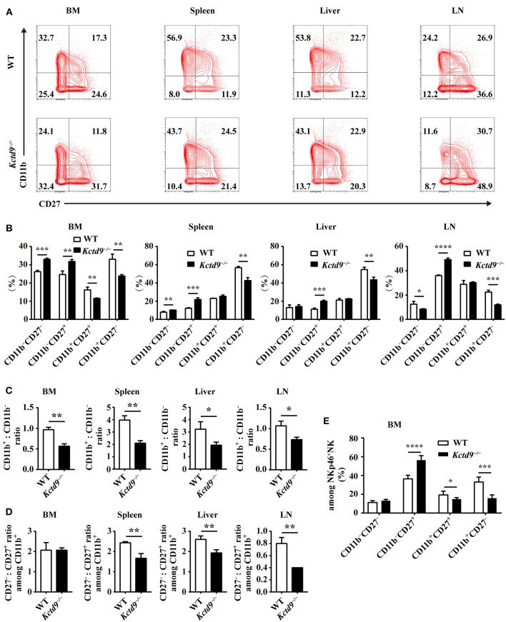
We previously showed that potassium channel tetramerization domain containing 9 (KCTD9) is aberrantly expressed in natural killer (NK) cells in patients with hepatitis B virus-associated acute-on-chronic liver failure and mice with experimental fulminant hepatitis. However, the mechanism underlying the regulation of NK cell function and fulminant hepatitis progression by KCTD9 is unknown. Here, we investigated the role of Kctd9 in regulation of early development, maturation, and function of NK cells using Kctd9-knockout mice. Compared to wild-type mice, Kctd9-deficient mice exhibited impaired NK cell lineage commitment, as evidenced by selective reduction in the refined NK progenitors, and incomplete NK cell maturation, as manifested by a higher proportion of CD11b- NK cells and a lower percentage of CD11b+ NK cells with high proliferative potential. Moreover, Kctd9-depleted NK cells displayed insufficient IFN-γ production, degranulation, and granzyme B production in response to cytokine stimulation, and attenuated cytotoxicity to tumor cells in vitro. The defect in NK cells was further supported by ameliorated liver damage and improved survival in Kctd9-deficient mice following murine hepatitis virus strain-3 (MHV-3) infection, which otherwise leads to immune-mediated fulminant hepatitis, a phenotype homologous to that caused by NK cell depletion in wild-type mice. Further investigation to identify the underlying mechanism revealed that Kctd9 deficiency hindered the expression of transcription factors, including Ets1, Nfil3, Eomes, and Id2 in NK cells. Collectively, our data reveal that Kctd9 acts as a novel regulator for NK cell commitment, maturation, and effector function.

Keywords: Kctd9; NK cells; development; fulminant hepatitis; liver damage.


Figures

Figure 1
Figure 1
Kctd9 deficiency ameliorated liver damage following MHV-3 infection. Wild-type (WT) and Kctd9−/− mice were injected intraperitoneally with MHV-3. (A) Serum alanine transaminase (ALT) and aspartate transaminase (AST) levels in WT and Kctd9−/− mice 24 h after MHV-3 infection. (B) Survival curve of WT and Kctd9−/− mice after MHV-3 infection. (C,D) Expression of Granzyme B (C) and IFN-γ (D) by CD3NKp46+DX5+ NK cells in liver from WT and Kctd9−/− mice 48 h after MHV-3 infection. (a) Flow cytometric analysis for Granzyme B (C) and IFN-γ (D). Plots represent CD3NKp46+DX5+ NK cells, and frequency of gated population is indicated. (b,c) Summary of percentage and intensity of expression of CD3NKp46+DX5+ NK cells positive for Granzyme B (C) and IFN-γ (D). All results were representative of three independent experiments. Ten mice of each genotype were used for measurement of ALT/AST levels, 15 Kctd9−/−mice and 20-21 WT mice were used for survival experiment, and 4–6 mice of each genotype were used for detection of Granzyme B and IFN-γ in each experiment. Comparison of survival curves: Log-rank (Mantel-Cox) test P = 0.0069, Gehan-Breslow-Wilcoxon test P = 0.0084; the median survival time: KO: WT 82 h vs. 76.5 h; the survival rate: KO: WT (1/15) vs. 0. Error bars indicate standard deviation. **P < 0.01, ***P < 0.001, and ****P < 0.0001.
Figure 2
Figure 2
Kctd9 selectively specifies rNKPs during NK cell commitment. (A,C) Flow cytometry gating strategies for identification of NK progenitors in BM from WT and Kctd9−/− mice. (A) Using CD135 and CD122 to identify CLP (CD135+CD122), pre-NKP (CD135CD122), and rNKP (CD135CD122+) among lineage (CD3/CD19/CD11b/Ly6d/DX5)CD27+CD244+CD127+ cells. (C) Using DX5 and CD11b to identify NKP (DX5CD11b), CD11b NK (DX5+CD11b), and CD11b+ NK (DX5+CD11b+) cells among lineage (CD3/CD19/CD4/CD8/Ter119)CD122+ cells. Frequency of gated population is indicated. (B,D) The total number of CLP, pre-NKP and rNKP (B) or NKP, CD11b NK, and CD11b+ NK cells (D) in BM. (E) The total number of CD3CD122+NKp46+ NK cells in the BM, spleen, liver, and mesenteric lymph node (LN) of WT and Kctd9−/− mice. All results were representative of three independent experiments. Six–eight mice of each genotype were used in each experiment. Error bars indicate standard deviation. *P < 0.05, **P < 0.01, and ***P < 0.001.
Figure 3
Figure 3
Kctd9 is required for NK cells maturation. (A) Flow cytometric analysis for expression of CD11b and CD27 by CD3DX5+ cells in four organs from WT and Kctd9−/− mice. Numbers in the quadrants indicate the percentage of subsets defined by CD11b/CD27 expression in CD3DX5+ cells. (B) Summary of the percentages of subsets in CD3DX5+ cells. (C) The ratio of the CD11b+ subset to the CD11b subset among CD3DX5+ cells in indicated organs. (D) The ratio of the CD27 subset to the CD27+ subset among CD3DX5+CD11b+ cells in indicated organs. (E) The percentages of subsets defined by CD11b/CD27 expression in CD3NKp46+ NK cells in BM from WT and Kctd9−/− mice. All results were representative of three independent experiments. Four to six mice of each genotype were used in each experiment. Error bars indicate standard deviation. *P < 0.05, **P < 0.01, ***P < 0.001, and ****P < 0.0001.
Figure 4
Figure 4
Kctd9 restricts the proliferation of NK cells. (A–D) Expression of Ki67 in NK cells and NK subsets in BM and spleen from WT and Kctd9−/− mice. (A,C) Representative flow cytometric analysis for Ki67 expression in BM (A) and spleen (C). Flow cytometric plots represent total CD3DX5+ NK cells (left), CD11bCD3DX5+ NK subset (middle), and CD11b+CD3DX5+ NK subset (right). Frequency of gated population is indicated. (B,D) Summary of the percentages of CD3DX5+ NK cells, CD11bCD3DX5+ subset, and CD11b+CD3DX5+ subset positive for Ki67 in indicated organs. (E) Flow cytometric analysis for CFSE concentration in purified splenic NK cells and CD11b NK subset and CD11b+ NK subset 72 h after initiation of culture in the presence of IL-15. (F) Summary of the percentages of NK cells and CD11b NK subset and CD11b+ NK subset with reduced concentration of CFSE. All results were representative of three independent experiments. Four mice of each genotype were used in each experiment. Error bars indicate standard deviation. *P < 0.05, **P < 0.01, ***P < 0.001, and ****P < 0.0001.
Figure 5
Figure 5
Kctd9 deficiency impairs NK cell effector functions. (A–C) Expression of IFN-γ, Granzyme B by NK cells and degranulation of NK cells from spleen of WT and Kctd9−/− mice. in vitro cell activation is described in Materials and Methods. (a) Flow cytometric analysis for expression of IFN-γ, Granzyme B, and CD107a by CD3DX5+ NK cells. Plots represent total CD3DX5+ NK cells. Frequency of gated population is indicated in plots. (b) The percentage of CD3DX5+ NK cells positive for IFN-γ (A), Granzyme B (B), and CD107a (C). (c) The expression intensity of IFN-γ (A), Granzyme B (B), and CD107a (C) of gated CD3DX5+ NK cells. (D) Apoptosis of Yac-1 cells after being co-cultured with purified NK cells from spleen of WT and Kctd9−/− mice. (a) Flow cytometric analysis for Annexin V on Yac-1 cells. Plots represent CFSE+ Yac-1 cells. (b) Summary of the percentage of apoptotic Yac-1 cells positive for Annexin V. All results were representative of three independent experiments. Four to six mice of each genotype were used in each experiment. Error bars indicate standard deviation. *p < 0.05, **p < 0.01.
Figure 6
Figure 6
Kctd9 depletion downregulated NK cell-related transcription factor expression. (A) Real-time PCR analysis for Ets1, Nfil3 (encodes E4bp4), Eomes, Tbx21 (encodes T-bet), Id2, and Tox expression by purified NK cells from spleen of WT and Kctd9−/− mice. (B) Expression of Eomes by rNKPs in BM from WT and Kctd9−/− mice. (a) Flow cytometric analysis for Eomes in rNKPs. Plots represent rNKPs. (b) Intensity of Eomes expression in total rNKPs. (C,D) Expression of Eomes by CD3NKp46+ NK cells (C) and subsets defined by CD11b/CD27 expression in CD3NKp46+ NK cells (D) from WT and Kctd9−/− BM. (a) Flow cytometric analysis for expression of Eomes. Plots represent CD3NKp46+ NK cells (C) or subsets of CD3NKp46+ NK cells (D). Frequency of gated population is indicated in plots. (b,c) The percentages and the intensity of Eomes expression of CD3NKp46+ NK cells (C) and subsets of CD3NKp46+ NK cells (D) positive for Eomes. All results were representative of three independent experiments. Four to six mice of each genotype were used in each experiment. Error bars indicate standard deviation. *P < 0.05, **p < 0.01.

References

    1. Geiger TL, Sun JC. Development and maturation of natural killer cells. Curr Opin Immunol. (2016) 39:82–9. 10.1016/j.coi.2016.01.007 - DOI - PMC - PubMed
    1. Rosmaraki EE, Douagi I, Roth C, Colucci F, Cumano A, Di Santo JP. Identification of committed NK cell progenitors in adult murine bone marrow. Eur J Immunol. (2001) 31:1900–9. 10.1002/1521-4141(200106)31:6<60;1900::AID-IMMU1900>62;3.0.CO;2-M - DOI - PubMed
    1. Carotta S, Pang SH, Nutt SL, Belz GT. Identification of the earliest NK-cell precursor in the mouse BM. Blood. (2011) 117:5449–52. 10.1182/blood-2010-11-318956 - DOI - PubMed
    1. Fathman JW, Bhattacharya D, Inlay MA, Seita J, Karsunky H, Weissman IL. Identification of the earliest natural killer cell-committed progenitor in murine bone marrow. Blood. (2011) 118:5439–47. 10.1182/blood-2011-04-348912 - DOI - PMC - PubMed
    1. Kim S, Iizuka K, Kang HS, Dokun A, French AR, Greco S, et al. . In vivo developmental stages in murine natural killer cell maturation. Nat Immunol. (2002) 3:523–8. 10.1038/ni796 - DOI - PubMed

Publication types

MeSH terms