. 2019 Apr 16:7:e6734.
doi: 10.7717/peerj.6734. eCollection 2019.

Characterization of the iPSC-derived conditioned medium that promotes the growth of bovine corneal endothelial cells

Affiliations

Characterization of the iPSC-derived conditioned medium that promotes the growth of bovine corneal endothelial cells

Qing Liu et al. PeerJ. .

Abstract

Corneal endothelial cells (CECs) maintain corneal transparency and visual acuity. However, the limited proliferative capability of these cells in vitro has prompted researchers to find efficient culturing techniques for them. The aim of our study was to evaluate the use of conditioned medium (CM) obtained from induced pluripotent stem cells (iPSCs) as a source for the effective proliferation of bovine CECs (B-CECs). In our study, the proliferative ability of B-CECs was moderately enhanced when the cells were grown in 25% iPSC conditioned medium (iPSC-CM). Additionally, hexagonal cell morphology was maintained until passage 4, as opposed to the irregular and enlarged shape observed in control corneal endothelial medium (CEM). B-CECs in both the 25% iPSC-CM and CEM groups expressed and Na+-K+-ATPase. The gene expression levels of NIFK, Na+-K+-ATPase, Col4A and Col8A and the percentage of cells entering S and G2 phases were higher in the iPSC-CM group. The number of apoptotic cells also decreased in the iPSC-CM group. In comparison to the control cultures, iPSC-CM facilitated cell migration, and these cells showed better barrier functions after several passages. The mechanism of cell proliferation mediated by iPSC-CM was also investigated, and phosphorylation of Akt was observed in B-CECs after exposure to iPSC-CM and showed sustained phosphorylation induced for up to 180 min in iPSC-CM. Our findings indicate that iPSC-CM may employ PI3-kinase signaling in regulating cell cycle progression, which can lead to enhanced cellular proliferation. Effective component analysis of the CM showed that in the iPSC-CM group, the expression of activin-A was significantly increased. If activin-A is added as a supplement, it could help to maintain the morphology of the cells, similar to that of CM. Hence, we conclude that activin-A is one of the effective components of CM in promoting cell proliferation and maintaining cell morphology.

Keywords: Conditioned medium; Corneal endothelial cells (CECs); Induction pluripotent stem cells.


Conflict of interest statement

Jiansu Chen and Zekai Cui are employed by Aier Eye Institute, Changsha, Hunan, China. The authors declare that they have no competing interests.

Figures

Figure 1
Figure 1. The proliferation of B-CECs treated with iPSC-CM was detected by CCK-8 analysis and live cell stain.
(A) Cell number in three different mediums. The B-CECs were seeded at a density of 1 × 10³ cells/well in 96-well plates. The CEM, iPSC-CM, and iPSC-M medium were added to passage 1 cells. The data are expressed as the mean ± SEM (n = 6). The results showed that the 25% iPSC-CM group had the best proliferation and had statistically significant differences to the other groups (*P < 0.05 was considered statistically significant; versus CEM group, n = 6). (B) The live cell count of the difference in the 25% iPSC-CM group was statistically significant (*P < 0.01 was considered significantly significant, versus CEM group, n = 3). Primary B-CECs live cell stain in CEM (C) and in 25% iPSC-CM (D) under phase-contrast microscopy. Scale bar: 200 μm.
Figure 2
Figure 2. The maintenance of CEC phenotype after treatment with iPSC-CM.
Morphological change of B-CECs in P0 (A, D), P2 (B, E), and P4 (C, F) under phase-contrast microscopy. Scale bar: 200 μm. (G–J) The protein expression of Na+-K+-ATPase and AQP1 by immunocytochemistry. Scale bar: 100 μm. Immunofluorescence staining showed that the 25% iPSC-CM group and the CEM group both expressed the same endothelial marker proteins, Na+/K+-ATPase, and AQP1. (K) The mRNA expression of ATP1A1, Col4A, Col8A, and NIFK by qPCR. The qPCR showed that the 25% iPSC-CM group had a higher expression than the CEM group, both in the endothelial marker genes ATP1A1, Col4A, Col8A and in the proliferation-related gene NIFK, and the difference was statistically significant (*P < 0.05 was considered statistically significant; versus CEM group, n = 3). Scale bar: (A) 100 μm; (B) 50 μm.
Figure 3
Figure 3. iPSC-CM treatment promoted antiapoptotic activity in cultured B-CECs.
(A) Cell cycle analysis of B-CECs between the 25% iPSC-CM group and CEM group. (B) Quantification of the cell cycle assays showed the percentage of cells entering the S and G2 phases were higher in 25% iPSC-CM than in CEM. (C) Annexin V and PI assay of the B-CECs in the 25% iPSC-CM group and CEM group. The Q1 area represents cell necrosis; Q2 represents late-apoptotic cells; Q3 represents viable cells; and Q4 represents early apoptotic cells. (D) Quantification of the Annexin V and PI assays. The percentage of apoptotic cells was calculated from Q2 and Q4. The number of apoptotic cells decreased in the 25% iPSC-CM group, which showed a statistically significant difference (*P < 0.05 was considered statistically significant; versus CEM group, n = 3).
Figure 4
Figure 4. Effect of iPSC-CM on the migration of B-CECs.
Wound healing was shown for cultured B-CECs in the 25% iPSC-CM group (B) and CEM group (A) (at 0, 9, and 24 h) under phase-contrast microscopy. (C) The percentage of wound areas were determined with ImageJ analysis software. A P-value of <0.05 was considered to be statistically significant (*P < 0.05 versus CEM group). Scale bar: 100 μm.
Figure 5
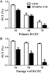
Figure 5. The percentage of fluorescein leakage was determined with ELISA.
(A) Quantification of FLK assays at P0 showed no significant difference (P > 0.05, n = 3) between the 25% iPSC-CM group and CEM group (at 3, 5, and 7 days). (B) Quantification of FLK assays at P4 showed a significant difference (*P < 0.001 versus CEM group, n = 3) between the 25% iPSC-CM group and CEM group (at 3, 5, and 7 days).
Figure 6
Figure 6. The effect of PI3-kinase in iPSC-CM on promoting B-CEC proliferation.
(A) Western blotting showed the pAkt levels in the B-CECs cultured in either 25% iPSC-CM or CEM for 30 min and 180 min, after starvation treatment. (B) Quantification of the western blot assays showed that the 25% iPSC-CM group had higher pAkt expression than the control group (at 30 and 180 min), and the difference was statistically significant (*P < 0.05 versus CEM group, n = 3). (C) ELISA detection showed that activin A expression was significantly higher in iPSC-CM medium than in normal iPSC medium, and the difference was statistically significant (*P < 0.01 versus CEM group, n = 3).
Figure 7
Figure 7. Morphology change of B-CECs with activin A.
iPSC-CM (A) and activin A (B) can restore the hexagonal paving stone shape of B-CECs. The cell morphological diversity could not be restored to its typical form in the control group (C). Scale bar: 50 μm.

References

    1. Avisar R, Weinberger D. Corneal endothelial morphologic features in toxic anterior segment syndrome. Cornea. 2010;29(3):251–253. doi: 10.1097/ICO.0b013e3181b11568. - DOI - PubMed
    1. Bader AM, Brodarac A, Klose K, Bieback K, Choi Y-H, Kang K-S, Kurtz A, Stamm C. Cord blood mesenchymal stromal cell-conditioned medium protects endothelial cells via STAT3 signaling. Cellular Physiology and Biochemistry. 2014;34(3):646–657. doi: 10.1159/000363030. - DOI - PubMed
    1. Beattie GM, Lopez AD, Bucay N, Hinton A, Firpo MT, King CC, Hayek A. Activin A maintains pluripotency of human embryonic stem cells in the absence of feeder layers. Stem Cells. 2005;23(4):489–495. doi: 10.1634/stemcells.2004-0279. - DOI - PubMed
    1. Bednarz J, Teifel M, Friedl P, Engelmann K. Immortalization of human corneal endothelial cells using electroporation protocol optimized for human corneal endothelial and human retinal pigment epithelial cells. Acta Ophthalmologica Scandinavica. 2000;78(2):130–136. doi: 10.1034/j.1600-0420.2000.078002130.x. - DOI - PubMed
    1. Byeon I-JL, Li H, Song H, Gronenborn AM, Tsai M-D. Sequential phosphorylation and multisite interactions characterize specific target recognition by the FHA domain of Ki67. Nature Structural & Molecular Biology. 2005;12(11):987–993. doi: 10.1038/nsmb1008. - DOI - PubMed

LinkOut - more resources