

Ultrasound imaging of abdominal sarcoidosis: State of the art
- PMID: 31024952
- PMCID: PMC6473121
- DOI: 10.12998/wjcc.v7.i7.809
Ultrasound imaging of abdominal sarcoidosis: State of the art
Abstract
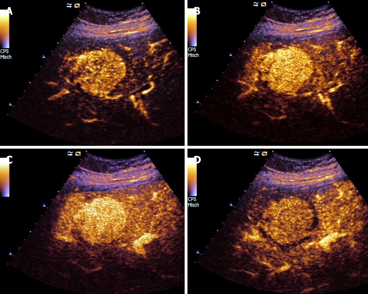
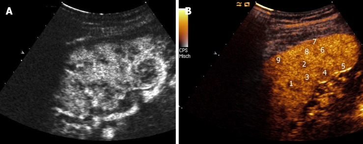
Since it has been recognized that sarcoidosis (SA) is not an exclusive disorder of the lungs but can also affect other organs such as the liver and spleen, efforts have been made to define specific imaging criteria for the diagnosis of the single organ involvement, and the concept has been reinforced that the exclusion of alternative causes is important to achieve the correct diagnosis. Ultrasound (US) is a useful tool to evaluate patients with suspected abdominal SA, such as of the liver, spleen, kidney, pancreas and other organs, showing findings such as organomegaly, focal lesions and lymphadenopathy. While the diagnosis of abdominal SA is more predictable in the case of involvement of other organs (e.g., lungs), the problem is more complex in the case of isolated abdominal SA. The recent use of contrast-enhanced ultrasound and endoscopic ultrasound elastography has provided additional information about the enhancement patterns and tissue rigidity in abdominal SA. Here we critically review the role of US in abdominal SA, reporting typical findings and limitations of current evidence and by discussing future perspectives of study.
Keywords: Contrast-enhanced ultrasound; Granulomatous disorders; Liver; Rare diseases; Sarcoid lesions; Sarcoidosis; Spleen; Ultrasound.
Conflict of interest statement
Conflict-of-interest statement: No potential conflicts of interest.
Figures
References
-
- Boeck C. Multiple benign sarcoid of the skin. J Cutan Genitourin Dis. 1899;17:543–550.
-
- Tchernev G, Cardoso JC, Chokoeva AA, Verma SB, Tana C, Ananiev J, Gulubova M, Philipov S, Kanazawa N, Nenoff P, Lotti T, Wollina U. The "mystery" of cutaneous sarcoidosis: facts and controversies. Int J Immunopathol Pharmacol. 2014;27:321–330. - PubMed
-
- Martin WJ 2nd, Iannuzzi MC, Gail DB, Peavy HH. Future directions in sarcoidosis research: summary of an NHLBI working group. Am J Respir Crit Care Med. 2004;170:567–571. - PubMed
-
- Mañá J, Rubio-Rivas M, Villalba N, Marcoval J, Iriarte A, Molina-Molina M, Llatjos R, García O, Martínez-Yélamos S, Vicens-Zygmunt V, Gámez C, Pujol R, Corbella X. Multidisciplinary approach and long-term follow-up in a series of 640 consecutive patients with sarcoidosis: Cohort study of a 40-year clinical experience at a tertiary referral center in Barcelona, Spain. Medicine (Baltimore) 2017;96:e7595. - PMC - PubMed
-
- Chen ES, Moller DR. Etiologies of Sarcoidosis. Clin Rev Allergy Immunol. 2015;49:6–18. - PubMed
Publication types
LinkOut - more resources
Full Text Sources
