Review
. 2019 Oct;60(4):347-360.
doi: 10.1002/mus.26497. Epub 2019 May 14.

Advancements in magnetic resonance imaging-based biomarkers for muscular dystrophy

Affiliations
Review

Advancements in magnetic resonance imaging-based biomarkers for muscular dystrophy

Doris G Leung. Muscle Nerve. 2019 Oct.

Abstract

Recent years have seen steady progress in the identification of genetic muscle diseases as well as efforts to develop treatment for these diseases. Consequently, sensitive and objective new methods are required to identify and monitor muscle pathology. Magnetic resonance imaging offers multiple potential biomarkers of disease severity in the muscular dystrophies. This Review uses a pathology-based approach to examine the ways in which MRI and spectroscopy have been used to study muscular dystrophies. Methods that have been used to quantitate intramuscular fat, edema, fiber orientation, metabolism, fibrosis, and vascular perfusion are examined, and this Review describes how MRI can help diagnose these conditions and improve upon existing muscle biomarkers by detecting small increments of disease-related change. Important challenges in the implementation of imaging biomarkers, such as standardization of protocols and validating imaging measurements with respect to clinical outcomes, are also described.

Keywords: magnetic resonance imaging; magnetic resonance spectroscopy; muscular dystrophy; outcome measures; quantitative imaging; skeletal muscle biomarkers.


Conflict of interest statement

Disclosure of Conflicts of Interest:

The author does not have any conflicts of interest to disclose.

Figures

Figure 1:
Figure 1:
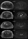
Axial T1-weighted (a, c, e) and STIR (b, d, f) images of the mid-thigh (a, b), mid-calf, (c, d) and shoulder girdle (e, f) in healthy volunteers. T1-weighted scans emphasize the contrast between muscle and fat, while STIR images highlight free water.
Figure 2:
Figure 2:
Representative T1-weighted (left) and STIR (right) axial cross-sections of the hip in multiple muscular dystrophies. The iliacus muscles (arrows) are shown to be severely atrophic in type 2 myotonic dystrophy (DM2), replaced by fat in limb-girdle muscular dystrophy type 2I (LGMD2I), and spared in facioscapulohumeral muscular dystrophy (FSHD). STIR hyperintensity is not observed in the imaged regions.
Figure 3:
Figure 3:
Dixon sequences acquire two sets of images: one with water and fat protons in the same phase and the other with fat and water protons in opposite phase. These images can be added or subtracted to generate water-only and fat-only images that can be used to estimate fat fractions. These axial cross-sections of the mid-thigh show extensive fat replacement of the quadriceps in a patient with DM2.
Figure 4:
Figure 4:
Using multivoxel 1H MRS, a 16×16 grid is positioned over an axial T1-weighted slice of the mid-thigh in a patient with facioscapulohumeral muscular dystrophy and both water suppressed and unsuppressed spectra are acquired from each voxel in the grid. A representative non-water-suppressed spectrum from a single voxel shows only water and lipid peaks. When the water signal is suppressed, additional peaks corresponding to creatine and trimethylamine (TMA) can be seen.
Figure 5:
Figure 5:
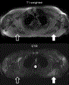
Representative T1-weighted and STIR images of the shoulder girdle in a patient with limb-girdle muscular dystrophy type 2I. There is STIR hyperintensity visible in the left infraspinatus muscle (white arrow) associated with moderate fat replacement on T1-weighted images. In the right infraspinatus (open arrow), the fat replacement is more extensive. Because fat signal is suppressed in STIR images, the right infraspinatus is not hyperintense.
Figure 6:
Figure 6:
Axial cross-sections of the mid-thigh in a patient with FSHD. Upper left: T2-weighted images. Upper right: Radiomic entropy images generated from the T2-weighted images showing changes associated with muscular dystrophy in the posterior thighs (white arrows). Lower left: Apparent diffusion coefficient (ADC) map from diffusion-weighted imaging. Lower right: Radiomic images generated from the ADC map. There is increased entropy (high intensity regions) within the fat-replaced posterior compartment muscles (white arrows) compared to the relatively unaffected anterior compartment muscles, which exhibit low entropy. Image provided by Dr. Michael A. Jacobs, Johns Hopkins University Radiology.

References

    1. Leung DG. Magnetic resonance imaging patterns of muscle involvement in genetic muscle diseases: a systematic review. J Neurol 2017;264:1320–1333. - PMC - PubMed
    1. Straub V, Murphy A, Udd B. 229th ENMC international workshop: Limb girdle muscular dystrophies - Nomenclature and reformed classification Naarden, the Netherlands, 17–19 March 2017. Neuromuscul Disord 2018;28:702–710. - PubMed
    1. Tordjman M, Dabaj I, Laforet P, Felter A, Ferreiro A, Biyoukar M, et al. Muscular MRI-based algorithm to differentiate inherited myopathies presenting with spinal rigidity. Eur Radiol 2018;28:5293–5303. - PubMed
    1. Mercuri E, Clements E, Offiah A, Pichiecchio A, Vasco G, Bianco F, et al. Muscle magnetic resonance imaging involvement in muscular dystrophies with rigidity of the spine. Ann Neurol 2010;67:201–208. - PubMed
    1. Barsotti S, Zampa V, Talarico R, Minichilli F, Ortori S, Iacopetti V, et al. Thigh magnetic resonance imaging for the evaluation of disease activity in patients with idiopathic inflammatory myopathies followed in a single center. Muscle Nerve 2016;54:666–672. - PubMed

Publication types

MeSH terms