

Diurnal change of retinal vessel density and mean ocular perfusion pressure in patients with open-angle glaucoma
- PMID: 31026291
- PMCID: PMC6485647
- DOI: 10.1371/journal.pone.0215684
Diurnal change of retinal vessel density and mean ocular perfusion pressure in patients with open-angle glaucoma
Abstract
Purpose: To investigate, in primary open-angle glaucoma (POAG) and healthy subjects, the pattern and magnitude of diurnal variation in optical coherence tomography angiography (OCTA) retinal vessel density (RVD).
Design: Prospective, observational cross-sectional study.
Participants: A prospective study was conducted on 20POAG patients and 19 healthy subjects.
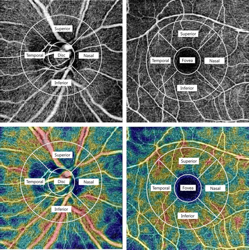
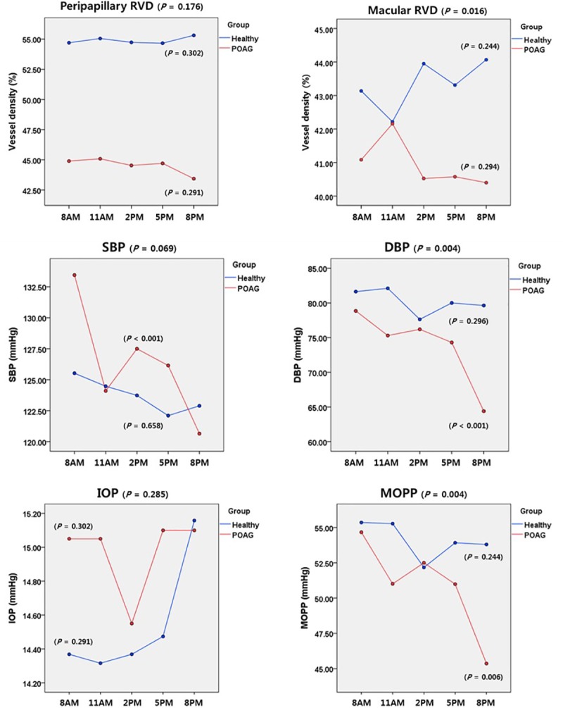
Methods: Peripapillary/macular RVD (using swept-source OCTA), intraocular pressure (IOP), and systemic blood pressure (BP) were measured five times a day (8 a.m., 11 a.m., 2 p.m.,5 p.m. and 8 p.m.). The magnitudes and patterns of diurnal changes in RVD, diastolic BP, and mean ocular-perfusion pressure (MOPP) were analyzed and compared between the POAG patients and the healthy subjects.
Main outcome measures: The patterns and magnitudes of diurnal RVD change in OCTA.
Results: Intra-visit repeatability (0.755-0.943) and inter-visit reproducibility (0.843-0.986) for the RVD measurements showed excellent reliability. In the POAG patients, the magnitude of diurnal change in peripapillary RVD (9.71±7.04%) and macular RVD (7.22±4.73%) were significantly greater than that in the healthy group (5.73±3.85%, P = 0.013 and 5.51±3.45%, P = 0.042, respectively). The magnitudes of diurnal variations of IOP and MOPP in the POAG group likewise were greater than those in the healthy group (P = 0.003 and 0.039). As for the patterns of diurnal RVD change, interestingly, at 8 p.m., the macular RVD of the healthy group increased to the highest level (44.12±2.95%) while that of the POAG group decreased to the lowest level (40.41±2.54%).
Conclusions: In POAG eyes, diurnal change of IOP, MOPP and RVD was significantly greater than in the healthy eyes. These findings suggest that diurnal RVD changes might reflect the hemodynamic variation of POAG.
Conflict of interest statement
The authors have declared that no competing interests exist.
Figures

References
-
- Flammer J, Orgül S, Costa VP, Orzalesi N, Krieglstein GK, Serra LM, et al. The impact of ocular blood flow in glaucoma. Prog Retin Eye Res. 2002;21:359–393. - PubMed
-
- Grunwald JE, Piltz J, Hariprasad SM, DuPont J. Optic nerve and choroidal circulation in glaucoma. Invest Ophthalmol Vis Sci. 1998; 39:2329–36. - PubMed
-
- Michelson G, Langhans MJ, Harazny J, Dichtl A. Visual field defect and perfusion of the juxtapapillary retina and the neuroretinal rim area in primary open-angle glaucoma. Graefes Arch Clin Exp Ophthalmol. 1998;236(2):80–5. - PubMed
Publication types
MeSH terms
LinkOut - more resources
Full Text Sources
Research Materials
