. 2019 Apr 26;12(9):1360.
doi: 10.3390/ma12091360.

Cell-Free Demineralized Bone Matrix for Mesenchymal Stem Cells Survival and Colonization

Affiliations

Cell-Free Demineralized Bone Matrix for Mesenchymal Stem Cells Survival and Colonization

Monica Mattioli-Belmonte et al. Materials (Basel). .

Abstract

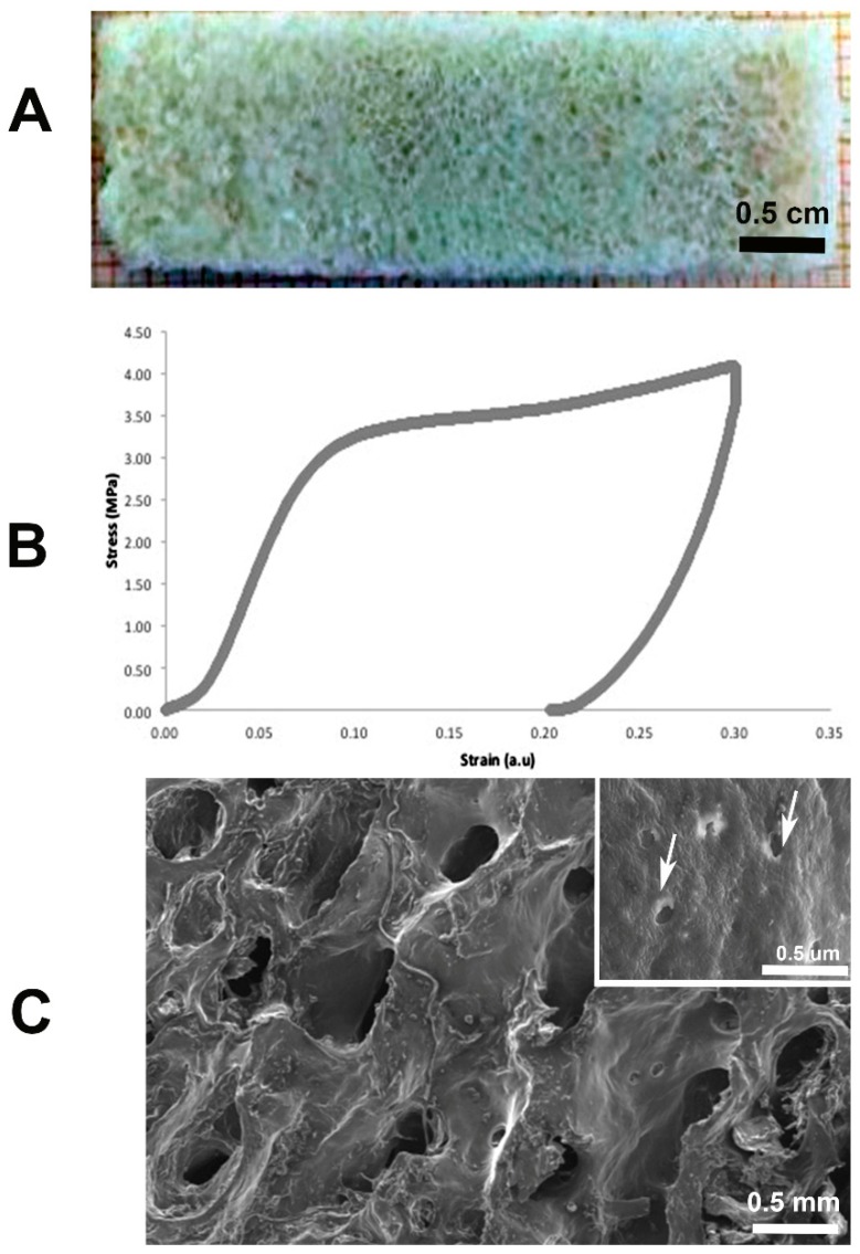
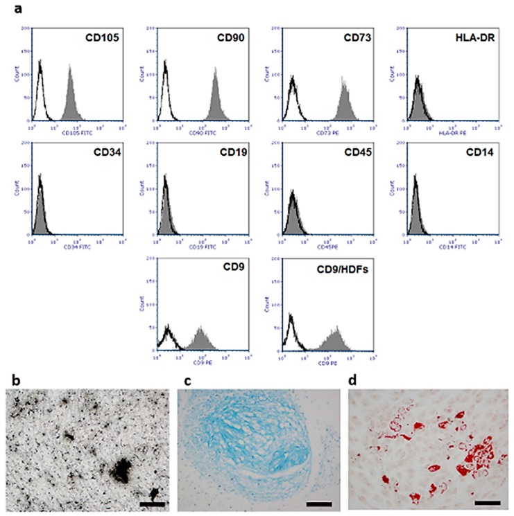
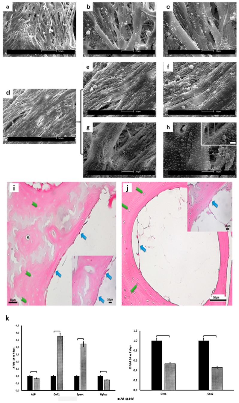
Decellularized bone matrix is receiving much attention as biological scaffolds and implantable biomaterials for bone tissue regeneration. Here, we evaluated the efficacy of a cell-free demineralized bone matrix on mesenchymal stem cells (MSCs) survival and differentiation in vitro. The seeding of human umbilical cord-derived MSCs (hUC-SCs) on decellularized bone matrices up to 14 days was exploited, assessing their capability of scaffold colonization and evaluating gene expression of bone markers. Light and Scanning Electron Microscopies were used. The obtained cell-free decalcified structures showed elastic moduli attributable to both topology and biochemical composition. Morphological observation evidenced an almost complete colonization of the scaffolds after 14 days of culture. Moreover, in hUC-SCs cultured on decalcified scaffolds, without the addition of any osteoinductive media, there was an upregulation of Collagen Type I (COL1) and osteonectin (ON) gene expression, especially on day 14. Modifications in the expression of genes engaged in stemness were also detected. In conclusion, the proposed decellularized bone matrix can induce the in vitro hUC-SCs differentiation and has the potential to be tested for in in vivo tissue regeneration.

Keywords: MSCs; decellularized bone matrix; gene expression; tissue engineering.


Conflict of interest statement

The authors declare no conflict of interest.

Figures

Figure 1
Figure 1
Representative: (A) Demineralized scaffold; (B) stress-strain curve of demineralized bone; (C) SEM observation with bony trabecular network and empty osteocytic lacunae (arrows).
Figure 2
Figure 2
hUC-SC flow cytometric phenotypic characterization and in vitro differentiation assays. (a) Characteristic single-variable histograms display negative control (black line) and the expression of cell surface antigens (grey) on hUC-SCs and Human Dermal Fibroblasts (HDFs); (b) osteogenic differentiation of hUC-SCs after 21 days of induction (Von Kossa staining); (c) chondrogenic differentiation of hUC-SCs after 14 days of induction (Alcian Blue staining); (d) adipogenic differentiation of hUC-SCs after 14 days of induction (Oil Red staining). Image magnification 20×, scale bars 50 μm.
Figure 3
Figure 3
SEM micrographs of scaffolds cultured with hUC-SCs for 7 (a–c) and 14 (dh) days at different magnifications. Cells were able to adhere and colonize the scaffolds. Note the presence of membrane blebs after 14 days of culture suggestive of active metabolizers. Light microscopy (LM) micrographs of cells on demineralized scaffold at 7 (i) and 14 (j) days of culture: H = Haversian canal; green arrows indicate osteocytic lacunae; blue arrows indicate hUC-SCs; (k) histograms depict changes in Alkaline Phosphatase (ALP), Collagen Type 1 (COL1), ostenectin (ON; also known as secreted protein acidic and rich in cysteine (SPARC)), osteocalcin (OC; also known as bone gamma-carboxyglutamic acid-containing protein (BGLAP), sex-determining region Y (SRY)-Box2 (Sox2), and octamer-binding transcription factor 4 (Oct4) mRNA expression in hUC-SCs. Data are expressed as fold-change (2−ΔΔCt) of the expression at 14 days culture over 7 days one: square brackets indicate significant differences (p < 0.05).

References

    1. Murphy W.L., McDevitt T.C., Engler A.J. Materials as stem cell regulators. Nat. Mater. 2014;13:547–557. doi: 10.1038/nmat3937. - DOI - PMC - PubMed
    1. Mattioli-Belmonte M., Vozzi G., Kyriakidou K., Pulieri E., Lucarini G., Vinci B., Pugnaloni A., Biagini G., Ahluwalia A. Rapid-prototyped and salt-leached PLGA scaffolds condition cell morpho-functional behavior. J. Biomed. Mater. Res. A. 2008;85:466–476. doi: 10.1002/jbm.a.31483. - DOI - PubMed
    1. Rossi F., Santoro M., Perale G. Polymeric scaffolds as stem cell carriers in bone repair. J. Tissue Eng. Regen. Med. 2015;9:1093–1119. - PubMed
    1. Ciardelli G., Gentile P., Chiono V., Mattioli-Belmonte M., Vozzi G., Barbani N., Giusti P. Enzymatically crosslinked porous composite matrices for bone tissue regeneration. J. Biomed. Mater. Res. A. 2010;92:137–151. doi: 10.1002/jbm.a.32344. - DOI - PubMed
    1. Jelen C., Mattei G., Montemurro F., De Maria C., Mattioli-Belmonte M., Vozzi G. Bone scaffolds with homogeneous and discrete gradient mechanical properties. Mat. Sci. Eng. C. 2013;3:28–36. doi: 10.1016/j.msec.2012.07.046. - DOI - PubMed

LinkOut - more resources