. 2019 May 7;116(19):9586-9591.
doi: 10.1073/pnas.1819965116. Epub 2019 Apr 26.

Dynamic super-resolution structured illumination imaging in the living brain

Affiliations

Dynamic super-resolution structured illumination imaging in the living brain

Raphaël Turcotte et al. Proc Natl Acad Sci U S A. .

Abstract

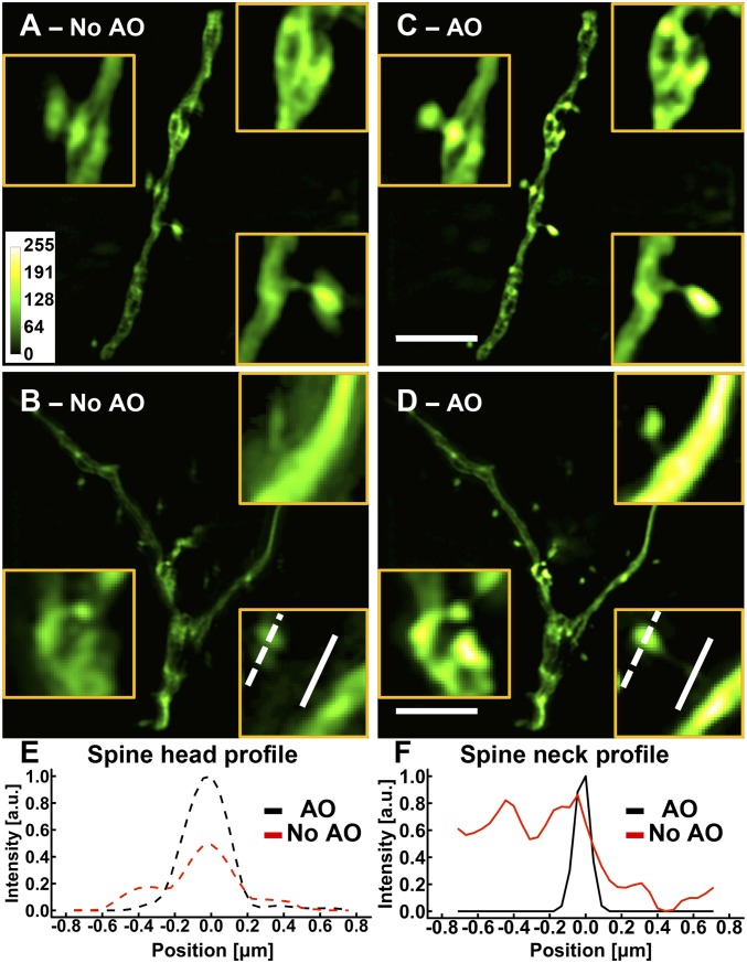
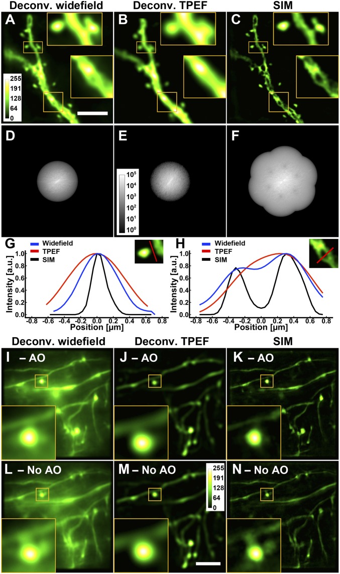
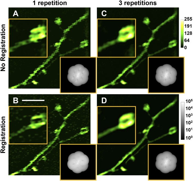
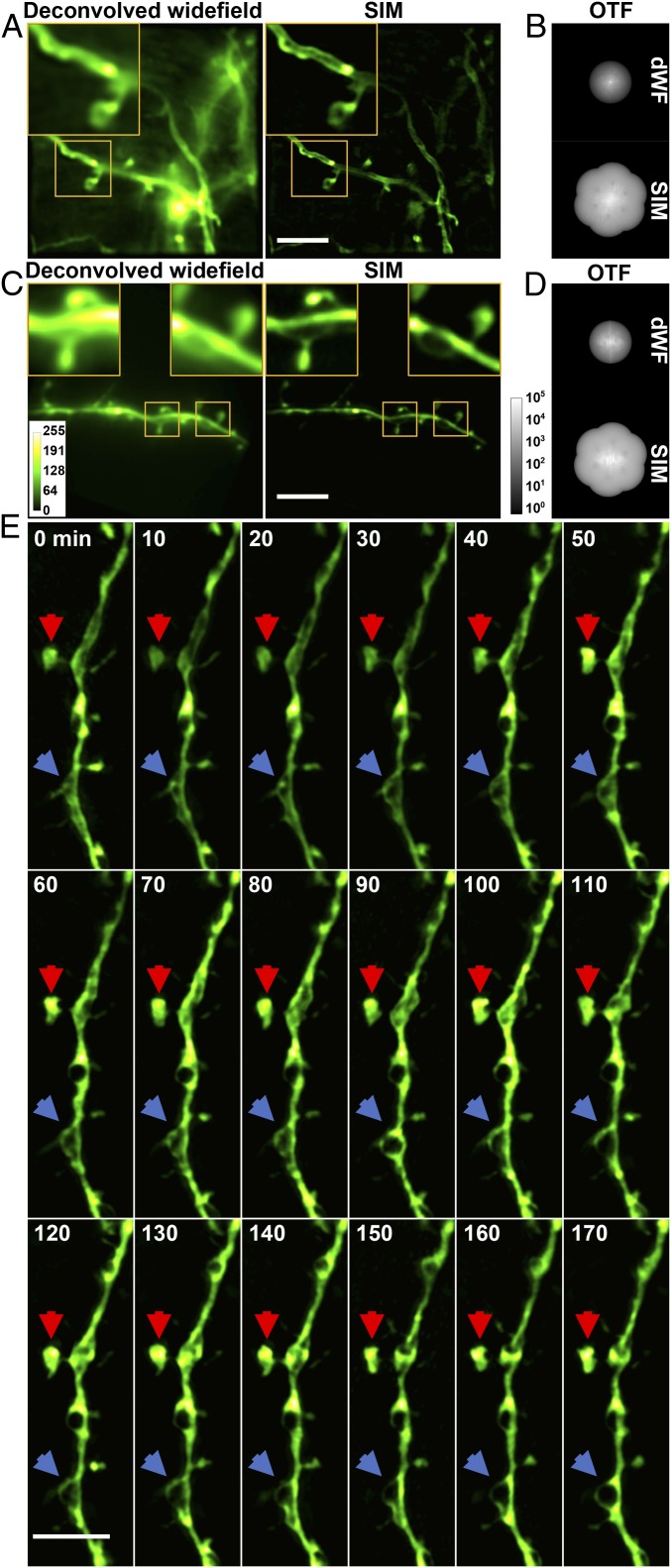
Cells in the brain act as components of extended networks. Therefore, to understand neurobiological processes in a physiological context, it is essential to study them in vivo. Super-resolution microscopy has spatial resolution beyond the diffraction limit, thus promising to provide structural and functional insights that are not accessible with conventional microscopy. However, to apply it to in vivo brain imaging, we must address the challenges of 3D imaging in an optically heterogeneous tissue that is constantly in motion. We optimized image acquisition and reconstruction to combat sample motion and applied adaptive optics to correcting sample-induced optical aberrations in super-resolution structured illumination microscopy (SIM) in vivo. We imaged the brains of live zebrafish larvae and mice and observed the dynamics of dendrites and dendritic spines at nanoscale resolution.

Keywords: adaptive optics; brain imaging; in vivo; super-resolution; synapses.


Conflict of interest statement

The authors declare no conflict of interest.

Figures

Fig. 1.
Fig. 1.
AO is essential for SIM imaging in brain tissue. (AD) Images of dendrites at a depth of 25 μm in a cortical slice of a Thy1-GFP line M mouse (A and B) without and (C and D) with AO. (Scale bars: 5 μm; Inset widths: A and C, 3 μm; B and D, 2 μm.) (E and F) Line profiles of (E) a spine head and (F) a spine neck with and without AO as identified by the lines in B and D. Images were normalized to the AO condition.
Fig. 2.
Fig. 2.
SIM yields spatial resolution superior to deconvolved widefield and TPEF microscopy both ex vivo and in vivo. (A–F) Images and corresponding OTFs of the same dendritic structure in a Thy1-GFP line M brain slice at a depth of 25 μm obtained with different imaging modalities, all with AO: (A and D) deconvolved widefield, (B and E) deconvolved TPEF, and (C and F) SIM. (Scale bar: 5 μm; Inset widths: 3 μm.) (G and H) Line profiles through a spine neck and a dendritic shaft, respectively. All deconvolutions were performed with Wiener filtering. (I–N) In vivo images of neurites in a larval zebrafish brain at a depth of 100 μm. Images of the same neurites obtained with (I and L) deconvolved widefield, (J and M) deconvolved TPEF, and (K and N) SIM with and without AO, respectively. Images were normalized independently. (Scale bar: 5 μm; Inset widths: 3 μm).
Fig. 3.
Fig. 3.
Strategies to combat motion-induced artifacts for in vivo SIM in the mouse brain. SIM images and OTFs reconstructed from raw data series (A) with one repetition and without raw image registration, (B) with one repetition and with registration, (C) with three repetitions and without registration, and (D) with three repetitions and with registration. Images were normalized independently. (Scale bar: 3 μm; Inset widths: 2.5 μm.)
Fig. 4.
Fig. 4.
In vivo SR imaging of the mouse brain with AO SIM. (A) Deconvolved widefield (dWF) and SIM images of dendrites expressing ChR2-GFP, a membrane label. (Scale bar: 5 μm; Inset width: 5 μm.) (B) OTFs of the SIM and dWF images in A. (C) dWF and SIM images of neurons expressing cytosolic GFP (Thy-1 line M mouse). (Scale bar: 5 μm; Inset width: 3 μm.) (D) OTFs of the SIM and dWF images in C. (E) Time-lapse in vivo SIM images showing structural dynamics of a dendrite at a depth of 25 μm in the brain of a Thy1-GFP line M mouse after KCl injection. Arrows point to highly dynamic structures. Images were normalized independently. (Scale bar: 4 μm.)

References

    1. Kandel ER, Schwartz JH, Jessell TM, Siegelbaum S, Hudspeth AJ, editors. Principles of Neural Science. 5th Ed McGraw-Hill Education/Medical; New York: 2012.
    1. Bethge P, Chéreau R, Avignone E, Marsicano G, Nägerl UV. Two-photon excitation STED microscopy in two colors in acute brain slices. Biophys J. 2013;104:778–785. - PMC - PubMed
    1. Tønnesen J, Nägerl UV. Superresolution imaging for neuroscience. Exp Neurol. 2013;242:33–40. - PubMed
    1. Tønnesen J, Katona G, Rózsa B, Nägerl UV. Spine neck plasticity regulates compartmentalization of synapses. Nat Neurosci. 2014;17:678–685. - PubMed
    1. Zhong H. Applying superresolution localization-based microscopy to neurons. Synapse. 2015;69:283–294. - PMC - PubMed

Publication types