Review
. 2019 Aug;60(8):1044-1052.
doi: 10.2967/jnumed.118.217901. Epub 2019 Apr 26.

Development of Dedicated Brain PET Imaging Devices: Recent Advances and Future Perspectives

Affiliations
Review

Development of Dedicated Brain PET Imaging Devices: Recent Advances and Future Perspectives

Ciprian Catana. J Nucl Med. 2019 Aug.

Abstract

Whole-body PET scanners are not optimized for imaging small structures in the human brain. Several PET devices specifically designed for this task have been proposed either for stand-alone operation or as MR-compatible inserts. The main distinctive features of some of the most recent concepts and their performance characteristics, with a focus on spatial resolution and sensitivity, are reviewed. The trade-offs between the various performance characteristics, desired capabilities, and cost that need to be considered when designing a dedicated brain scanner are presented. Finally, the aspirational goals for future-generation scanners, some of the factors that have contributed to the current status, and how recent advances may affect future developments in dedicated brain PET instrumentation are briefly discussed.

Keywords: PET; high spatial resolution; multimodal imaging; neuroimaging.


Figures

FIGURE 1.
FIGURE 1.
Examples of dedicated PET scanners with conventional geometries: HRRT (https://commons.wikimedia.org/wiki/File:HRRT_PET.JPG) (A), jPET-D4 (20) (B), NeuroPET/CT (22) (C), Hamamatsu (23) (D), BBX (http://prescient-imaging.com/products/bbx/) (E), PET-Hat (24) (F), Helmet-PET (27) (G), and Mind-Tracker (courtesy of Qiyu Peng, Lawrence Berkeley National Laboratory) (H).
FIGURE 2.
FIGURE 2.
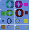
Schematic drawings (angled view) showing configuration of detector modules and cross-sectional detailed views (scale, 5:1) of scintillator arrays for dedicated PET scanners with conventional geometries: HRRT (A), jPET-D4 (B), NeuroPET/CT (C), Hamamatsu (D), BBX (E), PET-Hat (F), Helmet-PET (G), and Mind-Tracker (H). A 20-cm-diameter circle is shown inside each scanner. Scale bar represents 1 cm in detailed views.
FIGURE 3.
FIGURE 3.
Examples of dedicated PET scanners with unconventional geometries: Helmet-PET plus chin (33) (A), dodecahedral (courtesy of Qiyu Peng, Lawrence Berkeley National Laboratory) (B), and spherical (39) (C). NEMA = National Electrical Manufacturers Association.
FIGURE 4.
FIGURE 4.
BrainPET prototype installed inside 3-T MR scanner and representative images from studies performed at Athinoula A. Martinos Center using 11C-NNC112 (coronal view [top]), 18F-FDG (sagittal view [middle]), and 11C-temozolomide (transaxial view and fused with morphologic MR image [bottom]).
FIGURE 5.
FIGURE 5.
Schematic drawings (angled view) showing configuration of detector modules and cross-sectional detailed views (scale, 5:1) of scintillator arrays for dedicated MR-compatible PET inserts with conventional geometries: BrainPET (A), charge signal transmission (B), add-on PET insert with 4-layer DOI-capable detectors (C), radiofrequency-penetrable PET (D), MINDView (E), and TRIMAGE (F). A 20-cm-diameter circle is shown inside each scanner. Scale bar represents 1 cm in detailed views.
FIGURE 6.
FIGURE 6.
Possible geometry for 7-T MRI-compatible high-sensitivity brain PET scanner.

References

    1. Jones T, Rabiner EA. The development, past achievements, and future directions of brain PET. J Cereb Blood Flow Metab. 2012;32:1426–1454. - PMC - PubMed
    1. Jones T, Townsend D. History and future technical innovation in positron emission tomography. J Med Imaging (Bellingham). 2017;4:011013. - PMC - PubMed
    1. Thompson CJ, Yamamoto YL, Meyer E, Positome II: a high efficiency positron imaging device for dynamic brain studies. IEEE Trans Nucl Sci. 1979;26:583–589.
    1. Hoffman EJ, Phelps ME, Huang SC. Performance evaluation of a positron tomograph designed for brain imaging. J Nucl Med. 1983;24:245–257. - PubMed
    1. Freifelder R, Karp JS, Geagan M, Muehllehner G. Design and performance of the HEAD PENN-PET scanner. IEEE Trans Nucl Sci. 1994;41:1436–1440.

Publication types

MeSH terms