. 2019 May 31;294(22):8773-8778.
doi: 10.1074/jbc.AC119.007981. Epub 2019 Apr 26.

Mass spectrometry-based molecular mapping of native FXIIIa cross-links in insoluble fibrin clots

Affiliations

Mass spectrometry-based molecular mapping of native FXIIIa cross-links in insoluble fibrin clots

Lauren R Schmitt et al. J Biol Chem. .

Abstract

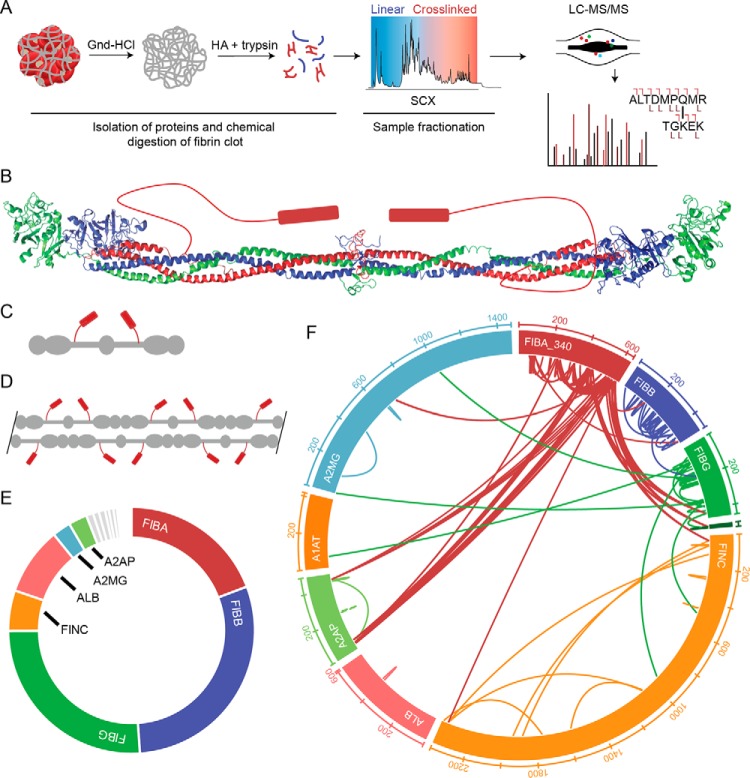
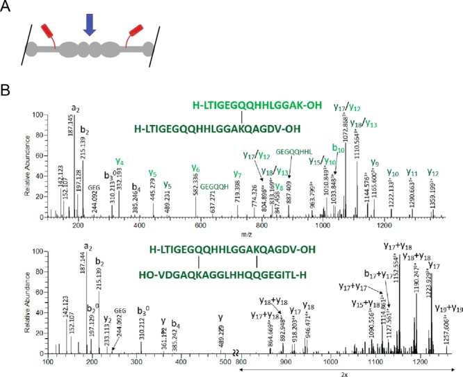
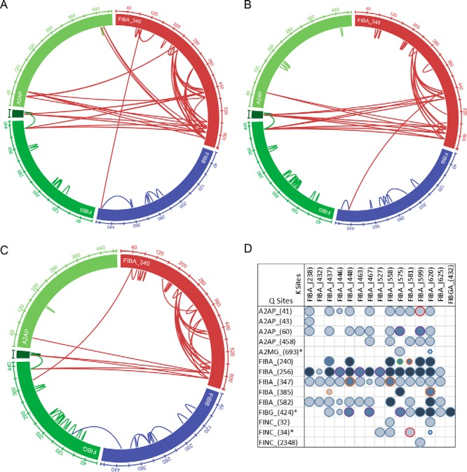
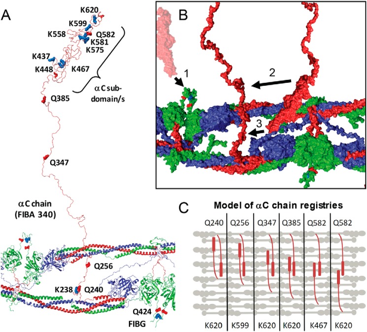
The roles of factor XIIIa-specific cross-links in thrombus formation, regression, or probability for embolization are largely unknown. A molecular understanding of fibrin architecture at the level of these cross-links could inform the development of therapeutic strategies to prevent the sequelae of thromboembolism. Here, we present an MS-based method to map native factor XIIIa cross-links in the insoluble matrix component of whole-blood or plasma-fibrin clots and in in vivo thrombi. Using a chaotrope-insoluble digestion method and quantitative cross-linking MS, we identified the previously mapped fibrinogen peptides that are responsible for covalent D-dimer association, as well as dozens of novel cross-links in the αC region of fibrinogen α. Our findings expand the known native cross-linked species from one to over 100 and suggest distinct antiparallel registries for interprotofibril association and covalent attachment of serpins that regulate clot dissolution.

Keywords: chemical digestion; factor XIII; fibrin; fibrinolysis; mass spectrometry (MS); protein cross-linking; thrombosis; transglutaminase.


Conflict of interest statement

The authors declare that they have no conflicts of interest with the contents of this article

Figures

Figure 1.
Figure 1.
Workflow and validation of clot proteomic method. A, outline of the clot processing and analysis workflow. After formation, the clot is washed extensively with the strong chaotrope guanidine (Gnd) HCl to remove components that are not covalently attached to the fibrin structure. The resulting pellet is subjected to chemical digestion with hydroxylamine (NH2OH; HA) followed by enzymatic digestion with trypsin. The resulting peptides are fractionated by SCX chromatography. Fractionated samples are analyzed by tandem MS. B, crystal structure of human fibrinogen with cartoon representation of the αC domain (PDB code 3GHG). C, cartoon model of fibrinogen structure shown in B. D, cartoon model of protofibril. E, area under the curve estimation of protein abundance in whole-blood clot. Proteomic characterization revealed 125 protein identifications at 1% FDR with the majority of signal arising from fibrin (70% total spectral counts, >80% AUC signal intensity). F, wheel diagram showing identifications and position within proteins of cross-links identified in whole-blood clot with expectation (E) value greater than 1.0E−5. A2MG, α2-macroglobulin; ALB, albumin; A1AT, α1-antitrypsin.
Figure 2.
Figure 2.
Identification of D-dimer cross-link. A, cartoon model of predicted D-dimer. The blue arrow indicates the D-dimer interface. B, the human FIBG-A isoform (P02769-2) C-terminal cross-linking sites shown for Gln-424–Lys-432 that correspond to the C-terminal γ–γ cross-linking site. Fragment ions between the cross-linked sites (di-cross-linked form; lower panel) are absent from the fragmentation spectra; these would require a minimum of two fragmentation events, which is highly unlikely.
Figure 3.
Figure 3.
In vivo thrombus and plasma clot analyses. A, wheel diagram showing identified cross-linked peptides within the fibrin and A2AP sequences for Plsm1. B, wheel diagram showing identifications and position within proteins of cross-links identified in Plsm2. C, wheel diagram showing identifications and position within proteins of cross-links identified in the in vivo thrombus (IVT). D, cross-linking profile showing the occurrence of spectral matches for prominent cross-link sites (93 unique cross-links in total). The symbol size indicates the minimum E value for a given cross-link across all data sets: E < 1E−4, large circle; E < 0.001, medium circle; E < 0.01, small circle. The symbol color represents the number of samples in which the cross-link was identified in in-depth analysis: dark blue, in all three clot samples (WB, Plsm2, and IVT); medium blue, in two samples; light blue, in a single sample. The symbol outline represents samples in which the cross-link was identified: purple, WB and Plsm2; dark orange, WB and IVT; green, Plsm2 and IVT; light blue, WB; red, Plsm2; light orange, IVT. A2MG, α2-macroglobulin.
Figure 4.
Figure 4.
Structural representation of FXIIIa fibrin cross-links. A, a modeled fibrin protofibril with αC chains used from Zhmurov et al. (S1.FP_9+10_AaBb.PDB PDB) (24) is shown with the αC linker and subdomain(s) labeled, consistent with the biochemical studies performed by Medved and co-workers (28). B, surface plot (using S1.FP_9+10_AaBb.PDB) showing the αC region from neighboring protofibrils with orientations for FIBG Gln-424–FIBA Lys-620 (1) cross-linking, the αC subdomain N terminus cross-linked to the C terminus of the γ-chain (2), and the αC subdomain cross-linking to the N-terminal side of the αC chain (3). C, simplified model showing multiple registries of antiparallel αC regions cross-linked with perpendicular orientation with respect to the fibrin fiber. Each panel represents a potential orientation of two αC regions spanning across several fibrin fibers.

References

    1. ISTH Steering Committee for World Thrombosis Day (2014) Thrombosis: a major contributor to global disease burden. Thromb. Res. 134, 931–938 10.1016/j.thromres.2014.08.014 - DOI - PubMed
    1. Collet J.-P., Moen J. L., Veklich Y. I., Gorkun O. V., Lord S. T., Montalescot G., and Weisel J. W. (2005) The αC domains of fibrinogen affect the structure of the fibrin clot, its physical properties, and its susceptibility to fibrinolysis. Blood 106, 3824–3830 10.1182/blood-2005-05-2150 - DOI - PMC - PubMed
    1. Litvinov R. I., and Weisel J. W. (2016) What is the biological and clinical relevance of fibrin? Semin. Thromb. Hemost. 42, 333–343 10.1055/s-0036-1571342 - DOI - PMC - PubMed
    1. Byrnes J. R., and Wolberg A. S. (2016) Newly-recognized roles of factor XIII in thrombosis. Semin. Thromb. Hemost. 42, 445–454 10.1055/s-0036-1571343 - DOI - PMC - PubMed
    1. van Giezen J. J., Minkema J., Bouma B. N., and Jansen J. W. (1993) Cross-linking of α2-antiplasmin to fibrin is a key factor in regulating blood clot lysis: species differences. Blood Coagul. Fibrinolysis 4, 869–875 10.1097/00001721-199312000-00002 - DOI - PubMed

Publication types

Associated data

LinkOut - more resources