Review
. 2019 Sep;16(9):519-537.
doi: 10.1038/s41569-019-0200-7.

Molecular mechanisms of arrhythmogenic cardiomyopathy

Affiliations
Review

Molecular mechanisms of arrhythmogenic cardiomyopathy

Karyn M Austin et al. Nat Rev Cardiol. 2019 Sep.

Abstract

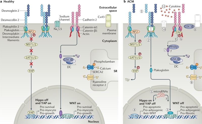
Arrhythmogenic cardiomyopathy is a genetic disorder characterized by the risk of life-threatening arrhythmias, myocardial dysfunction and fibrofatty replacement of myocardial tissue. Mutations in genes that encode components of desmosomes, the adhesive junctions that connect cardiomyocytes, are the predominant cause of arrhythmogenic cardiomyopathy and can be identified in about half of patients with the condition. However, the molecular mechanisms leading to myocardial destruction, remodelling and arrhythmic predisposition remain poorly understood. Through the development of animal, induced pluripotent stem cell and other models of disease, advances in our understanding of the pathogenic mechanisms of arrhythmogenic cardiomyopathy over the past decade have brought several signalling pathways into focus. These pathways include canonical and non-canonical WNT signalling, the Hippo-Yes-associated protein (YAP) pathway and transforming growth factor-β signalling. These studies have begun to identify potential therapeutic targets whose modulation has shown promise in preclinical models. In this Review, we summarize and discuss the reported molecular mechanisms underlying the pathogenesis of arrhythmogenic cardiomyopathy.


Figures

Fig. 1 |
Fig. 1 |. Cellular components implicated in ACM.
The intercalated disc of cardiomyocytes contains the area composita, which is an intermixed architectural and signalling structure that includes components of the desmosome, adherens junction and ion channels. Mutations in cellular components of the intercalated disc, as well as in intracellular structures, have been identified in arrhythmogenic cardiomyopathy (ACM). Categories of protein in which ACM-causing mutations occur are labelled accordingly. (1) Components of the desmosome, including desmocollin 2, desmoglein 2, junction plakoglobin, plakophilin 2 and desmoplakin. (2) Components of the adherens junction, including cadherin 2 and catenin-α3. (3) Contributors to calcium handling, including phospholamban and ryanodine receptor 2 located in the membrane of the sarcoplasmic reticulum (SR). (4) Intracellular structural proteins, including desmin, titin and filamin C. (5) The sodium channel and transforming growth factor-β3 (TGFβ3). (6) Nuclear envelope proteins transmembrane protein 43 and lamin A. SERCA2, sarcoplasmic/endoplasmic reticulum calcium ATPase 2.
Fig. 2 |
Fig. 2 |. Cross and histological features of ACM.
a | Illustration depicting the most commonly affected ventricular regions in arrhythmogenic cardiomyopathy (ACM). Right ventricular disease predominantly affects the inflow tract, apex and infundibulum, known as the triangle of dysplasia (dashed triangle). Left-dominant disease commonly affects the inferior and inferolateral walls (dashed rectangle). b | Gross images of the right ventricle (RV) and left ventricle (LV), highlighting epicardial fat deposition (black arrowheads). c | Histological features of ACM including adipogenesis and cardiomyocyte replacement (left; trichrome stain), fibrosis (middle; trichrome stain) and inflammation juxtaposed to myocardial tissue (right; haematoxylin and eosin stain). ENDO, endocardium; EPI, epicardium.
Fig. 3 |
Fig. 3 |. Proposed molecular mechanisms contributing to the pathogenesis of ACM.
a | In the healthy cardiomyocyte, both desmosomes and adherens junctions in the area composita form strong intercellular connections with neighbouring cells. Likewise, both the gap junction, formed by connexin 43 (Cx43), and the sodium channel (Nav1.5) are appropriately positioned as a result of coordinated trafficking and membrane tethering. Catenin-β1 has a structural function in adherens junctions as well as a role in modifying transcriptional activity through activation of WNT-dependent gene expression. Cytoplasmic catenin-β1 is quickly degraded through proteosomal targeting by the destruction complex (DC), which contains glycogen synthase kinase 3β (GSK3β). The Hippo pathway is appropriately ‘off’, allowing for transcription of genes that promote cardiomyocyte survival, function (‘pro-myocyte’) and growth. Calcium flux is well regulated in the sarcoplasmic reticulum (SR) through functioning sarcoplasmic/endoplasmic reticulum calcium ATPase 2 (SERCA2), phospholamban and ryanodine receptor 2. b | In arrhythmogenic cardiomyopathy (ACM), multiple signalling pathways seem to be perturbed. (1) Disruption of the desmosomes and adherens junctions leads to increased mechanical stress on the cardiomyocyte. (2) Plakoglobin can dissociate from the desmosome, further destabilizing the intercalated disc and (3) inhibiting WNT-dependent gene transcription. (4) Activation of the Hippo pathway, potentially through neurofibromin 2 (NF2), results in inhibition of gene targets and promotes a pro-apoptotic and adipogenic phenotype. (5) Likewise, microRNAs can modulate both Hippo and WNT signalling. (6) Active Hippo signalling leads to phosphorylation of Yes-associated protein (YAP), which potentially associates with both plakoglobin and catenin-β1 at the plasma membrane, sequestering catenin-β1 and further inhibiting canonical WNT signalling. (7) Increased peroxisome proliferator-activated receptor-γ (PPARγ) expression has been associated with WNT inhibition, potentially through a direct relationship that promotes catenin-β1 degradation. (8) GSK3β translocates to the plasma membrane, although the relevance of this change in localization is uncertain. (9) Dysregulation of calcium handling in the SR is thought to contribute to arrhythmogenesis in a subset of patients. (10) Abnormal shuttling and tethering of both the sodium channel and gap junction components (Cx43) have been suspected to be involved in arrhythmogenesis. (ii) Increased pro-inflammatory and profibrotic cytokine production, including transforming grown factor-β1 (TGFβ1) and TGFβ3, is thought to contribute to the pathogenesis of ACM via canonical and non-canonical pathways. LATS, large tumour suppressor homologue; MST, mammalian STE20-like protein kinase; TEAD, transcriptional enhancer factor TEF.

References

    1. Sen-Chowdhry S & McKenna WJ Reconciling the protean manifestations of arrhythmogenic cardiomyopathy. Circ. Arrhythm. Electrophysiol 3, 566–570 (2010). - PubMed
    1. Marcus FI et al. Right ventricular dysplasia: a report of 24 adult cases. Circulation 65, 384–398 (1982). - PubMed
    1. Thiene G, Nava A, Corrado D, Rossi L & Pennelli N Right ventricular cardiomyopathy and sudden death in young people. N. Engl. J. Med 318, 129–133 (1988). - PubMed
    1. Sen-Chowdhry S, Morgan RD, Chambers JC & McKenna WJ Arrhythmogenic cardiomyopathy: etiology, diagnosis, and treatment. Annu. Rev. Med 61, 233–253 (2010). - PubMed
    1. Peters S, Trummel M & Meyners W Prevalence of right ventricular dysplasia-cardiomyopathy in a non-referral hospital. Int. J. Cardiol 97, 499–501 (2004). - PubMed

Publication types

MeSH terms

LinkOut - more resources