

Intercostal artery pseudoaneurysm following thoracentesis: multi-modal imaging and treatment
- PMID: 31029094
- PMCID: PMC6487039
- DOI: 10.1186/s12880-019-0333-5
Intercostal artery pseudoaneurysm following thoracentesis: multi-modal imaging and treatment
Abstract
Background: A pseudoaneurysm occurs as the result of a contained rupture of an arterial wall, yielding a perfused sac that communicates with the arterial lumen. Pseudoaneurysm of an intercostal artery is an extremely rare event but it carries with it a significant risk of rupture and subsequent hemothorax. It must be considered as a potential complication of thoracentesis.
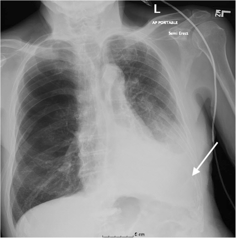
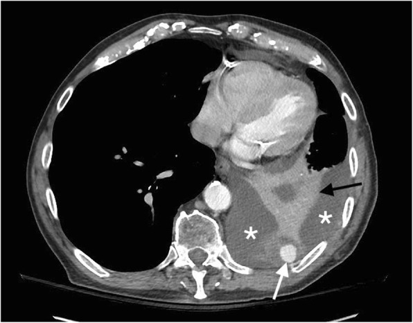
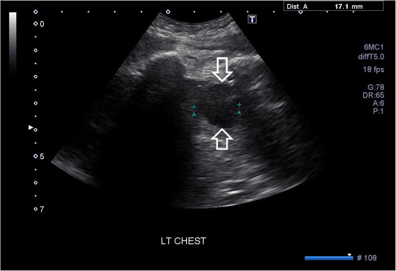
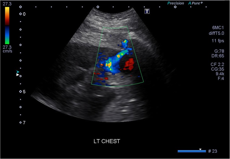
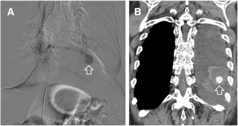
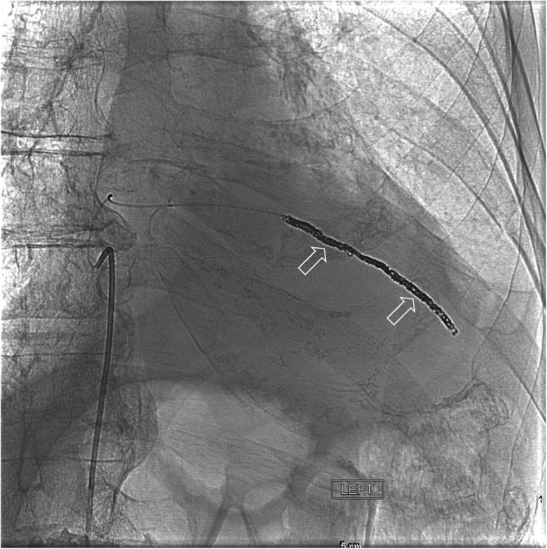
Case presentation: Here, we report a rare case of an intercostal artery pseudoaneurysm following thoracentesis in an 82-year old male. The patient presented with respiratory distress 1 day after a therapeutic thoracentesis had been performed. Computed tomography (CT) with contrast revealed a left intercostal pseudoaneurysm with hemothorax and adjacent compressive atelectasis. Doppler ultrasound revealed bidirectional blood flow in the pseudoaneurysm sac. An intercostal arteriogram and thoracic aortogram aided in confirmation of the pseudoaneurysm and successful treatment with coil embolization.
Conclusions: An intercostal pseudoaneurysm complication following thoracentesis is very rare but important to rule out as a possible cause of hemothorax after the procedure. Capturing this finding with the aid of multiple imaging modalities allowed for diagnostic certainty and rapid treatment with coil embolization, leading to a successful patient recovery.
Keywords: Coil embolism; Hemothorax; Intercostal artery; Multi-modal imaging; Pseudoaneurysm; Thoracentesis.
Conflict of interest statement
Authors’ information
KPC is a medical student in the WWAMI Medical Education Program at the University of Washington School of Medicine, 1959 NE Pacific St, Seattle, WA, 98195. PJS is a diagnostic radiologist and vascular and interventional radiologist at St Joseph Regional Medical Center, 415 6th St, Lewiston, ID, 83501. DCP is a professor in the Department of Biological Sciences and the WWAMI Medical Education Program at the University of Idaho, 875 Perimeter Drive, Moscow, ID, 83844–3051, USA.
Ethics approval and consent to participate
Not applicable.
Consent for publication
The patient has provided written informed consent to the clinical details including images of the case being submitted and published as a case report.
Competing interests
The authors declare they have no competing interests.
Publisher’s Note
Springer Nature remains neutral with regard to jurisdictional claims in published maps and institutional affiliations.
Figures
References
-
- Schwartz LB, Clark ET, Gewertz BL. Anastomotic and other pseudoaneurysms. In: Rutherford RB, editor. Vascular surgery. 5. Philadelphia: Saunders; 2000. pp. 752–763.
-
- Saad NEA, Saad WEA, Davies MG, Waldman DL, Fultz PJ, Rubens DJ. Pseudoaneurysms and the role of minimally invasive techniques in their management. RadioGraphics. 2005;25:S173–S189. - PubMed
-
- Casas JD, Perendreu J, Gallart A, Muchart J. Intercostal artery pseudoaneurysm after a percutaneous biliary procedure: diagnosis with CT and treatment with transarterial embolization. J Comput Assist Tomogr. 1997;21:729–730. - PubMed
Publication types
MeSH terms
Substances
LinkOut - more resources
Full Text Sources
