. 2019 Apr 27;50(1):28.
doi: 10.1186/s13567-019-0648-9.

UBXN1 interacts with the S1 protein of transmissible gastroenteritis coronavirus and plays a role in viral replication

Affiliations

UBXN1 interacts with the S1 protein of transmissible gastroenteritis coronavirus and plays a role in viral replication

Peng Yuan et al. Vet Res. .

Abstract

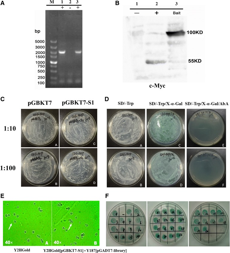
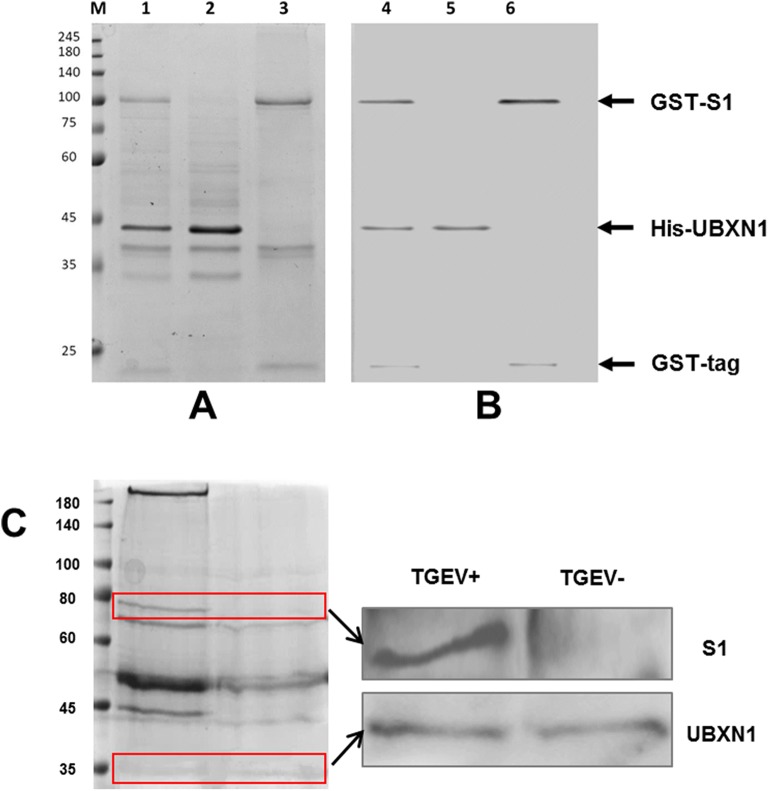
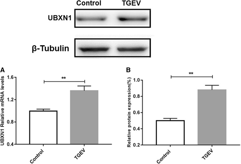
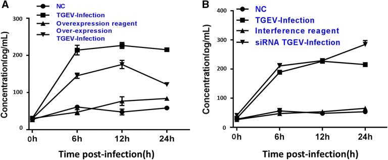
Transmissible gastroenteritis coronavirus (TGEV) is an enteropathogenic coronavirus that causes diarrhea in pigs and is associated with high morbidity and mortality in sucking piglets. S1 is one of two protein domains in the spike (S) glycoprotein and is responsible for enteric tropism, sialic acid recognition, and host receptor binding. Although there has been extensive research on the S1 protein of TGEV, little is known about the intracellular role of TGEV-S1. In the present study, we used yeast two-hybrid screening of a cDNA library from porcine intestinal cells to identify proteins that interact with TGEV-S1. Among 120 positive clones from the library, 12 intracellular proteins were identified after sequencing and a BLAST search. These intracellular proteins are involved in protein synthesis and degradation, biological signal transduction, and negative control of signaling pathways. Using a glutathione-S-transferase (GST) pulldown assay and Co-IP, we found that UBXN1 interacts with the S1 protein. Here, we observed that TGEV infection led to increased UBXN1 expression levels during the late phase of infection in IPEC-J2 cells. Inhibition of UBXN1 in IPEC-J2 cells via siRNA interference significantly decreased the viral titer and downregulated the expression of S1. UBXN1 overexpression significantly increased the viral copy number. Additionally, we provided data suggesting that UBXN1 negatively regulates IFN-β expression after TGEV infection. Finally, our research indicated that UBXN1 plays a vital role in the process of TGEV infection, making it a candidate target for the development of a novel antiviral method.


Figures

Figure 1
Figure 1
Testing bait protein expression, autoactivation, and toxicity. A Yeast colony identification by PCR. M: DNA marker DL5000; 1–2: PCR of colonies transformed with pGBKT7-S1; −: blank control. B Expression of S1 in yeast by Western blotting. 1: pGBKT7 negative control; 2: pGBKT7-53 positive control; 3: pGBKT7-S1 expressed protein. C Toxicity assessment of the pGBKT7-S1 bait plasmid. Yeast cells harboring pGBKT7-S1 and the pGBKT7 control plasmid were spread at two dilutions (1:10 and 1:100) on SD/Trp plates, and the size and number of the colonies were compared to evaluate the toxicity of the bait plasmid. D Identification of autoactivation of the pGBKT7-S1 bait plasmid. Yeast cells harboring pGBKT7-S1 were spread at 1/10 and 1/100 dilutions on SD/−Trp, SD/−Trp/X-α-Gal, and SD/−Trp/X-α-Gal/AbA plates for autoactivation testing. E A yeast two-hybrid system was used to screen for S1-interacting proteins. Diploids after yeast two-hybrid screening. The zygotes typically displayed a three-lobed structure similar to a “Mickey Mouse” face at 40× magnification. F The third screen was conducted using SD/−Trp/−Leu/−Ade/−His/X-α-Gal/AbA selective medium.
Figure 2
Figure 2
SDS-PAGE (A) and Western blotting (B) results of the glutathione-S-transferase (GST) pulldown assay and CO-IP assay. The GST-S1 and His-UBXN1 fusion proteins were used in the GST pulldown assay to verify the interaction between S1 and UBXN1. Lanes 1 and 4: result of S1 and UBXN1 pulldown; lanes 2 and 5: His-UBXN1 fusion protein (42 kDa); lanes 3 and 6: GST-S1 fusion protein (97 kDa) and GST tag blank control (20 kDa). C Co-IP assay for the interaction between S1 and UBXN1 in vivo. IPEC-J2 cells were infected with TGEV Miller for 36 h and were then subjected to a Co-IP assay. The coimmunoprecipitated proteins were detected by SDS-PAGE (left) and Western blotting (right). +: virus-infected group; −: normal group.
Figure 3
Figure 3
TGEV infection led to increased UBXN1 expression levels. Cell and protein samples from the group infected with TGEV for 24 h and the blank control group were collected, and the mRNA and protein levels of UBXN1 were determined. A Relative mRNA levels of UBXN1. B Relative protein expression of UBXN1. Control: blank group; TGEV: TGEV-infected group.
Figure 4
Figure 4
Interference and overexpression of UBXN1. A, B siRNA interference fragments reduced the expression of UBXN1. The relative expression of UBXN1 was assessed by Western blotting after 24 h of interference. Control: normal group; NC1: transfection reagent group; siRNA: fragment 1. B The lentiviral plasmid overexpressed UBXN1. The plasmid was transfected into cells for 36 h, and the relative expression of UBXN1 was assessed by Western blotting. Control: normal group; NC2: transfection reagent group; Overexpression: transfection plasmid group. C Molecular modulators of UBXN1 do not affect cell viability. Cell viability was determined by an MTT assay after treatment with the interference fragment (SiRNA) or overexpression plasmid (Plenti-CMV-GFP-2A-Puro-UBXN1) for 48 h. The data are the means ± SEMs of three independent experiments (t-test, #p > 0.05). Control: normal group; IF1: interference fragment 1 group; TP: transfection plasmid group.
Figure 5
Figure 5
UBXN1 mediated the infection and replication of TGEV. A, B Generation of the viral proliferation curves in IPEC-J2 cells under different conditions. The cells were transfected with siRNA interference fragments and overexpression plasmids for 24 h and were then infected with virus to measure the TCID50 and copy numbers. C, D Quantitative real-time reverse transcription PCR results. At 12 h after the cells were incubated with TGEV Miller, qRT-PCR was performed to measure the mRNA expression levels of TGEV N, UBXN1 and β-actin. The standard curve based on N was used to examine the TGEV copy number. TGEV-infection: virus-infected group; siRNA: virus-infected siRNA interference group. Overexpression: virus-infected overexpression group. NC: blank group. EG Western blotting results after siRNA interference and viral infection. At 24 h after siRNA interference, the cells were incubated with TGEV Miller. After 48 h, the cells were harvested and assessed by Western blotting for UBXN1, S1, and M protein expression. TGEV: virus-infected group; siRNA TGEV: virus-infected siRNA interference group.
Figure 6
Figure 6
IFN ELISA results. A IFN production was measured at 0 h, 6 h, 12 h and 24 h after TGEV infection in cells overexpressing UBXN1. B IFN production was measured at 0 h, 6 h, 12 h and 24 h after TGEV infection in cells transfected with UBXN1 siRNA. NC: normal group; TGEV-infected: virus-infected group; siRNA: virus-infected siRNA interference group; Overexpression: virus-infected overexpression group; Overexpression reagent: group with UBXN1 overexpression without TGEV infection; Interfering reagent: group with treated with UBXN1 siRNA without TGEV infection.

Similar articles

Cited by

References

    1. Weiwei H, Qinghua Y, Liqi Z, Haofei L, Shanshan Z, Qi G, Kongwang H, Qian Y. Complete genomic sequence of the coronavirus transmissible gastroenteritis virus SHXB isolated in China. Arch Virol. 2014;159:2295–2302. doi: 10.1007/s00705-014-2080-9. - DOI - PMC - PubMed
    1. Zou H, Zarlenga DS, Sestak K, Suo S, Ren X. Transmissible gastroenteritis virus: identification of M protein-binding peptide ligands with antiviral and diagnostic potential. Antivir Res. 2013;99:383–390. doi: 10.1016/j.antiviral.2013.06.015. - DOI - PMC - PubMed
    1. Yin J, Glende J, Schwegmann-Wessels C, Enjuanes L, Herrler G, Ren X. Cholesterol is important for a post-adsorption step in the entry process of transmissible gastroenteritis virus. Antivir Res. 2010;88:311–316. doi: 10.1016/j.antiviral.2010.10.002. - DOI - PMC - PubMed
    1. Ahn DJ, Youm JW, Kim SW, Yoon WK, Kim HC, Hur TY, Joung YH, Jeon JH, Kim HS. Expression of the S glycoprotein of transmissible gastroenteritis virus (TGEV) in transgenic potato and its immunogenicity in mice. Korean J Vet Res. 2013;53:217–224. doi: 10.14405/kjvr.2013.53.4.217. - DOI
    1. Belouzard S, Millet JK, Licitra BN, Whittaker GR. Mechanisms of coronavirus cell entry mediated by the viral spike protein. Viruses. 2012;4:1011–1033. doi: 10.3390/v4061011. - DOI - PMC - PubMed

MeSH terms

Substances