. 2019 Apr 27;10(1):126.
doi: 10.1186/s13287-019-1229-6.

Silk fibroin scaffolds seeded with Wharton's jelly mesenchymal stem cells enhance re-epithelialization and reduce formation of scar tissue after cutaneous wound healing

Affiliations

Silk fibroin scaffolds seeded with Wharton's jelly mesenchymal stem cells enhance re-epithelialization and reduce formation of scar tissue after cutaneous wound healing

José E Millán-Rivero et al. Stem Cell Res Ther. .

Abstract

Background: The treatment of extensive and/or chronic skin wounds is a widespread and costly public health problem. Mesenchymal stem cells (MSCs) have been proposed as a potential cell therapy for inducing wound healing in different clinical settings, alone or in combination with biosynthetic scaffolds. Among them, silk fibroin (SF) seeded with MSCs has been shown to have increased efficacy in skin wound healing experimental models.

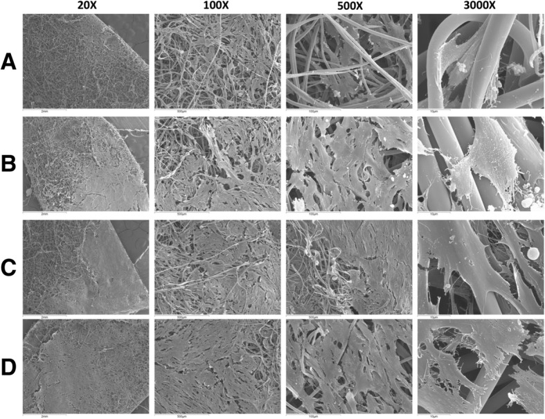
Methods: In this report, we investigated the wound healing effects of electrospun SF scaffolds cellularized with human Wharton's jelly MSCs (Wj-MSCs-SF) using a murine excisional wound splinting model.

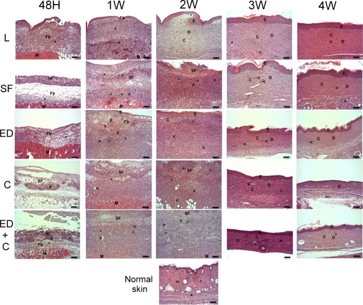
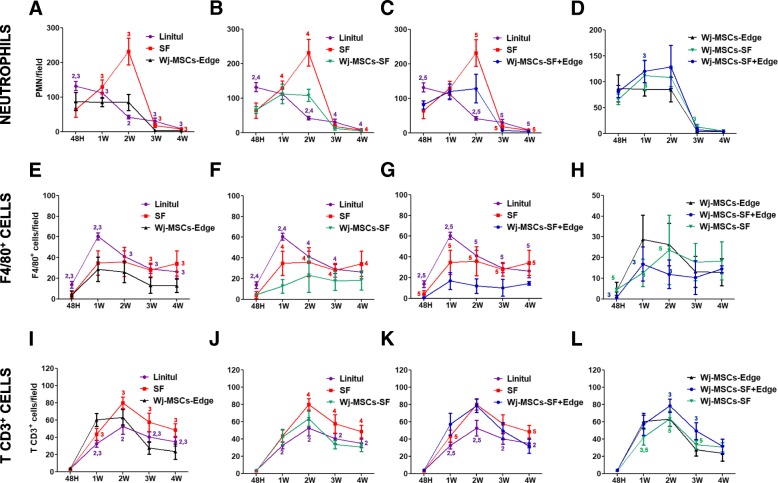
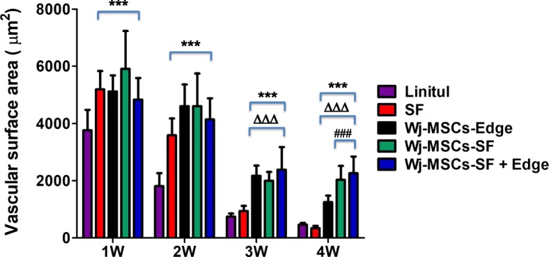
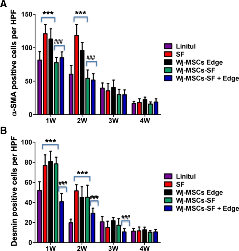
Results: Immunohistopathological examination after transplant confirmed the presence of infiltrated human fibroblast-like CD90-positive cells in the dermis of the Wj-MSCs-SF-treated group, yielding neoangiogenesis, decreased inflammatory infiltrate and myofibroblast proliferation, less collagen matrix production, and complete epidermal regeneration.

Conclusions: These findings indicate that Wj-MSCs transplanted in the wound bed on a silk fibroin scaffold contribute to the generation of a well-organized and vascularized granulation tissue, enhance reepithelization of the wound, and reduce the formation of fibrotic scar tissue, highlighting the potential therapeutic effects of Wj-MSC-based tissue engineering approaches to non-healing wound treatment.

Keywords: Mesenchymal stem cells; Silk fibroin; Wharton’s jelly; Wound healing.


Conflict of interest statement

Ethics approval and consent to participate

This study was approved by the ethics committee of Hospital Clinico Universitario Virgen de la Arrixaca (Murcia, Spain). Umbilical cord donors provided written and informed consent in accordance with the Declaration of Helsinki. All procedures involving animals were previously approved by the University of Murcia animal care committee and conducted in accordance with the national guidelines on animal care.

Consent for publication

Not applicable

Competing interests

The authors declare that they have no competing interests.

Publisher’s Note

Springer Nature remains neutral with regard to jurisdictional claims in published maps and institutional affiliations.

Figures

Fig. 1
Fig. 1
Proliferation kinetics of Wj-MSCs and BM-MSCs overextended in vitro propagation. a Number of cumulative population doubling level (PD) as a function of time in culture. b Cell population doubling time (PDT) (hours) after sequential passages. Statistically significant differences using one-way ANOVA, N = 3; *p < 0.05, **p < 0.01, ***p < 0.001. c The capacity of BM- or Wj-MSCs to inhibit the proliferation of stimulated peripheral blood T cells was analyzed. BM- or Wj-MSCs were cultured with 1 × 105 MNCs in different ratios and stimulated with CD3/CD28 beads for 5 days. After, the proliferation of the T cells was measured by thymidine (3H-Thy) incorporation. MSCs from both sources significantly inhibited the proliferation of T cells in a dose-dependent manner (***p < 0.001). The proliferation of T cells in the presence of Wj-MSCs was significantly lower than that obtained using BM-MSCs at the same ratio (##p < 0.01). All data are presented as mean ± SD. N = 3. d Immunophenotypical analysis of Wj-MSCs by flow cytometry. Wj-MSCs were seeded at a density of 5 × 104 cells/cm2 on plastic culture plates (control) (upper panels) or SF patches (bottom panels) for 4 days. After, cells were detached and labeled with specific antibodies for the indicated markers or their control isotypes. Histograms show representative flow cytometry results obtained from N = 3 independent experiments. e To evaluate Wj-MSCs multipotent differentiation properties, cells were cultured in adipogenic, osteogenic, and chondrogenic differentiation media for 14–21 days. After, differentiation was evaluated by staining of lipid droplets with Oil Red O (adipogenic, right), by detection of calcium depositions and alkaline phosphatase activity by Alizarin Red and BCIP-NBT staining (osteogenic, middle left and middle right, respectively), or by detection of glycosaminoglycans by Alcian blue staining (chondrogenic, right). Images shown are representative of N = 3 independent experiments. Scale bar 200 μm. Abbreviations: PD population doubling level, PDT population doubling time, Wj-MSCs Wharton’s jelly MSCs, BM-MSCs bone marrow MSCs
Fig. 2
Fig. 2
Scanning electron microscopy micrographs of Wj-MSCs growing on electrospun SF scaffold at different cell densities. SF patches were seeded at a density of 3 × 104cells/cm2 (a), 4 × 104cells/cm2 (b), 5 × 104cells/cm2 (c), and 6 × 104cells/cm2 (d) and cultured for 4 days. Images at a magnification of × 20, × 100, × 500, and × 3000 are shown. N = 3
Fig. 3
Fig. 3
Histopathological assessment of wound healing by hematoxylin and eosin (H&E) staining. Control groups: L (wounds covered with Linitul) and SF (wounds covered with SF scaffolds). Treated groups: ED (wounds treated with Wj-MSCs injected at the edge), C (wounds covered with SF patches cellularized with Wj-MSCs), and ED+C (wounds with SF patches cellularized with Wj-MSCs over the wound bed combined with Wj-MSCs injected at the edge). Abbreviations: Fb fibrin, “*” inflammatory infiltrate and edema, “>” blood vessels, E epidermis, M muscular layer, D dermis, C collagen, SF silk fibroin scaffold, PD papillary dermis, RD reticular dermis. Scale bar 100 μm
Fig. 4
Fig. 4
Influx of inflammatory cells into wounded tissues: polymorphonuclear neutrophils (ad), macrophages (eh), and T cells (il). Experimental groups: wounds covered with Linitul (purple line), wounds covered with SF scaffolds (red line), Wharton’s jelly MSCs injected at the wound edge (Wj-MSCs-Edge) (black line), wounds covered with SF scaffolds cellularized with Wharton’s jelly MSCs (Wj-MSCs-SF) (green line), and wounds treated with Wharton’s jelly MSCs injected at the wound edge and cellularized SF scaffolds onto the wound bed (Wj-MSCs-SF+Edge) (blue line). Statistically significant differences (p < 0.05) compared to Linitul (value 1), scaffold (value 2), Wj-MSCs-Edge (value 3), Wj-MSCs-SF (value 4), and Wj-MSCs-SF+Edge (value 5), according to one-way ANOVA
Fig. 5
Fig. 5
Vascular surface area (expressed in square micrometers per field) determined by immunohistochemical analysis of CD31 expression in wound sections. Vascularized area was significantly increased compared to the untreated group (Linitul) (***p < 0.001), SF group (∆∆∆p < 0.001), or Wj-MSCs-Edge-treated group (###p < 0.001), respectively, according to one-way ANOVA. Results are shown as mean ± SD of the most vascularized areas measured in three different mice of each group, corresponding to images made at × 400 magnification. Experimental groups: SF (wounds covered with silk fibroin scaffold), Wj-MSCs-Edge (Wj-MSCs injected at the wound edge), Wj-MSCs-SF (wounds covered with silk fibroin scaffold cellularized with Wj-MSCs), and Wj-MSCs-SF+Edge (wounds treated with Wj-MSCs injected at the wound edge and cellularized silk fibroin scaffold onto the wound bed)
Fig. 6
Fig. 6
Immunohistochemical analysis of α-SMA expression and desmin in wound sections. For quantitative assessment of myofibroblast proliferation, the number of positive cells for α-SMA (a) or desmin (b) was determined in ten random sections at × 400 magnification from three different mice of each group. Results are shown as mean ± SD. Myofibroblast proliferation was significantly increased compared to the untreated group (Linitul) (***p < 0.001) or significantly decreased compared to the Wj-MSCs-Edge-treated group (###p < 0.001), respectively, according to one-way ANOVA. Experimental groups: SF (wounds covered with silk fibroin scaffold), Wj-MSCs-Edge (Wj-MSCs injected at the wound edge), Wj-MSCs-SF (wounds covered with silk fibroin scaffold cellularized with Wj-MSCs), and Wj-MSCs-SF+Edge (wounds treated with Wj-MSCs injected at the wound edge and cellularized silk fibroin scaffold onto the wound bed)
Fig. 7
Fig. 7
ae Expression of the human mesenchymal stem cell marker CD90 on mouse dermal wounds. Experimental groups: L (wounds covered with Linitul), SF (wounds covered with SF scaffolds), ED (wounds with Wj-MSCs injected at the edge), C (wounds covered with SF patches cellularized with Wj-MSCs), and ED+C (wounds with SF patches cellularized with Wj-MSCs over the wound bed and Wj-MSCs injected at the edge). Immunostaining with the anti-human CD90 antibody of a mouse skin section and a pellet made of Wj-MSCs served as negative and positive controls, respectively (f). Scale bar 50 μm

Similar articles

Cited by

References

    1. Singer AJ, Clark RA. Cutaneous wound healing. N Engl J Med. 1999;341:738–746. doi: 10.1056/NEJM199909023411006. - DOI - PubMed
    1. Coutinho P, Qiu C, Frank S, Tamber K, Becker D. Dynamic changes in connexin expression correlate with key events in the wound healing process. Cell Biol Int. 2003;27:525–541. doi: 10.1016/S1065-6995(03)00077-5. - DOI - PubMed
    1. Martin P. Wound healing-aiming for perfect skin regeneration. Science. 1997;276:75–81. doi: 10.1126/science.276.5309.75. - DOI - PubMed
    1. Falanga V. Wound healing and its impairment in the diabetic foot. Lancet. 2005;366:1736–1743. doi: 10.1016/S0140-6736(05)67700-8. - DOI - PubMed
    1. Kirby GT, Mills SJ, Cowin AJ, Smith LE. Stem cells for cutaneous wound healing. Biomed Res Int. 2015;2015:285869. doi: 10.1155/2015/285869. - DOI - PMC - PubMed

Publication types