. 2019 Jul;96(1):80-93.
doi: 10.1016/j.kint.2019.01.037. Epub 2019 Feb 27.

Novel parietal epithelial cell subpopulations contribute to focal segmental glomerulosclerosis and glomerular tip lesions

Affiliations

Novel parietal epithelial cell subpopulations contribute to focal segmental glomerulosclerosis and glomerular tip lesions

Christoph Kuppe et al. Kidney Int. 2019 Jul.

Abstract

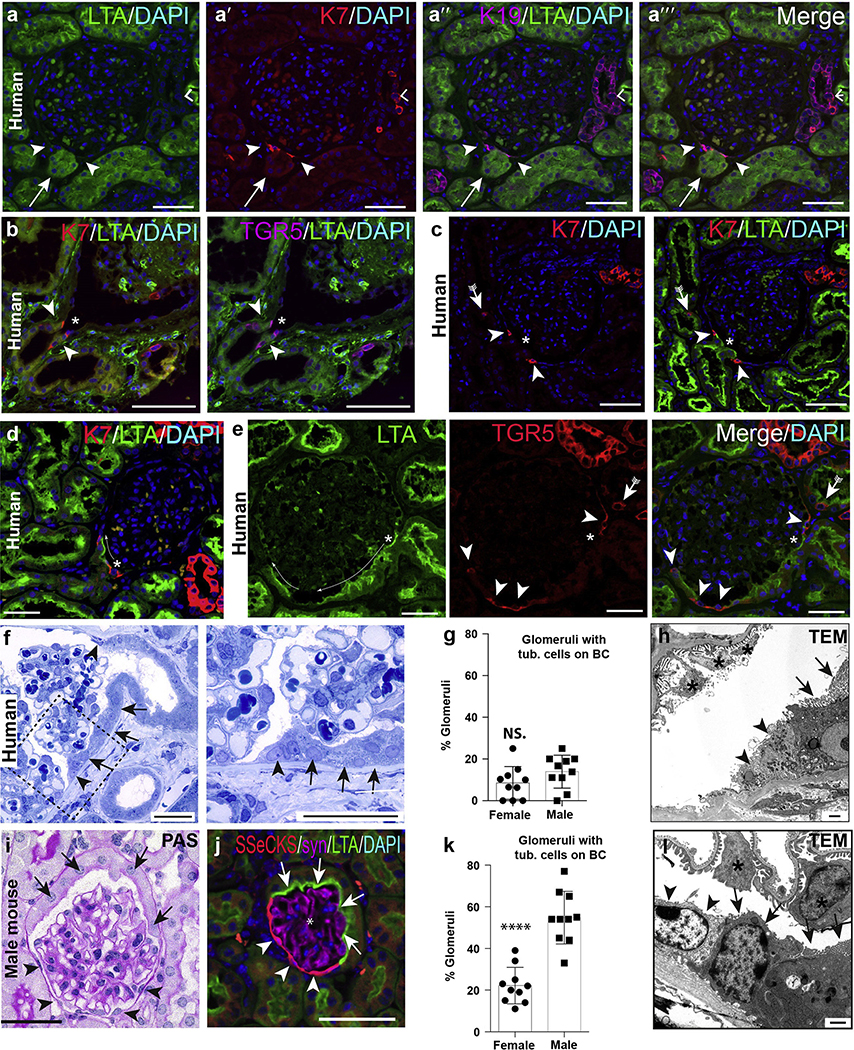
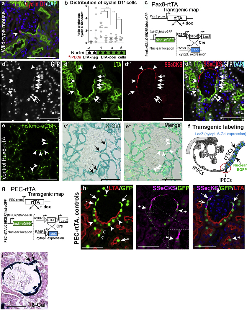
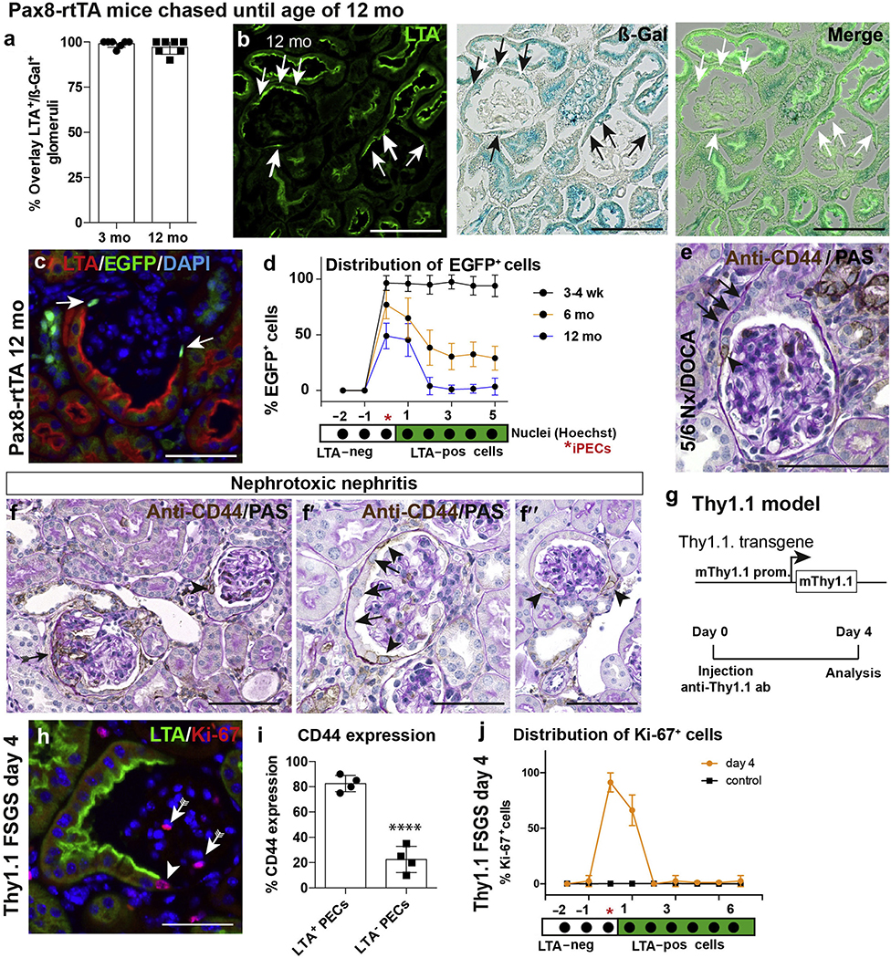
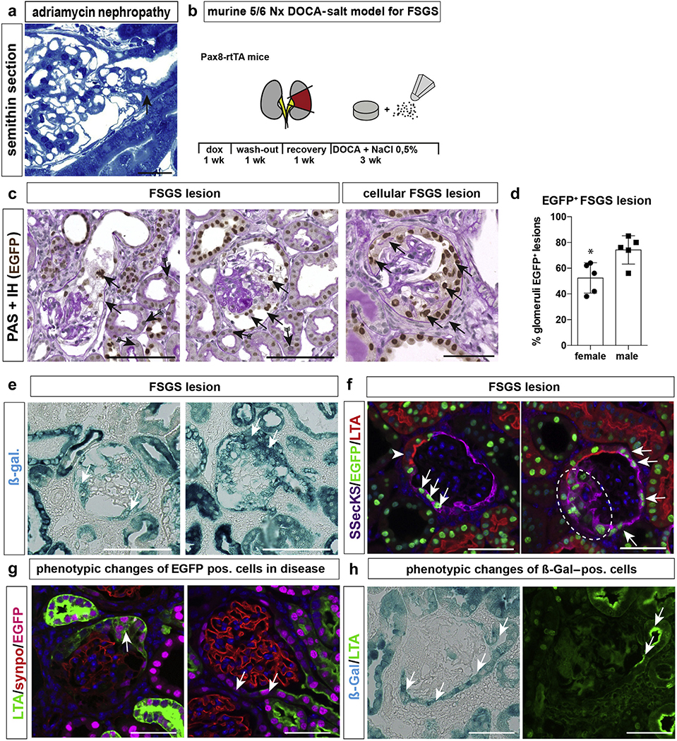
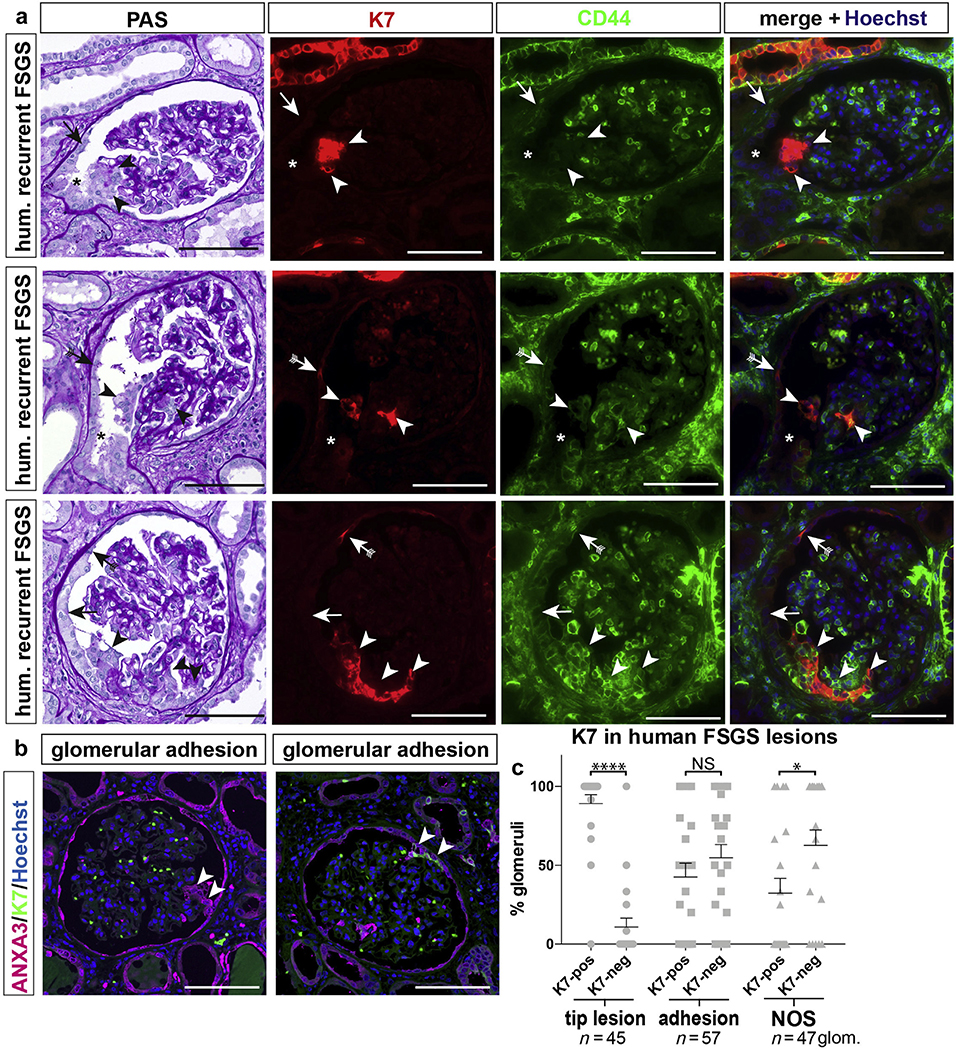
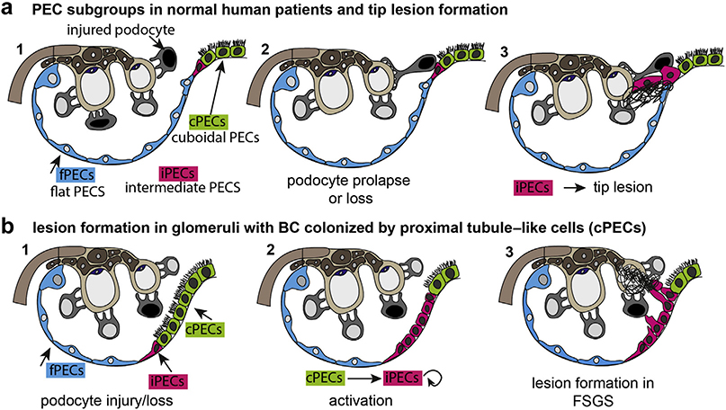
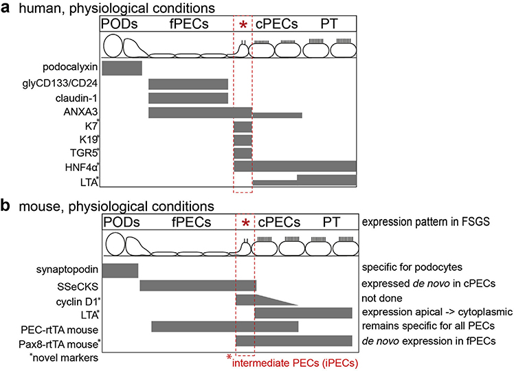
Beside the classical flat parietal epithelial cells (PECs), we investigated proximal tubular epithelial-like cells, a neglected subgroup of PECs. These cells, termed cuboidal PECs, make up the most proximal part of the proximal tubule and may also line parts of Bowman's capsule. Additionally, a third intermediate PEC subgroup was identified at the junction between the flat and cuboidal PEC subgroups at the tubular orifice. The transgenic mouse line PEC-rtTA labeled all three PEC subgroups. Here we show that the inducible Pax8-rtTA mouse line specifically labeled only cuboidal and intermediate PECs, but not flat PECs. In aging Pax8-rtTA mice, cell fate mapping showed no evidence for significant transdifferentiation from flat PECs to cuboidal or intermediate PECs or vice versa. In murine glomerular disease models of crescentic glomerulonephritis, and focal segmental glomerulosclerosis (FSGS), intermediate PECs became more numerous. These intermediate PECs preferentially expressed activation markers CD44 and Ki-67, suggesting that this subgroup of PECs was activated more easily than the classical flat PECs. In mice with FSGS, cuboidal and intermediate PECs formed sclerotic lesions. In patients with FSGS, cells forming the tip lesions expressed markers of intermediate PECs. These novel PEC subgroups form sclerotic lesions and were more prone to cellular activation compared to the classical flat PECs in disease. Thus, colonization of Bowman's capsule by cuboidal PECs may predispose to lesion formation and chronic kidney disease. We propose that tip lesions originate from this novel subgroup of PECs in patients with FSGS.

Keywords: Columbia classification; FSGS; glomerular disease; parietal epithelial cells; tip lesions.


Conflict of interest statement

DISCLOSURE

All the authors declared no competing interests.

Figures

Figure 1 |
Figure 1 |. Parietal epithelial cell (PEC) subpopulations in humans and mice.
(a–e) Immunofluorescence stainings of normal human kidney shows co-expression of keratin 7 (K7), K19, and G protein–coupled bile acid receptor 1 (TGR5) in intermediate PECs at the tubular (tub.) orifice (arrowheads) and within cells of the proximal (scattered tubular cells, arrows with tails) and distal tubule (right angles). *Podocytes (a,h). As shown in the examples (c) to (e), lotus tetragonolobus agglutinin (LTA)+ proximal tubular-like cells may extend onto Bowman’s capsule (BC; cuboidal PECs, long thin arrows). Bars = 100 μm. (f–h) Cuboidal PECs with brush border in human biopsy kidney section (arrows). In addition, intermediate PECs (arrowheads) between flat and cuboidal PECs (semithin section). Bar = 50 μm. (g) Quantification of cuboidal PECs in tumor nephrectomies of human male and female subjects (1 dot equals mean of 40–50 glomeruli per patient, n = 10 each, unpaired t-test; not significant [n.s.]). (h) Transmission electron microscopy (TEM) showing intermediate PECs (arrowheads) and cuboidal PECs on Bowman’s capsule in human kidney. Bar = 1 μm. (il) PEC subpopulations in healthy mice. (i) Periodic acid–Schiff (PAS)–staining. Bar = 50 μm. (j) Immunofluorescent staining for src-suppressed C-kinase (SSeCKS; flat PECs, arrowheads), LTA (brush border of cuboidal PECs, arrows), and synaptopodin (*podocytes). (k) Quantification of glomerular cross sections with LTA+ cells on Bowman’s capsule (****P < 0.0001, n = 10 mice/sex, 25 glomerular cross sections per mouse). (l) TEM of Bowman’s capsule in mice. PECs show brush border at the apical membrane (arrows with tails). Another population shows an intermediate cell phenotype with an absent brush border and reduced amount of mitochondria (arrowheads). Bar = 1000 nm. DAPI, 4′,6-diamidino-2-phenylindole. To optimize viewing of this image, please see the online version of this article at www.kidney-international.org.
Figure 2 |
Figure 2 |. Markers for intermediate parietal epithelial cells (iPECs) in mice (a,b) and lineage tracing and immunolabeling of PEC populations (d–i).
(a) Distribution of cyclin D1+ cells on Bowman’s capsule in wild-type mice. (Immunofluorescent staining for cyclin D1, red and lotus tetragonolobus agglutinin [LTA], green.) Note that cyclin D1 showed the highest expression level in presumptive iPECs between flat PECs (fPECs) and cuboidal PECs (cPECs). (b) Analysis of distribution of cyclin D1 on Bowman’s capsule (n = 10; shown are the mean and SDs, analysis of variance; *P < 0.1, **P < 0.01). (c–i) Pax8-rtTA transgenic reporter mice label cuboidal and intermediate PECs on Bowman’s capsule. (c) Transgenic map of Pax8-rtTA mice. (d) Quadruple immunofluorescent staining (src-suppressed C-kinase [SSeCKS], enhanced green fluorescent protein [EGFP], LTA, and 4′,6-diamidino-2-phenylindole [DAPI]) in Pax8-rtTA mice shows no labeling with EGFP in fPECs (arrowheads), whereas cPECs stained positive (pos) for EGFP. Note that SSeCKS marks fPECs and iPECs are marked by both EGFP and SSeCKS (arrows with tails). Bars = 75 μm (a,d). (e) Co-staining of histone-eGFP and X-gal staining. EGFP (arrowheads) staining was only positive in ß-Gal+ cPECs (arrows). Bars = 100 μm. (f) Schematic of the labeling pattern in Pax8-rtTA quadruple transgenic mice. (g) Transgenic map of the PEC-rtTA mouse. (h) It labels SSeCKS+ fPECs (arrows), iPECs (arrows with circles), and LTA+ cPECs (arrowheads) including the proximal part of the proximal tubule (right angles). Intermediate PECs are highlighted in the inset; note that SSeCKS staining is on both sides of the green nucleus (arrows). As described previously, proximal tubular cells in the scattered tubular cell phenotype (LTA-negative) are labeled by the PEC-rtTA mouse as well (arrow with tails). Bars = 50 μm. (i) Labeling of the proximal part of the proximal tubule by the PEC-rtTA mouse visualized by the irreversible reporter β-galactosidase (β-Gal) (arrows). Bar = 50 μm. cytopl, cytoplasmic; dox, doxycycline; neg, negative; prom, promoter. To optimize viewing of this image, please see the online version of this article at www.kidney-international.org.
Figure 3 |
Figure 3 |. Lineage tracing of Pax8-rtTA mice (a–d) and preferential activation of intermediate parietal epithelial cells (iPECs) and cuboidal PECs (e–j).
(a) Lotus tetragonolobus agglutinin (LTA)+ PECs were nearly always positive (pos) for β-galactosidase (ß-gal) at 3 months of age. (b) Co-staining of ß-gal and LTA in PAX8-rtTA mice at 12 months of age shows co-expression of both markers (arrows). Bars = 100 μm. (c,d) Analysis of enhanced green fluorescent protein (EGFP) in kidneys of aged Pax8-rtTA mice. PECs located between flat PECs and cuboidal PECs showed preferentially a positive staining for EGFP (arrows). Bar = 50 μm. (d) Analysis of distribution of EGFP signal on Bowman’s capsule. n = 5 per time point. (e) CD44 immunostaining of kidneys of mice subjected to 5/6 nephrectomy (Nx) + deoxycorticosterone acetate (DOCA)–salt treatment as outlined in Figure 4b and (f) of mice treated with nephrotoxic nephritis serum to induce crescentic glomerulonephritis for 8 days. In sporadic glomeruli without lesions, CD44 was positive on Bowman’s capsule in a subset of iPECs (arrowheads) preferentially at the interface between flat PECs and cuboidal PECs (arrows in f,h). Bars = 100 μm (e,f) and 50 μm (f′,f′′). (g) Schematic summary of the Thy1.1 focal segmental glomerulosclerosis (FSGS) mouse model, which expresses the Thy1.1 antigen in podocytes. Acute podocyte injury and FSGS can be induced after injection of Thy1 antibody (ab) 19XE5. (h) Exemplary immunofluorescence staining of Ki-67 and LTA 4 days after induction of FSGS demonstrates preferential expression of proliferation marker Ki-67 in iPECs (arrowhead). Bar = 50 μm. (i) Analysis of de novo expression of CD44 in LTA-negative (neg) versus LTA+ PECs (i.e., flat vs. cuboidal PECs, respectively) in Thy1.1 mice (n = 4; 50 glomerular cross sections per mouse, unpaired t-test; ****P < 0.0001). (j) Analysis of the distribution of Ki-67 expression in PECs on Bowman’s capsule in Thy1.1 mice 4 days after induction. PAS, periodic acid–Schiff; prom, promoter. To optimize viewing of this image, please see the online version of this article at www.kidney-international.org.
Figure 4 |
Figure 4 |. Tubularized parietal epithelial cells (PECs) contribute to focal segmental glomerulosclerosis (FSGS) lesions in mice.
(a) FSGS tip lesion (arrow) in mice treated with adriamycin to induce FSGS (arrow, semithin sections, 2 weeks after injection). Bar = 20 μm. (b) Schematic of 5/6 nephrectomy (Nx)+ deoxycorticosterone acetate (DOCA)–salt model. (c) Exemplary images of FSGS lesions show green fluorescent protein (GFP)+ cells within sclerotic lesions (arrows). Histone-eGFP is lost in some of the proximal tubular cells (arrows with tails; periodic acid–Schiff [PAS]-stainings and enhanced GFP (eGFP) immunostaining in Pax8rtTA mice). (d) Quantification of eGFP+ (pos) FSGS lesion (*P = 0.0165, n = 5). (e) β-galactosidase (ß-gal) staining of kidneys from Pax8-rtTA mice in the 5/6 nephrectomy and DOCA-salt FSGS model. (f) Presumptive FSGS lesions in the early stage (left panel) or late stage (right panel; dashed circle marks sclerotic lesion); eGFP+ cells express src-suppressed C-kinase (SSeCKS) de novo (arrows; cuboidal PECs, arrowhead). (g) Immunofluorescence of lotus tetragonolobus agglutinin (LTA) (green), eGFP (magenta), and synaptopodin (synpo; red) shows loss of polarized expression of LTA in eGFP+ PECs within a proliferative lesion (left) and complete loss of expression in eGFP+ cells on Bowman’s capsule (right panel). (h) Co-staining of ß-gal and LTA. LTA-negative ß-gal+ PECs can be observed (arrows), indicating that cuboidal PECs have lost their brush border in FSGS. Bars = 50 μm. dox, doxycycline; IH, immunohistochemistry. To optimize viewing of this image, please see the online version of this article at www.kidney-international.org.
Figure 5 |
Figure 5 |. Tip lesions contain keratin 7 (K7)+ (pos) cells.
Analysis of human (hum) glomerular (glom) tip lesions, adhesions, or not otherwise specified (NOS) sclerotic lesions in transplant patients with early recurrence of focal segmental glomerulosclerosis (FSGS). (a) Serial sections stained with periodic acid-Schiff (PAS) or immunofluorescent anti-K7 (red) and anti-CD44 (green). Cells within tip lesions were K7+ (arrowheads) but not always CD44+. K7+ cells were observed also on Bowman’s capsule (presumptive iPECs, arrows with tails). CD44 was expressed by tubular cells in the scattered tubular cell phenotype and inflammatory cells. Additional autofluorescent staining comes from erythrocytes within the glomerular tuft (green). Asterisks indicate the tubular outlet. (b,c) Frequency of K7+ lesions in FSGS. Exemplary images of a K7-negative (neg) (left panel) or pos (right panel) glomerular adhesion (arrowheads), visualized by the global PEC marker annexin A3 (ANXA3). (c) ANXA3+ tip lesions contained K7+ cells in 89.2% in 23 patients diagnosed with the tip variant of FSGS (*P < 0.001, ****P < 0.0001, n = 45 glomeruli). This difference was less pronounced in other adhesions away from the tubular outlet or FSGS lesions not otherwise specified. Bar = 100 μm. NS, not significant. To optimize viewing of this image, please see the online version of this article at www.kidney-international.org.
Figure 6 |
Figure 6 |. Schematic summary and proposed pathways of early lesion formation.
(a) Parietal epithelial cell (PEC) subgroups are highlighted in color. Upon glomerular injury, podocytes are injured and partially lost. Some prolapse toward the tubular outlet (black nucleus) and come into contact with intermediate PECs (iPECs) at the epithelial interface. iPECs migrate onto the glomerular tuft and deposit their matrix (tip lesion). (b) In male mice and in some human patients, proximal tubule cells extend onto Bowman’s capsule (BC) and are known as cuboidal PECs (cPECs). In glomerular disease, cellular activation results in proliferation of iPECs. In addition, cPECs may transition into the intermediate phenotype. iPECs form adhesions that are not immediately at the tubular orifice. As demonstrated previously, flat PECs (fPECs) may also become activated and may proliferate and participate in lesion formation (not shown in this schematic). FSGS, focal segmental glomerulosclerosis.
Figure 7 |
Figure 7 |. Summary of markers of this study.
Markers employed in this study to identify specific subgroups of cells within the human (a) or murine (b) glomerulus under physiological conditions. Novel markers are marked with an asterisk; the expression pattern of the markers is noted in mice in experimental focal segmental glomerulosclerosis (FSGS). ANXA3, annexin A3; cPECs, cuboidal PECs; fPECs, focal PECs; glyCD133/CD24, glycosylated CD133 and CD24; HNF4α, hepatocyte nuclear factor-4; iPECs, intermediate PECs; K7, keratin 7; LTA, lotus tetragonolobus agglutinin; PEC, parietal epithelial cell; PODs, podocytes; PT, proximal tubule; SSECKS, src-suppressed C-kinase; TGR5, G protein-coupled bile acid receptor 1.

Comment in

Similar articles

Cited by

References

    1. Levey AS, Coresh J. Chronic kidney disease. Lancet. 2012;379:165–180. - PubMed
    1. Wiggins RC. The spectrum of podocytopathies: a unifying view of glomerular diseases. Kidney Int. 2007;71:1205–1214. - PubMed
    1. Smeets B, Kuppe C, Sicking EM, et al. Parietal epithelial cells participate in the formation of sclerotic lesions in focal segmental glomerulosclerosis. J Am Soc Nephrol. 2011;22:1262–1274. - PMC - PubMed
    1. Smeets B, Uhlig S, Fuss A, et al. Tracing the origin of glomerular extracapillary lesions from parietal epithelial cells. J Am Soc Nephrol. 2009;20:2604–2615. - PMC - PubMed
    1. Bollee G, Flamant M, Schordan S, et al. Epidermal growth factor receptor promotes glomerular injury and renal failure in rapidly progressive crescentic glomerulonephritis. Nat Med. 2011;17:1242–1250. - PMC - PubMed

Publication types

MeSH terms

LinkOut - more resources