. 2019 Aug:100:92-101.
doi: 10.1016/j.ssnmr.2019.04.003. Epub 2019 Apr 17.

A biradical-tagged phospholipid as a polarizing agent for solid-state MAS Dynamic Nuclear Polarization NMR of membrane proteins

Affiliations

A biradical-tagged phospholipid as a polarizing agent for solid-state MAS Dynamic Nuclear Polarization NMR of membrane proteins

Daryl B Good et al. Solid State Nucl Magn Reson. 2019 Aug.

Abstract

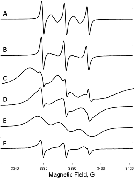
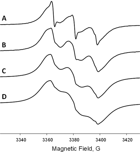
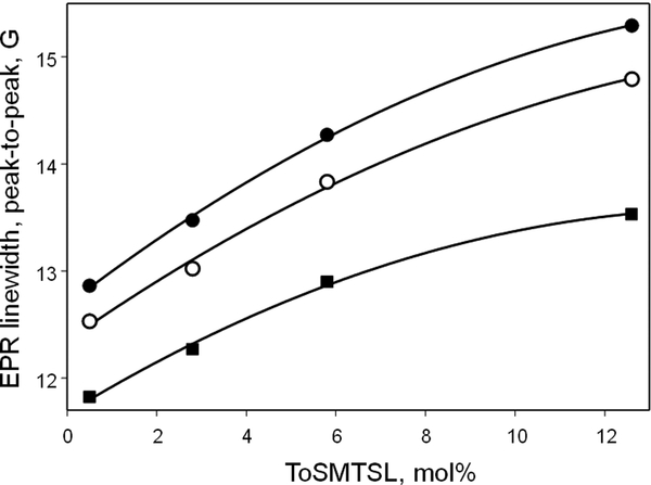
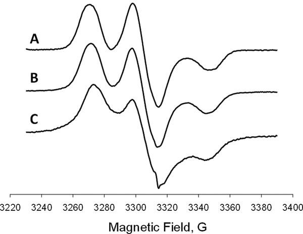
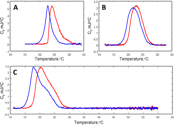
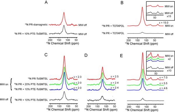
A novel Dynamic Nuclear Polarization (DNP) NMR polarizing agent ToSMTSL-PTE representing a phospholipid with a biradical TOTAPOL tethered to the polar head group has been synthesized, characterized, and employed to enhance solid-state Nuclear Magnetic Resonance (SSNMR) signal of a lipid-reconstituted integral membrane protein proteorhodopsin (PR). A matrix-free PR formulation for DNP improved the absolute sensitivity of NMR signal by a factor of ca. 4 compared to a conventional preparation with TOTAPOL dispersed in a glassy glycerol/water matrix. DNP enhancements measured at 400 MHz/263 GHz and 600 MHz/395 GHz showed a strong field dependence but remained moderate at both fields, and comparable to those obtained for PR covalently modified with ToSMTSL. Additional continuous wave (CW) X-band electron paramagnetic resonance (EPR) experiments with ToSMTSL-PTE in solutions and in lipid bilayers revealed that an unfavorable conformational change of the linker connecting mononitroxides could be one of the reasons for moderate DNP enhancements. Further, differential scanning calorimetry (DSC) and CW EPR experiments indicated an inhomogeneous distribution and/or a possibility of a partial aggregation of ToSMTSL-PTE in DMPC:DMPA bilayers when the concentration of the polarizing agent was increased to 20 mol% to maximize the DNP enhancement. Thus, conformational changes and an inhomogeneous distribution of the lipid-based biradicals in lipid bilayers emerged as important factors to consider for further development of this matrix-free approach for DNP of membrane proteins.

Keywords: Bilayer; Biradical; Dynamic nuclear polarization; Electron spin resonance; Lipid; Membrane protein; Nuclear magnetic resonance; Signal enhancement.


Conflict of interest statement

Authors declare no competing financial interest.

Figures

Figure 1.
Figure 1.
Chemical structure of ToSMTSL-PTE – an unnatural phospholipid with a biradical tethered to the lipid polar head synthesized and characterized in this work.
Figure 2.
Figure 2.
Room temperature (23 °C) X-band (9.5 GHz) CW EPR spectra of nitrogen equilibrated 1 mM chloroform solutions of ToSMTSL (A) and ToSMTSL-PTE (B). EPR spectra of aqueous suspension of multilamellar lipid vesicles (10 w% of lipids, 24 mM CHES buffer, 10 mM NaCl, pH=9.0) prepared from the mixture of DMPC:DMPA (9:1 w:w) and ToSMTSL-PTE at 0.5 mol% measured at 23 °C (C) and 55 °C (D). (E) is a simulated broad component attributable to ToSMTSL-PTE and (F) is the difference spectrum characteristic of a nitroxide biradical partitioning between the lipid and the aqueous bilayer phases. All spectra are normalized by peak-to-peak amplitude and magnetic field positions are adjusted by g-factors.
Figure 3.
Figure 3.
X-band (9.5 GHz) EPR spectra of lipid vesicles (aqueous suspension of 10 w% of lipids in 24 mM CHES buffer, 10 mM NaCl, pH=9.0) prepared from the mixture of DMPC:DMPA (9:1 w:w) and doped with ToSMTSL-PTE at 0.5 mol% (A), 2.8 mol% (4.5 mM) (B), 5.8 mol% (8.9 mM) (C), and 12.6 mol% (17.9 mM) (D) and measured at 55 °C. All spectra are normalized by peak-to-peak amplitude and magnetic field positions are adjusted by g-factors.
Figure 4.
Figure 4.
Best-fit peak-to-peak EPR linewidths of each of the three nitrogen hyperfine coupling components of X-band (9.5 GHz) CW EPR spectra of lipid vesicles (aqueous suspension of 10 w% of lipids, in 24 mM CHES buffer, 10 mM NaCl, pH=9.0) prepared from the mixture of DMPC:DMPA as a function of ToSMTSL-PTE concentration. All spectra were measured at 55 °C. The solid lines are quadratic regressions provided to guide the eye. See text for details.
Figure 5.
Figure 5.
Experimental rigid limit (T = 77 K) X-band (9.5 GHz) EPR spectra of (A) 0.8 mM solution of TOTAPOL in water:glycerol (80:20 v/v), (B) 0.8 mM solution of TOTAPOL in water:glycerol (80:20 v/v) when mixed with DMPC:DMPA lipid bilayers, and (C) lipid bilayers (dispersion 10 w% of total lipids in 24 mM CHES buffer, 10 mM NaCl, pH=9.0) composed of DMPC:DMPA and doped with 0.5 mol% of ToSMTSL-PTE. All spectra are normalized by peak-to-peak amplitude and magnetic field positions are adjusted by g-factors.
Figure 6.
Figure 6.
DSC data for aqueous dispersions (10% of total lipids) of multilamellar vesicles composed of pure DMPC:DMPA (A), and doped with ToSMTSL-PTE at 5.8 mol% (B) and 12.6 mol% (C) of total lipids. Red lines represent heating temperature scan while the cooling scans are colored in blue.
Figure 7.
Figure 7.
Comparison of CPMAS NMR spectra of different 15N-labeled PR sample preparations measured with mm-wave on and off. (A) 15N spectra of diamagnetic PR (no biradicals present) and PR with 10% ToSMTSL-PTE collected at a 1H Larmor frequency of 600 MHz in a H2O-based buffer. (B) 15N spectra PR demonstrating DNP enhancements from TOTAPOL. Inset shows the region of arginine and lysine side chains. (C) Spectra collected on PR a 1H Larmor frequency of 600 MHz in a H2O-based buffer. (D) The same as in C, but samples were resuspended in a D2O-based buffer. Reduced absolute intensity in these samples is due to the lower CP efficiency. (E) Spectra collected at the magnetic field strength of 400 MHz on samples in a D2O-based buffer. Inset shows the region of arginine and lysine side chains. All spectra were processed with 100 Hz exponential line broadening.

References

    1. Lilly Thankamony AS, Wittmann JJ, Kaushik M, Corzilius B, Dynamic nuclear polarization for sensitivity enhancement in modern solid-state NMR, Prog Nucl Magn Reson Spectrosc 102–103 (2017) 120–195. - PubMed
    1. Song CS, Hu KN, Joo CG, Swager TM, Griffin RG, TOTAPOL: A biradical polarizing agent for dynamic nuclear polarization experiments in aqueous media, J. Am. Chem. Soc 128(35) (2006) 11385–11390. - PubMed
    1. Hu KN, Polarizing agents and mechanisms for high-field dynamic nuclear polarization of frozen dielectric solids, Solid State Nucl Magn Reson 40(2) (2011) 31–41. - PMC - PubMed
    1. Rogawski R, McDermott AE, New NMR tools for protein structure and function: Spin tags for dynamic nuclear polarization solid state NMR, Archives of biochemistry and biophysics 628 (2017) 102–113. - PMC - PubMed
    1. Jagtap AP, Geiger MA, Stoppler D, Orwick-Rydmark M, Oschkinat H, Sigurdsson ST, bcTol : a highly water-soluble biradical for efficient dynamic nuclear polarization of biomolecules, Chem Commun (Camb) 52(43) (2016) 7020–3. - PubMed

Publication types

MeSH terms