

Gait in patients with symptomatic osteoporotic vertebral compression fractures over 6 months of recovery
- PMID: 31030421
- PMCID: PMC7033073
- DOI: 10.1007/s40520-019-01203-9
Gait in patients with symptomatic osteoporotic vertebral compression fractures over 6 months of recovery
Abstract
Background: One factor related to disability in people with spinal deformity is decreased postural control and increased risk of falling. However, little is known about the effect of osteoporotic vertebral compression fractures (OVCFs) and their recovery on gait and stability. Walking characteristics of older adults with and without vertebral fractures have not yet been compared.
Aims: The purpose of the current study was to examine the spatiotemporal gait parameters and their variability in patients with an OVCF and healthy participants during treadmill walking at baseline and after 6 months of recovery.
Methods: Twelve female patients suffering a symptomatic OVCF were compared to 11 matched controls. Gait analysis was performed with a dual-belt instrumented treadmill with a 180° projection screen providing a virtual environment (computer-assisted rehabilitation environment). Results of patients with an OVCF and healthy participants were compared. Furthermore, spatiotemporal gait parameters were assessed over 6 months following the fracture.
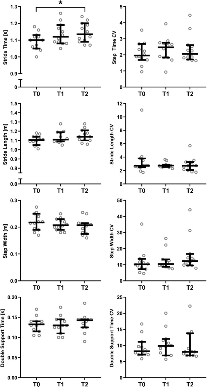
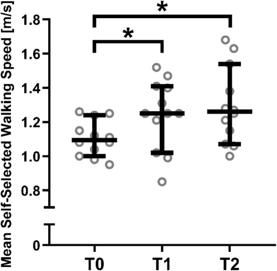
Results: Patients suffering from an OVCF appeared to walk with significantly shorter, faster and wider strides compared to their healthy counterparts. Although stride time and length improved over time, the majority of the parameters analysed remained unchanged after 6 months of conservative treatment.
Discussion: Since patients do not fully recover to their previous level of mobility after 6 months of conservative treatment for OVCF, it appears of high clinical importance to add balance and gait training to the treatment algorithm of OVCFs.
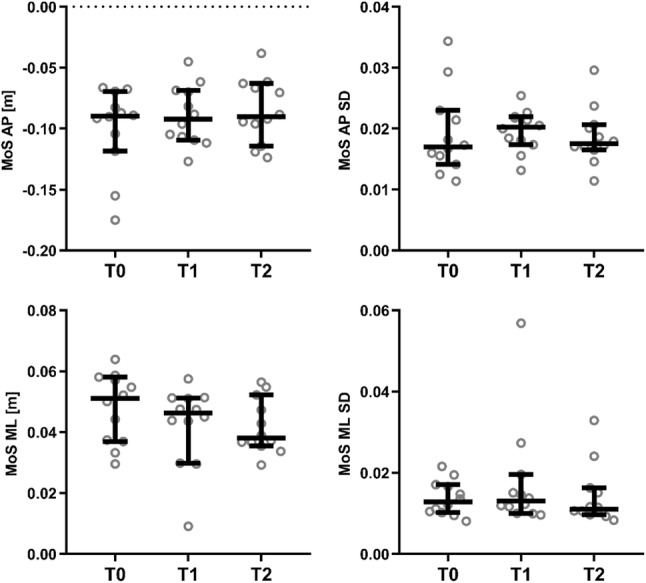
Conclusions: Patients suffering from an OVCF walk with shorter, faster and wider strides compared to their healthy counterparts adopt a less stable body configuration in the anterior direction, potentially increasing their risk of forward falls if perturbed. Although stride time and stride length improve over time even reaching healthy levels again, patients significantly deviate from normal gait patterns (e.g. in stability and step width) after 6 months of conservative treatment.
Keywords: Ageing spine; Gait analysis; OVCF; Spatiotemporal gait parameters; Vertebral fracture.
Conflict of interest statement
The authors declare that they have no conflict of interest.
Figures
References
MeSH terms
LinkOut - more resources
Full Text Sources
Medical
