. 2019 May 21;116(10):1960-1969.
doi: 10.1016/j.bpj.2019.03.037. Epub 2019 Apr 8.

Force-Regulated Refolding of the Mechanosensory Domain in the Platelet Glycoprotein Ib-IX Complex

Affiliations

Force-Regulated Refolding of the Mechanosensory Domain in the Platelet Glycoprotein Ib-IX Complex

X Frank Zhang et al. Biophys J. .

Abstract

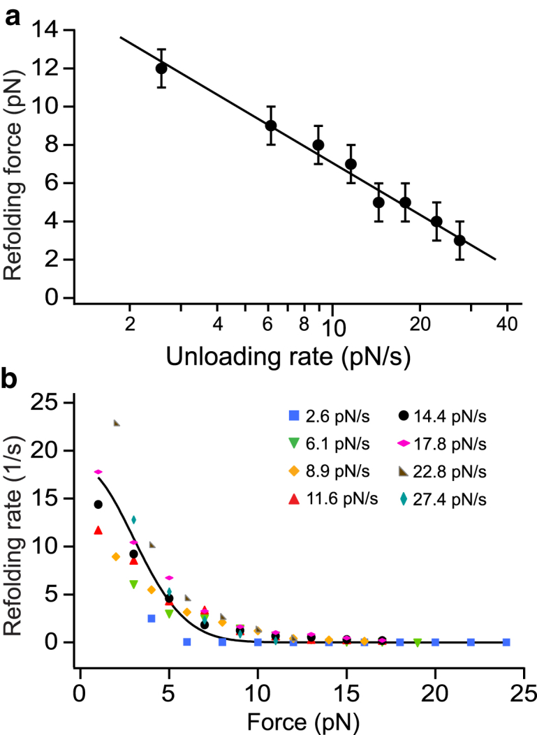
In platelets, the glycoprotein (GP) Ib-IX receptor complex senses blood shear flow and transmits the mechanical signals into platelets. Recently, we have discovered a juxtamembrane mechanosensory domain (MSD) within the GPIbα subunit of GPIb-IX. Mechanical unfolding of the MSD activates GPIb-IX signaling into platelets, leading to their activation and clearance. Using optical tweezer-based single-molecule force measurement, we herein report a systematic biomechanical characterization of the MSD in its native, full-length receptor complex and a recombinant, unglycosylated MSD in isolation. The native MSD unfolds at a resting rate of 9 × 10⁻³ s-1. Upon exposure to pulling forces, MSD unfolding accelerates exponentially over a force scale of 2.0 pN. Importantly, the unfolded MSD can refold with or without applied forces. The unstressed refolding rate of MSD is ∼17 s-1 and slows exponentially over a force scale of 3.7 pN. Our measurements confirm that the MSD is relatively unstable, with a folding free energy of 7.5 kBT. Because MSD refolding may turn off GPIb-IX's mechanosensory signals, our results provide a mechanism for the requirement of a continuous pulling force of >15 pN to fully activate GPIb-IX.


Figures

Figure 1
Figure 1
Single-molecule force measurement of pulling mAb on GPIb-IX to induce the unfolding of MSD. (a) A diagram of the GPIb-IX construct used in this study is shown. The biotag that is biotinylated by E. coli biotin ligase was appended to the GPIX cytoplasmic domain. The binding epitope of WM23 is indicated. (b) WM23 Fab was covalently attached to a DNA handle. The other end of the DNA handle was immobilized to a 2-μm polystyrene bead via biotin-streptavidin linkage, which was controlled by the optical trap. The biotinylated GPIb-IX was captured by another streptavidin bead. This bead was held by a fixed micropipette. (c) Successive cycles of stretching and relaxation with MSD unfolding and refolding events was arrowed, with the pulling speed at 150 nm/s. To see this figure in color, go online.
Figure 2
Figure 2
Unfolding properties of MSD. (a) Shown is an overlay of 53 consecutive pulling-relaxation traces of stretching a GPIb-IX complex from one single tether by mAb WM23. Inset: a zoomed-in force-trap position traces show the force-induced unfolding and refolding of MSD from pulling at 150 nm/s. (b) Superimposed plots of unfolding force versus unfolding extension data and their fits to the WLC model is shown (dashed line): F(x)LpkBT=14(1xLc)214+xLc where F(x) is the applied force on the polymer, x is the end-to-end distance, Lc is the contour length, and Lp is the persistence length of the polymer. The unfolding extension is defined as the increase in end-to-end length of MSD when it unfolds. A histogram of unfolding extension (inset) was used to find peak extension. The fit and data plot of A1 pulling on GPIb-IX was from an earlier study (20). Error bars are one SD for force and half width of the Gaussian fit for extension. (c) Shown are plots of the most probable unfolding force of the MSD in GPIb-IX and of a recombinant isolated MSD (cSc) as a function of loading rates. Unfolding forces at different loading rates were plotted as histograms (Fig. S1). The most probable unfolding forces were identified from these histograms and plotted against their corresponding loading rates. The error bars are the half bin width. The solid line is a linear fit of the data to the Bell-Evans model (Eq. 3). (d) Shown are plots of unfolding force versus unfolding extension data for the MSD and cSc and their fits to the WLC model (dashed line). Error bars are one SD for force and half width of the Gaussian fit for extension. MSD unfolding data was obtained from 32 tethers, which generated 636 unfolding forces. cSc unfolding data was obtained from 57 tethers and a total of 410 unfolding forces.
Figure 3
Figure 3
Refolding properties of MSD. (a) The plot of most probable refolding forces as a function of unloading rates is shown. The most probable refolding forces were obtained from refolding force histograms of eight different unloading rates (Fig. S3). The error bars are the half bin width. The solid line is a linear fit of the data to the modified Bell-Evans model to describe protein refolding (Eq. 4). (b) The plot of refolding rates kf as a function of pulling force is shown. The kf values were calculated using Eq. 6 from the refolding force distributions (Fig. S3). The force dependence of refolding was fitted to a protein refolding model developed by Evans et al. (33) that takes into account the soft compliance of the unfolded state. The refolding data was generated from 38 tethers and 867 refolding curves. To see this figure in color, go online.
Figure 4
Figure 4
MSD refolding weakens platelet activation signals. (a) Shown is the overlaid flow cytometry histograms comparing the binding of 5G6 Fab to platelets under the indicated conditions, in the presence of botrocetin (1 μg·mL−1). (bd) Shown is the percentage of platelets positive for (b) P-selectin exposure, (c) phosphatidylserine (PS) exposure, or (d) both on human platelets treated with control IgG, 11A8, or AK2 under 20 dyn/cm2 of shear force at the indicated time points. n = 3; significance determined by one-way analysis of variance with Tukey test for multiple comparisons. p < 0.05; ∗∗p < 0.01; ∗∗∗p < 0.001; ∗∗∗∗p < 0.0001. To see this figure in color, go online.
Figure 5
Figure 5
Comparison of lifetimes and MSD-unfolded fraction as a function of force. (a) Shown is a comparison of bond/folded lifetime as a function of the force of folded MSD, A1-GPIbα interactions, and interactions between GPIbα and activated A1 (e.g., by botrocetin) or plasma autoantibodies found in immune thrombocytopenia against Ibα. The force-dependent lifetimes (i.e., the reciprocal dissociation rate constant) were determined by Eq. 1, using the Bell-Evans model parameters determined for MSD unfolding from the current study or from literature using optical tweezers (OT) (20, 47) or a biomembrane force probe (BFP) (35). (b) Shown is the unfolding fraction of MSD at equilibrium as a function of force. The unfolded fraction is estimated using kunfold/krefold/(1 + kunfold/krefold).

References

    1. Moake J.L., Turner N.A., Hellums J.D. Involvement of large plasma von Willebrand factor (vWF) multimers and unusually large vWF forms derived from endothelial cells in shear stress-induced platelet aggregation. J. Clin. Invest. 1986;78:1456–1461. - PMC - PubMed
    1. Peterson D.M., Stathopoulos N.A., Moake J.L. Shear-induced platelet aggregation requires von Willebrand factor and platelet membrane glycoproteins Ib and IIb-IIIa. Blood. 1987;69:625–628. - PubMed
    1. Kroll M.H., Harris T.S., Schafer A.I. von Willebrand factor binding to platelet GpIb initiates signals for platelet activation. J. Clin. Invest. 1991;88:1568–1573. - PMC - PubMed
    1. Springer T.A. von Willebrand factor, Jedi knight of the bloodstream. Blood. 2014;124:1412–1425. - PMC - PubMed
    1. Savage B., Saldívar E., Ruggeri Z.M. Initiation of platelet adhesion by arrest onto fibrinogen or translocation on von Willebrand factor. Cell. 1996;84:289–297. - PubMed

Publication types

MeSH terms

Substances

LinkOut - more resources