. 2019 May 2;104(5):815-834.
doi: 10.1016/j.ajhg.2019.03.022. Epub 2019 Apr 25.

Mutations in ACTL6B Cause Neurodevelopmental Deficits and Epilepsy and Lead to Loss of Dendrites in Human Neurons

Scott Bell  1 Justine Rousseau  2 Huashan Peng  1 Zahia Aouabed  1 Pierre Priam  3 Jean-Francois Theroux  1 Malvin Jefri  1 Arnaud Tanti  1 Hanrong Wu  1 Ilaria Kolobova  1 Heika Silviera  1 Karla Manzano-Vargas  1 Sophie Ehresmann  2 Fadi F Hamdan  2 Nuwan Hettige  1 Xin Zhang  1 Lilit Antonyan  1 Christina Nassif  2 Lina Ghaloul-Gonzalez  4 Jessica Sebastian  4 Jerry Vockley  4 Amber G Begtrup  5 Ingrid M Wentzensen  5 Amy Crunk  5 Robert D Nicholls  4 Kristin C Herman  6 Joshua L Deignan  7 Walla Al-Hertani  8 Stephanie Efthymiou  9 Vincenzo Salpietro  9 Noriko Miyake  10 Yoshio Makita  11 Naomichi Matsumoto  10 Rune Østern  12 Gunnar Houge  13 Maria Hafström  12 Emily Fassi  14 Henry Houlden  9 Jolien S Klein Wassink-Ruiter  15 Dominic Nelson  16 Amy Goldstein  17 Tabib Dabir  18 Julien van Gils  19 Thomas Bourgeron  19 Richard Delorme  20 Gregory M Cooper  21 Jose E Martinez  22 Candice R Finnila  21 Lionel Carmant  22 Anne Lortie  23 Renske Oegema  24 Koen van Gassen  24 Sarju G Mehta  25 Dagmar Huhle  25 Rami Abou Jamra  26 Sonja Martin  26 Han G Brunner  27 Dick Lindhout  28 Margaret Au  29 John M Graham Jr  29 Christine Coubes  30 Gustavo Turecki  1 Simon Gravel  15 Naguib Mechawar  1 Elsa Rossignol  2 Jacques L Michaud  2 Julie Lessard  3 Carl Ernst  31 Philippe M Campeau  32
Affiliations

Mutations in ACTL6B Cause Neurodevelopmental Deficits and Epilepsy and Lead to Loss of Dendrites in Human Neurons

Scott Bell et al. Am J Hum Genet. .

Abstract

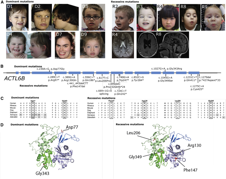
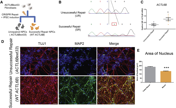
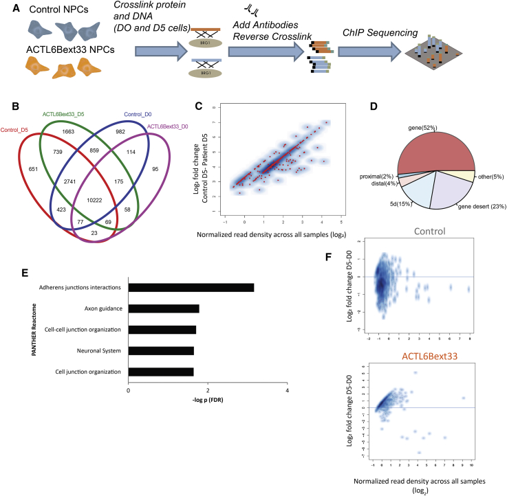
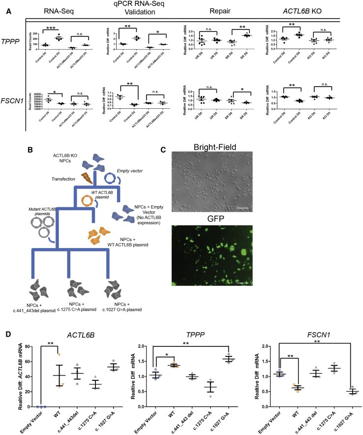
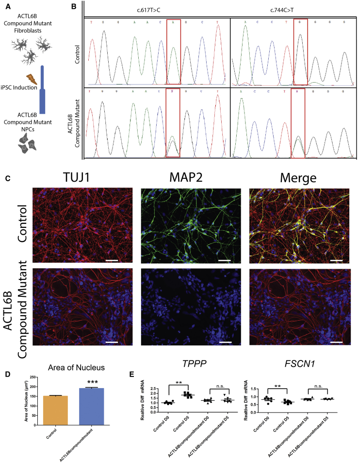
We identified individuals with variations in ACTL6B, a component of the chromatin remodeling machinery including the BAF complex. Ten individuals harbored bi-allelic mutations and presented with global developmental delay, epileptic encephalopathy, and spasticity, and ten individuals with de novo heterozygous mutations displayed intellectual disability, ambulation deficits, severe language impairment, hypotonia, Rett-like stereotypies, and minor facial dysmorphisms (wide mouth, diastema, bulbous nose). Nine of these ten unrelated individuals had the identical de novo c.1027G>A (p.Gly343Arg) mutation. Human-derived neurons were generated that recaptured ACTL6B expression patterns in development from progenitor cell to post-mitotic neuron, validating the use of this model. Engineered knock-out of ACTL6B in wild-type human neurons resulted in profound deficits in dendrite development, a result recapitulated in two individuals with different bi-allelic mutations, and reversed on clonal genetic repair or exogenous expression of ACTL6B. Whole-transcriptome analyses and whole-genomic profiling of the BAF complex in wild-type and bi-allelic mutant ACTL6B neural progenitor cells and neurons revealed increased genomic binding of the BAF complex in ACTL6B mutants, with corresponding transcriptional changes in several genes including TPPP and FSCN1, suggesting that altered regulation of some cytoskeletal genes contribute to altered dendrite development. Assessment of bi-alleic and heterozygous ACTL6B mutations on an ACTL6B knock-out human background demonstrated that bi-allelic mutations mimic engineered deletion deficits while heterozygous mutations do not, suggesting that the former are loss of function and the latter are gain of function. These results reveal a role for ACTL6B in neurodevelopment and implicate another component of chromatin remodeling machinery in brain disease.

Keywords: ACTL6B; genetic engineering; intellectual disability; neurodevelopment; seizure; stem cells.


Figures

Figure 1
Figure 1
Location of Mutations in ACTL6B Found in Individuals with Potential Recessive or Dominant Disease-Causing Mutations (A) Photos of individuals with ACTL6B mutations. Note broad mouth of individuals D1, D2, D3, and D7, diastema in D1, D3, D7, bulbous tip of the nose in all D individuals, and hypertelorism with telecanthi in individual D8. Lower right: MRI images of individuals with recessive ACTL6B mutations. For individual R4, note white matter T2 hyperintensity (arrows). For individual R8, note enlarged lateral ventricles and asymmetric gyral pattern (left, arrows). On the right, note thin corpus callosum (arrows). (B) Linear graph of mutations in ACTL6B (introns not drawn to scale). (C) Conservation of the residues affected by amino acid substitutions. (D) 3D model generated with SWISSMODEL based on S. cerevisiae Arp4 (yeast homolog of ACTL6B), visualized with Swiss-PdbViewer showing that recessive mutations are not focused in one region. Note, however, that the dominant mutations seem to lie at the periphery of the protein and thus they might affect protein-protein interactions.
Figure 2
Figure 2
Generation of iPSC-Derived Neurons for BAF53 Studies (A) Representative images of quality control staining done on iPSCs. (B) Representative quality control staining on NPC cultures. (C) Representative staining of control cells for TUJ1 and MAP2 at D15 of differentiation. (D) Representative trace of miniature EPSCs from D25 neurons held at −40 mv. (E) Representative recordings showing spontaneous activity of D25 neurons in current-clamp mode. (F) Trace of a hyperpolarizing pulse showing a depolarizing sag followed by multiple rebound action potentials. The first action potential is shown at a higher temporal resolution. All scale bars represent 40 μm.
Figure 3
Figure 3
Comparison of Control and ACTL6Bext33 before and after Expression of ACTL6B (A) Diagram illustrating the production of control and ACTL6Bext33 iPSC-derived NPCs from fibroblasts. (B) ACTL6B expression normalized to GAPDH expression plotted against number of days of differentiation of NPCs. n = 4, error bars represent standard error around the mean. (C) Expression of key genes in the SWI/SWF complex in 706 ACTL6Bext33 and control NPCs in proliferating and post-mitotic states. Genes are normalized to GAPDH expression. n ≥ 3; Student’s t test, p < 0.05, ∗∗p < 0.01. (D) Western blots assessing the level of proteins encoded by the genes displayed in (C).
Figure 4
Figure 4
Generation and Characterization of ACTL6B KO Neurons Reveals a Loss of Dendrites (A) Diagram of the experimental approach taken to generate ACTL6B KO NPCs. (B) Sanger sequencing traces of two ACTL6B KO lines. (C) ACTL6B expression in control and ACTL6B KO NPCs at a D0 and D5 time point (n ≥ 3). (D) Western blots assessing the protein levels of BAF53A/B in ACTL6B KO lines. (E) Representative TUJ1 and MAP2 staining of control and ACTL6B KO D15 immature forebrain neurons. (F) Quantification of the surface area of the nucleus in the cell lines shown in (E) (n > 50). Student’s t test, p < 0.05, ∗∗p < 0.01.
Figure 5
Figure 5
Repair of the ACTL6B c.1279del Mutation Restores Morphological and Dendritic Deficits (A) Schematic detailing ACTL6B CRISPR repair. (B) Sanger sequencing traces of a successful repair (SR) and unrepaired (UR) cell line generated from ACTL6Bext33 cell line. (C) ACTL6B expression in SR and UR NPCs at a D5 time point (n = 6). (D) Representative TUJ1 and MAP2 staining taken from SR and UR forebrain immature neurons at D15. Scale bars represent 40 μm. (E) Quantification of the surface area of the nucleus and soma and the length of projections in the cell lines shown in (D) (n > 50). Student’s t test, p < 0.05, ∗∗p < 0.01, ∗∗∗p < 0.001.
Figure 6
Figure 6
ACTL6Bext33 Variant Leads to Increased Binding of BRG1-BAF Complex to the Genome (A) Diagram illustrating the ChIP-seq experiment. (B) Venn diagram showing overlap of genes that the BRG1 complex is bound to. (C) Decreased binding at all 382 FDR significant sites in control cells compared to ACTL6Bext33 cells (pink dots are significant, while blue dots are not). (D) Proportion of BRG1 binding sites found in relation to their proximity to a gene. (E) Gene ontology analysis of differentially bound regions. (F) Within-subjects differential binding across developmental stages (D0 and D5) showing decreased binding in D0 compared to D5 in ACTL6Bext33 cells (pink dots are significant, while blue dots are not). Genes showing a significant difference (FDR-adjusted p values [Benjamini-Hochberg] ≤ 0.05) in D5 relative to D0 using a GLM as implemented in DESeq2.
Figure 7
Figure 7
External Validity in Multiple ACTL6B Mutant Models in Human Neurons (A) TPPP and FSCN1 expression in initial RNA-seq (n ≥ 4) and qPCR (n ≥ 3) data (ACTL6Bext33 versus control); unrepaired (UR) ACTL6Bext33 versus ACTL6Bext33 successful repair (SR) (n = 6); and ACTL6B KO versus isogenic control cells (n = 5). Results are represented as mean ± SEM. Student’s t test, p < 0.05, ∗∗p < 0.01, ∗∗∗p < 0.001. (B) Experimental plan for generation of multiple human neuronal cell lines expressing various mutant ACTL6B constructs. (C) Brightfield and GFP images demonstrating high transfection of ACTL6B constructs. (D) mRNA expression in transfected ACTL6B KO NPCs at D5 time points of ACLT6B, TPPP, and FSCN1 (n = 3). Results are represented as mean ± SEM. Student’s t test, p < 0.05, ∗∗p < 0.01, ∗∗∗p < 0.001.
Figure 8
Figure 8
Neurons Derived from an Individual with a Compound Mutation in ACTL6B Show a Similar Phenotype to ACTL6Bext33 and ACTL6B KO Neurons (A) Schematic showing generation of ACTL6B compound mutant NPCs. (B) Sanger sequencing traces of ACTL6B compound mutant and control cell line at both identified point mutations in ACTL6B. (C) Representative TUJ1 and MAP2 staining of control and ACTL6B compound mutation immature forebrain neurons. (D) Quantification of the surface area of the nucleus in the cell lines shown in (E). (E) TPPP and FSCN1 expression in ACTL6B compound mutant versus control cells at mitotic (D0) and post-mitotic (D5) time points (n > 50). Student’s t test, p < 0.05, ∗∗p < 0.01.

References

    1. Meagher R.B., Kandasamy M.K., Deal R.B., McKinney E.C. Actin-related proteins in chromatin-level control of the cell cycle and developmental transitions. Trends Cell Biol. 2007;17:325–332. - PubMed
    1. Biggar S.R., Crabtree G.R. Continuous and widespread roles for the Swi-Snf complex in transcription. EMBO J. 1999;18:2254–2264. - PMC - PubMed
    1. Lessard J., Wu J.I., Ranish J.A., Wan M., Winslow M.M., Staahl B.T., Wu H., Aebersold R., Graef I.A., Crabtree G.R. An essential switch in subunit composition of a chromatin remodeling complex during neural development. Neuron. 2007;55:201–215. - PMC - PubMed
    1. Wu J.I., Lessard J., Olave I.A., Qiu Z., Ghosh A., Graef I.A., Crabtree G.R. Regulation of dendritic development by neuron-specific chromatin remodeling complexes. Neuron. 2007;56:94–108. - PubMed
    1. Peterson C.L. Multiple SWItches to turn on chromatin? Curr. Opin. Genet. Dev. 1996;6:171–175. - PubMed

Publication types

MeSH terms