. 2019 Jun 11;12(6):1212-1222.
doi: 10.1016/j.stemcr.2019.03.014. Epub 2019 Apr 25.

The Cell-Surface Marker Sushi Containing Domain 2 Facilitates Establishment of Human Naive Pluripotent Stem Cells

Affiliations

The Cell-Surface Marker Sushi Containing Domain 2 Facilitates Establishment of Human Naive Pluripotent Stem Cells

Nicholas Bredenkamp et al. Stem Cell Reports. .

Abstract

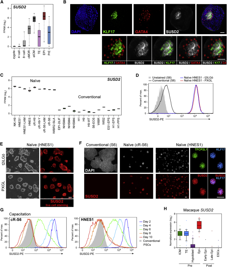
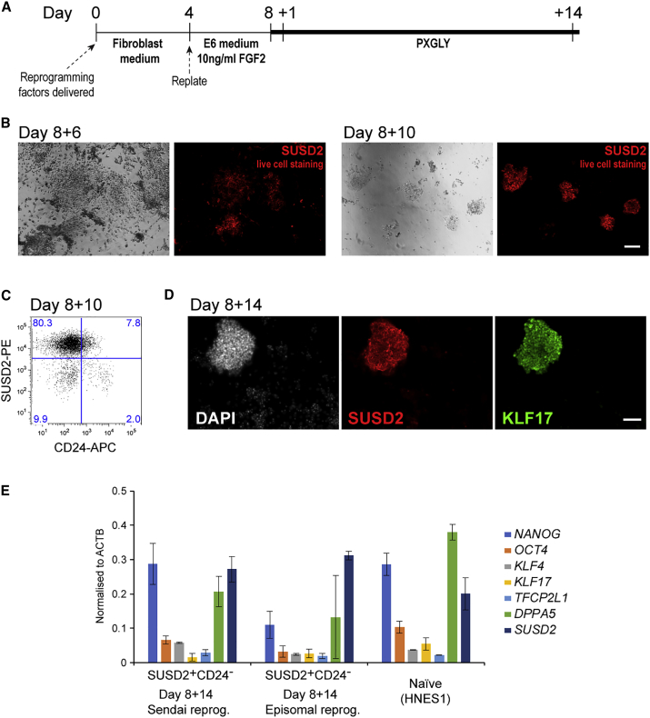
Recently naive human pluripotent stem cells (hPSCs) have been described that relate to an earlier stage of development than conventional hPSCs. Naive hPSCs remain challenging to generate and authenticate, however. Here we report that Sushi Containing Domain 2 (SUSD2) is a robust cell-surface marker of naive hPSCs in the embryo and in vitro. SUSD2 transcripts are enriched in the pre-implantation epiblast of human blastocysts and immunostaining shows localization of SUSD2 to KLF17-positive epiblast cells. SUSD2 mRNA is strongly expressed in naive hPSCs but is negligible in other hPSCs. SUSD2 immunostaining of live or fixed cells provides unambiguous discrimination of naive versus conventional hPSCs. SUSD2 staining or flow cytometry enable monitoring of naive hPSCs in maintenance culture, and their isolation and quantification during resetting of conventional hPSCs or somatic cell reprogramming. Thus SUSD2 is a powerful non-invasive tool for reliable identification and purification of the naive hPSC phenotype.

Keywords: SUSD2; cell-surface marker; chemical resetting; human naive pluripotent stem cell; somatic cell reprogramming.


Figures

Figure 1
Figure 1
SUSD2 Is Expressed by Human Naive Pluripotent Cells in the Embryo and in Culture (A) SUSD2 transcript levels in human pre-implantation embryos at different stages and lineages, extracted from integrated single-cell RNA-sequencing data (Stirparo et al., 2018). (B) Immunostaining for KLF17, GATA4, and SUSD2 in the E7 human blastocyst. Scale bars, 50 μm. (C) SUSD2 transcript levels in naive and conventional hPSCs (Stirparo et al., 2018). (D) Flow-cytometry analysis of SUSD2 in conventional and naive cells. (E) Images of bright-field and SUSD2 immunostaining using a SUSD2-PE antibody. Scale bar, 50 μm. (F) Immunostaining for SUSD2, TFCP2L1, and KLF17 in conventional and naive (cR-S6 and HNES1) cells. Scale bars, 100 μm. (G) Flow-cytometry analysis of SUSD2 expression during capacitation of cR-S6 and HNES1 cells. (H) SUSD2 transcript levels in Macaca fascicularis embryos (Nakamura et al., 2016). cMOR, compacted morula; eICM, early inner cell mass; TE, trophectoderm; Epi, epiblast; PrE, primitive endoderm. See also Figures S1–S3.
Figure 2
Figure 2
SUSD2 Identifies and Purifies Reset Naive hPSCs (A) Schematic of the chemical resetting protocol. HDACi, histone deacetylase inhibitor. (B) Images of cultures at day 10 of resetting. Scale bar, 50 μm. (C and D) Flow-cytometry analysis of SUSD2 and CD24 expression in conventional and naive hPSCs (C) and during resetting (D). (E) Flow-cytometry analysis of GFP and SSEA4 on SUSD2+CD24 cells at day 14 of resetting. (F) Immunostaining for SUSD2, NANOG, and KLF17 at day 14 of resetting. Scale bar, 100 μm. (G) qRT-PCR analysis of sorted SUSD2+CD24 and SUSD2CD24+ cells at day 10 and day 14 of resetting. Error bars indicate SD of three independent experiments. (H) Flow cytometry sort plot at day 14 of resetting. (I) Flow-cytometry analysis of SUSD2 and CD24 expression on cell populations sorted in (H), 5 days after sorting. SUSD2+, SUSD2+CD24; SUSD2, SUSD2CD24+. (J) Bright-field and GFP images of cell populations sorted in (H) at passages 1 and 5 after sorting. Scale bar, 50 μm. See also Figure S4.
Figure 3
Figure 3
SUSD2 Identifies Reset Naive hPSCs upon Resetting of Multiple Cell Lines (A) Flow-cytometry analysis of SUSD2 and CD24 expression during resetting of two human ESC lines (H1 and H7) and two iPSC lines (MECP2-clone17 and NCRM-2). (B) qRT-PCR analysis of marker expression in sorted SUSD2+CD24 and SUSD2CD24+ cells at passage 3 (P3). Note that no SUSD2CD24+ population is evident for the cR-H1 line. DPPA5 expression is shown at 0.5× actual expression. Error bars indicate SD of two independent experiments. (C) Flow-cytometry analysis of SUSD2 and CD24 expression and bright-field images of reset cultures at P5. Top row: unsorted reset cultures; bottom row: cultures sorted for SUSD2+CD24 at P3. Scale bar, 50 μm. (D) Immunostaining for SUSD2, NANOG, and KLF17 at P6 for cultures that were sorted for SUSD2+CD24 at P3. Scale bar, 100 μm. (E) Immunostaining for NANOG, MECP2, and SUSD2 on reset (P6) and parental MECP2-clone17 cells. Scale bar, 100 μm.
Figure 4
Figure 4
SUSD2 Identifies Naive hPSCs during Somatic Cell Reprogramming (A) Schematic of the reprogramming protocol. (B–D) Sendai vector reprogramming of human dermal fibroblasts. (B) Images of bright-field and SUSD2 live staining. Scale bar, 50 μm. (C) Flow-cytometry analysis of SUSD2 and CD24 expression. (D) Immunostaining for SUSD2, NANOG, and KLF17. Scale bar, 50 μm. (E) qRT-PCR analysis of markers on sorted SUSD2+CD24 at day 8 + 14 for Sendai vector- and episomal-mediated reprogramming, and on established naive hPSCs. Bars indicate the SD of two independent experiments. See also Figure S4.

References

    1. Blakeley P., Fogarty N.M.E., del Valle I., Wamaitha S.E., Hu T.X., Elder K., Snell P., Christie L., Robson P., Niakan K.K. Defining the three cell lineages of the human blastocyst by single-cell RNA-seq. Development. 2015;142:3151–3165. - PMC - PubMed
    1. Chen G., Gulbranson D.R., Hou Z., Bolin J.M., Ruotti V., Probasco M.D., Smuga-Otto K., Howden S.E., Diol N.R., Propson N.E. Chemically defined conditions for human iPSC derivation and culture. Nat. Methods. 2011;8:424–429. - PMC - PubMed
    1. Collier A.J., Panula S.P., Schell J.P., Chovanec P., Plaza Reyes A., Petropoulos S., Corcoran A.E., Walker R., Douagi I., Lanner F. Comprehensive cell surface protein profiling identifies specific markers of human naive and primed Pluripotent states. Cell Stem Cell. 2017;20:874–890.e7. - PMC - PubMed
    1. Davidson K.C., Mason E.A., Pera M.F. The pluripotent state in mouse and human. Development. 2015;142:3090–3099. - PubMed
    1. Fusaki N., Ban H., Nishiyama A., Saeki K., Hasegawa M. Efficient induction of transgene-free human pluripotent stem cells using a vector based on Sendai virus, an RNA virus that does not integrate into the host genome. Proc. Jpn. Acad. Ser. B Phys. Biol. Sci. 2009;85:348–362. - PMC - PubMed

Publication types

MeSH terms