

Pulmonary amyloidosis: A case series
- PMID: 31031344
- PMCID: PMC6503711
- DOI: 10.4103/lungindia.lungindia_205_18
Pulmonary amyloidosis: A case series
Abstract
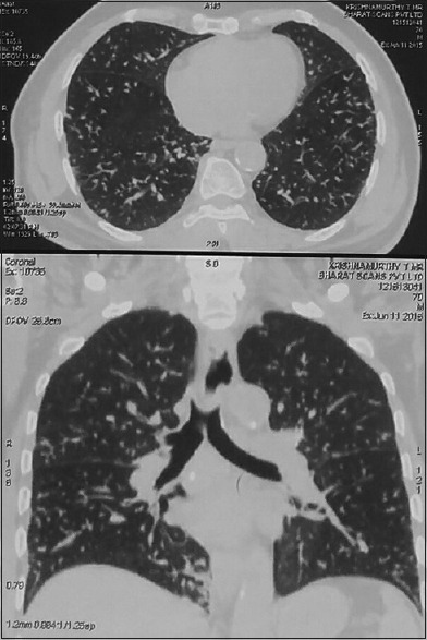
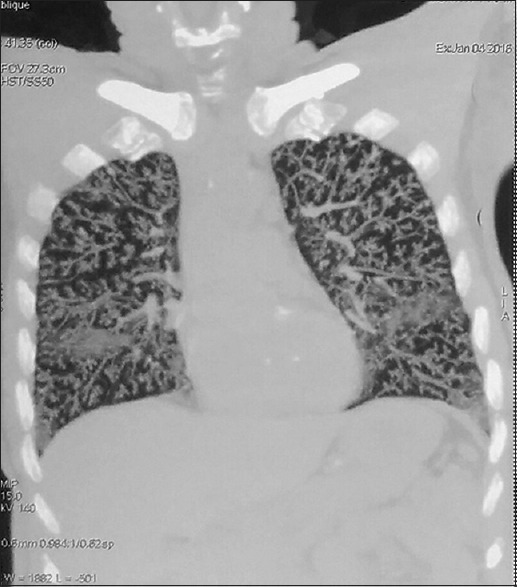
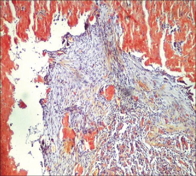
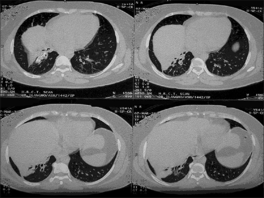
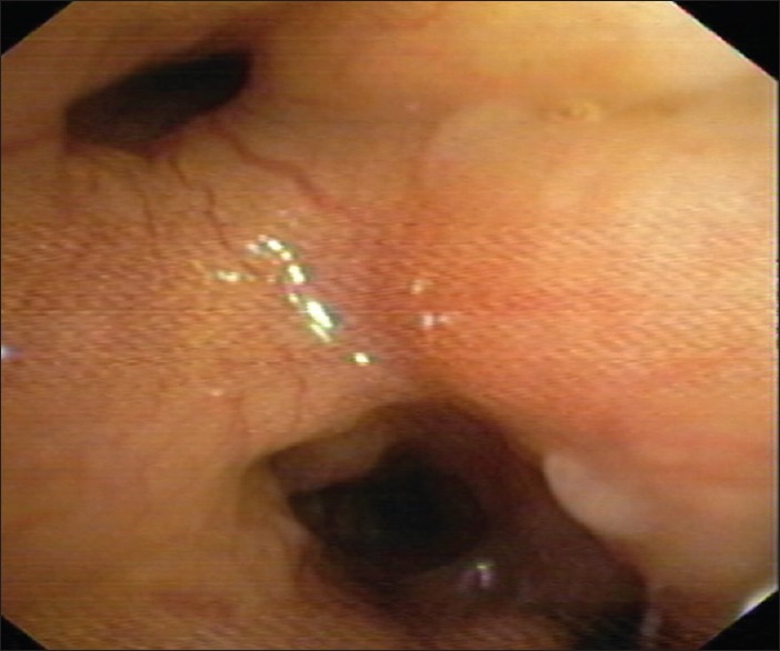
Amyloidosis is a spectrum of diseases, in which various proteins which are usual components of plasma are deposited as insoluble beta-pleated sheets extracellularly, disrupting function of various organs. Amyloid light-chain amyloidosis occurs due to the deposition of proteins, derived from immunoglobulin light chains, routinely manifesting with multisystem involvement. Pulmonary involvement is seen in about 50% of cases. Three common patterns of pulmonary amyloidosis on computed tomography (CT) chest are tracheobronchial, nodular parenchymal, and diffuse alveolar septal variety. We hereby report two cases of pulmonary amyloidosis, one being a case of diffuse alveolar septal pulmonary amyloidosis, which is an extremely rare pattern of involvement, with a very poor prognosis, and the other one being tracheobronchial pattern of involvement, which usually results due to the localized deposition of amyloid in the tracheobronchial tree. Knowledge about pulmonary amyloidosis is important due to its poor prognosis and nonspecific findings in CT chest.
Keywords: Amyloid light-chain amyloidosis; apple-green birefringence; chemotherapy; diffuse alveolar septal pattern; diffuse parenchymal pattern; pulmonary amyloidosis; tracheobronchial amyloidosis; video-assisted thoracoscopic surgery-guided lung biopsy.
Conflict of interest statement
None
Figures
References
-
- Urban BA, Fishman EK, Goldman SM, Scott WW, Jr, Jones B, Humphrey RK, et al. CT evaluation of amyloidosis: Spectrum of disease. Radiographics. 1993;13:1295–308. - PubMed
-
- Berk JL, O'Regan A, Skinner M. Pulmonary and tracheobronchial amyloidosis. Semin Respir Crit Care Med. 2002;23:155–65. - PubMed
-
- Kyle RA, Greipp PR. Amyloidosis (AL).Clinical and laboratory features in 229 cases. Mayo Clin Proc. 1983;58:665–83. - PubMed
-
- Kyle RA, Gertz MA. Primary systemic amyloidosis: Clinical and laboratory features in 474 cases. Semin Hematol. 1995;32:45–59. - PubMed
Publication types
LinkOut - more resources
Full Text Sources
