. 2019 Apr 29:25:3146-3153.
doi: 10.12659/MSM.916254.

Estrogen Modulates Cartilage and Subchondral Bone Remodeling in an Ovariectomized Rat Model of Postmenopausal Osteoarthritis

Affiliations

Estrogen Modulates Cartilage and Subchondral Bone Remodeling in an Ovariectomized Rat Model of Postmenopausal Osteoarthritis

Xiao Xu et al. Med Sci Monit. .

Abstract

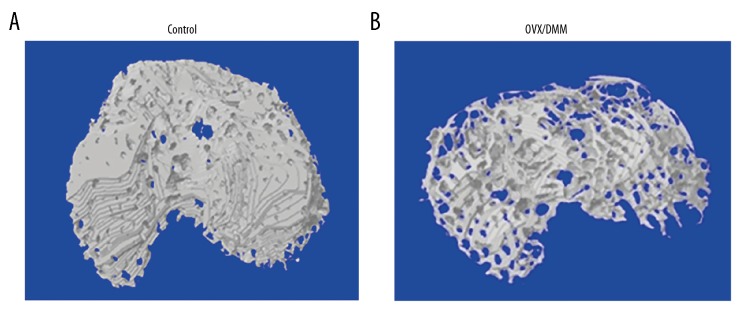
BACKGROUND Estrogen levels regulate changes in osteoarthritis (OA) by inhibiting degradation of the extracellular matrix. Recent in vitro studies have also shown the role of microRNA-140-5p (miR-140-5p). This study aimed to investigate the role of estrogen deficiency, selective modulation of expression of the estrogen receptor (ER), and expression of miR-140-5p in cartilage and subchondral bone remodeling in an ovariectomized rat model of postmenopausal OA. MATERIAL AND METHODS Female Sprague-Dawley rats included two model groups, ovariectomized (OVX) rats and rats with destabilization of the medial meniscus (DMM) rats. Two months after surgery, estrogen levels were measured by the enzyme-linked immunosorbent assay (ELISA). Three-dimensional (3D) micro-computed tomography (micro-CT) was used to image the knee joints. Rats were treated with subcutaneous injection of estrogen (E2) or the selective estrogen receptor modulator (SERM), raloxifene (RAL), for one month. Quantitative real-time polymerase chain reaction (qRT-PCR) was used to detect miR-140-5p in serum, and histology of the knee joint cartilage and bone was performed. RESULTS In the ovariectomized rat model of OA, estrogen therapy reduced the degree of cartilaginous degeneration, while treatment with raloxifene showed no significant effect. Expression levels of miR-140-5p in the OA model group were significantly lower than the control group. Micro-CT showed that in the model group, anterior cruciate ligament dislocation and subchondral bone density were significantly reduced. CONCLUSIONS In an ovariectomized rat model of postmenopausal OA, estrogen deficiency resulted in resorption of subchondral bone and degeneration of articular cartilage.


Figures

Figure 1
Figure 1
Enzyme-linked immunosorbent assay (ELISA) for the detection of changes in serum estrogen levels in the rats. One month after injection, the serum levels of estrogen are compared between the ovariectomized/destabilization of the medial meniscus (OVX/DMM) rat group and the normal rat group (** P<0.01).
Figure 2
Figure 2
Three-dimensional (3D) micro-computed tomography (micro-CT) images of the rat knee joints. (A) The normal rat knee joint. (B–D) The rat knee joints after ovariectomy. Osteophyte formation and joint space narrowing are seen in the joints.
Figure 3
Figure 3
The representative three-dimensional (3D) micro-computed tomography (micro-CT) images of the control rat group and the ovariectomized/destabilization of the medial meniscus (OVX/DMM) rat group. (A) Images of the knee joints from the control group induced by the sham operation. The structural integrity of subchondral bone is not changed. (B) Images of the knee joints from the ovariectomized/destabilization of the medial meniscus (OVX/DMM) rat group. Osteoporosis of the subchondral bone and destroyed areas of the cartilage are shown.
Figure 4
Figure 4
Three-dimensional (3D) micro-computed tomography (micro-CT) images of the subchondral bone in the ovariectomized/destabilization of the medial meniscus (OVX/DMM) rat group with and without estrogen (E2) and raloxifene (RAL) treatment and the control group. (A) Severe bone erosion, cyst formation, and bone sclerosis are shown in the OVX/DMM group. (B) Near-normal subchondral bone in the OVX/DMM+E2 group. (C) The subchondral bone shows osteoporosis and the bone structure is destroyed in the OVX/DMM+RAL group. (D–F) The degree of osteoporosis in the subchondral bone was increased.
Figure 5
Figure 5
The bone mineral density (BMD) of the subchondral bone at 0, 2, and 4 weeks after surgery. Compared with the control group, the ovariectomized/destabilization of the medial meniscus (OVX/DMM) rat group, the OVX/DMM+estrogen (E2) group, and the raloxifene (RAL) treated group all show a loss of bone mass. Maintenance of bone mass showed no significant difference between the OA group and the OVX/DMM+RAL group, but the OVX/DMM+E2 group show significant improvement in bone mass (* P<0.05, ** P<0.01).
Figure 6
Figure 6
The expression of microRNA-140-5p (miR-140-5p) determined by quantitative real-time polymerase chain reaction (qRT-PCR). Compared with the control group, the expression level of miR-140-5p is significantly reduced in the OVX/DMM and the OVX/DMM+E2 group. The expression level of miR-140-5p in the OVX/DMM+E2 group is significantly greater than in the OVX/DMM group (** P<0.01, *** P<0.001).
Figure 7
Figure 7
Photomicrographs of the histology of the rat knee joints in the groups treated with estrogen (E2) and raloxifene (RAL). (A) The histology of the knee joint in the normal control rat group. Hematoxylin and eosin (H&E) ×100. (B) The histology of the knee joint in the ovariectomized/destabilization of the medial meniscus (OVX/DMM) rat group. H&E ×100. (C) The histology of the knee joint in the estrogen (E2)-treated rat group. H&E ×100. (D) The histology of the knee joint in the raloxifene (RAL)-treated rat group, H&E ×100.

References

    1. Li S, Ou Y, Zhang H, et al. Vitamin D status and its relationship with body composition, bone mineral density and fracture risk in urban central South Chinese postmenopausal women. Ann Nutr Metab. 2014;64:13–19. - PubMed
    1. Helmick CG, Felson DT, Lawrence RC, et al. Estimates of the prevalence of arthritis and other rheumatic conditions in the United States: Part I. Arthritis Rheum. 2008;58:15–25. - PubMed
    1. World Health Organization (WHO) Chronic Rheumatic Conditions. Available at [URL]: http://www.who.int/chp/topics/rheumatic/en/
    1. Bijlsma JW, Berenbaum F, Lafeber FP. Osteoarthritis: An update with relevance for clinical practice. Lancet. 2011;377:2115–26. - PubMed
    1. Cooper C, Adachi JD, Bardin T, et al. How to define responders in osteoarthritis. Curr Med Res Opin. 2013;29:719–29. - PMC - PubMed

MeSH terms