

Mycolactone as Analgesic: Subcutaneous Bioavailability Parameters
- PMID: 31031626
- PMCID: PMC6473063
- DOI: 10.3389/fphar.2019.00378
Mycolactone as Analgesic: Subcutaneous Bioavailability Parameters
Abstract
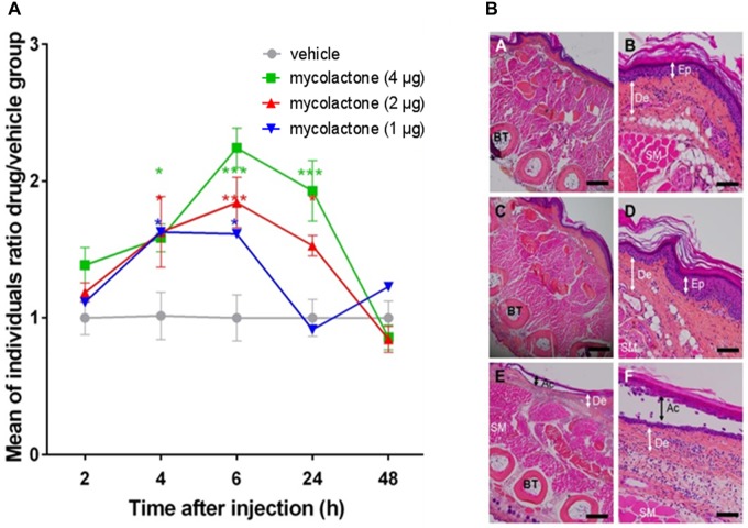
Mycobacterium ulcerans is the bacillus responsible for Buruli ulcer, an infectious disease and the third most important mycobacterial disease worldwide, after tuberculosis and leprosy. M. ulcerans infection is a type of panniculitis beginning mostly with a nodule or an oedema, which can progress to large ulcerative lesions. The lesions are caused by mycolactone, the polyketide toxin of M. ulcerans. Mycolactone plays a central role for host colonization as it has immunomodulatory and analgesic effects. On one hand, mycolactone induces analgesia by targeting type-2 angiotensin II receptors (AT2R), causing cellular hyperpolarization and neuron desensitization. Indeed, a single subcutaneous injection of mycolactone into the mouse footpad induces a long-lasting hypoesthesia up to 48 h. It was suggested that the long-lasting hypoesthesia may result from the persistence of a significant amount of mycolactone locally following its injection, which could be probably due to its slow elimination from tissues. To verify this hypothesis, we investigated the correlation between hypoesthesia and mycolactone bioavailability directly at the tissue level. Various quantities of mycolactone were then injected in mouse tissue and hypoesthesia was recorded with nociception assays over a period of 48 h. The hypoesthesia was maximal 6 h after the injection of 4 μg mycolactone. The basal state was reached 48 h after injection, which demonstrated the absence of nerve damage. Surprisingly, mycolactone levels decreased strongly during the first hours with a reduction of 70 and 90% after 4 and 10 h, respectively. Also, mycolactone did not diffuse in neighboring skin tissue and only poorly into the bloodstream upon direct injection. Nevertheless, the remaining amount was sufficient to induce hypoesthesia during 24 h. Our results thus demonstrate that intact mycolactone is rapidly eliminated and that very small amounts of mycolactone are sufficient to induce hypoesthesia. Taken together, our study points out that mycolactone ought to be considered as a promising analgesic.
Keywords: Mycobacterium ulcerans; analgesia; bioavailability; biological action; mycolactone.
Figures
Similar articles
-
Could Mycolactone Inspire New Potent Analgesics? Perspectives and Pitfalls.Toxins (Basel). 2019 Sep 4;11(9):516. doi: 10.3390/toxins11090516. Toxins (Basel). 2019. PMID: 31487908 Free PMC article. Review.
-
Mycolactone is responsible for the painlessness of Mycobacterium ulcerans infection (buruli ulcer) in a murine study.Infect Immun. 2008 May;76(5):2002-7. doi: 10.1128/IAI.01588-07. Epub 2008 Mar 3. Infect Immun. 2008. PMID: 18316387 Free PMC article.
-
[Role of mycolactone in the nerve damage of Buruli ulcer (Mycobacterium ulcerans infection)].Nihon Hansenbyo Gakkai Zasshi. 2011 Feb;80(1):5-10. doi: 10.5025/hansen.80.5. Nihon Hansenbyo Gakkai Zasshi. 2011. PMID: 21404590 Review. Japanese.
-
Mycolactone-mediated neurite degeneration and functional effects in cultured human and rat DRG neurons: Mechanisms underlying hypoalgesia in Buruli ulcer.Mol Pain. 2016 Jun 20;12:1744806916654144. doi: 10.1177/1744806916654144. Print 2016. Mol Pain. 2016. PMID: 27325560 Free PMC article.
-
A Bacterial Toxin with Analgesic Properties: Hyperpolarization of DRG Neurons by Mycolactone.Toxins (Basel). 2017 Jul 18;9(7):227. doi: 10.3390/toxins9070227. Toxins (Basel). 2017. PMID: 28718822 Free PMC article.
Cited by
-
The Angiotensin AT2 Receptor: From a Binding Site to a Novel Therapeutic Target.Pharmacol Rev. 2022 Oct;74(4):1051-1135. doi: 10.1124/pharmrev.120.000281. Pharmacol Rev. 2022. PMID: 36180112 Free PMC article. Review.
-
Could Mycolactone Inspire New Potent Analgesics? Perspectives and Pitfalls.Toxins (Basel). 2019 Sep 4;11(9):516. doi: 10.3390/toxins11090516. Toxins (Basel). 2019. PMID: 31487908 Free PMC article. Review.
-
Mycolactone: A Broad Spectrum Multitarget Antiviral Active in the Picomolar Range for COVID-19 Prevention and Cure.Int J Mol Sci. 2023 Apr 12;24(8):7151. doi: 10.3390/ijms24087151. Int J Mol Sci. 2023. PMID: 37108313 Free PMC article.
-
Toward Understanding the Mechanism of Client-Selective Small Molecule Inhibitors of the Sec61 Translocon.J Mol Recognit. 2025 Jan;38(1):e3108. doi: 10.1002/jmr.3108. Epub 2024 Oct 12. J Mol Recognit. 2025. PMID: 39394908 Free PMC article. Review.
-
Mycolactone toxin induces an inflammatory response by targeting the IL-1β pathway: Mechanistic insight into Buruli ulcer pathophysiology.PLoS Pathog. 2020 Dec 18;16(12):e1009107. doi: 10.1371/journal.ppat.1009107. eCollection 2020 Dec. PLoS Pathog. 2020. PMID: 33338061 Free PMC article.
References
-
- Converse P. J., Xing Y., Kim K. H., Tyagi S., Li S. Y., Almeida D. V., et al. (2014). Accelerated detection of mycolactone production and response to antibiotic treatment in a mouse model of Mycobacterium ulcerans disease. PLoS Negl. Trop. Dis. 8:e2618. 10.1371/journal.pntd.0002618 - DOI - PMC - PubMed
LinkOut - more resources
Full Text Sources
Other Literature Sources
Molecular Biology Databases