

Characterizing the Penumbras of White Matter Hyperintensities and Their Associations With Cognitive Function in Patients With Subcortical Vascular Mild Cognitive Impairment
- PMID: 31031687
- PMCID: PMC6474292
- DOI: 10.3389/fneur.2019.00348
Characterizing the Penumbras of White Matter Hyperintensities and Their Associations With Cognitive Function in Patients With Subcortical Vascular Mild Cognitive Impairment
Abstract
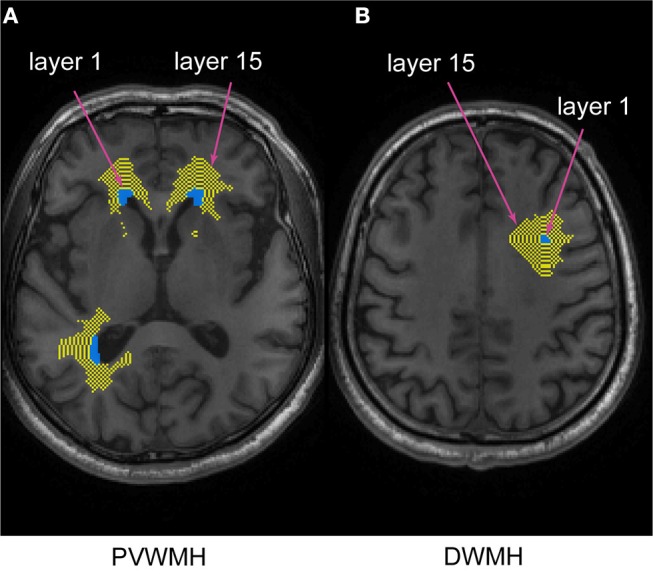
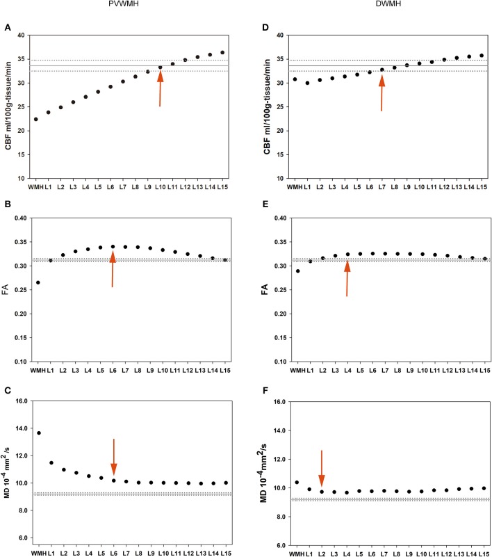
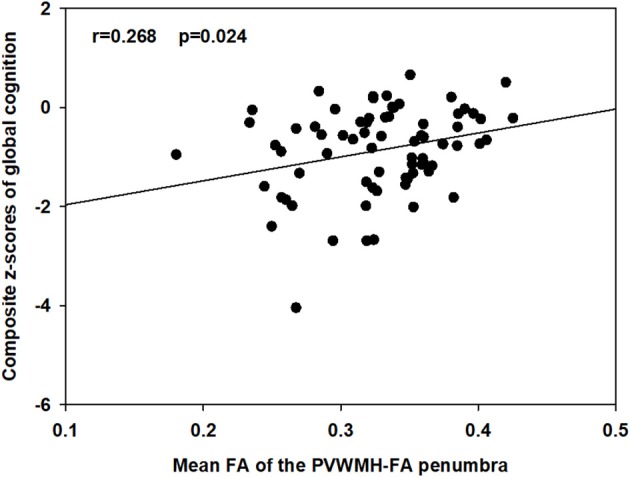
Normal-appearing white matter (NAWM) surrounding white matter hyperintensities (WMHs), frequently known as the WMH penumbra, is associated with subtle white matter injury and has a high risk for future conversion to WMHs. The goal of this study was to define WMH penumbras and to further explore whether the diffusion and perfusion parameters of these penumbras could better reflect cognitive function alterations than WMHs in subjects with subcortical vascular mild cognitive impairment (svMCI). Seventy-three svMCI subjects underwent neuropsychological assessments and 3T MRI scans, including diffusion tensor imaging (DTI) and arterial spin labeling (ASL). To determine the extent of cerebral blood flow (CBF) and DTI penumbras. A NAWM layer mask was generated for periventricular WMHs (PVWMHs) and deep WMHs (DWMHs) separately. Mean values of CBF, fractional anisotropy (FA), mean diffusivity (MD) within the WMHs and their corresponding NAWM layer masks were computed and compared using paired t-tests. Pearson's partial correlations were used to assess the relations of the mean CBF, FA, and MD values within the corresponding penumbras with composite z-scores of global cognition and four cognitive domains controlling for age, sex, and education. For both PVWMHs and DWMHs, the CBF penumbras were wider than the DTI penumbras. Only the mean FA value of the PVWMH-FA penumbra was correlated with the composite z-scores of global cognition before correction (r = 0.268, p = 0.024), but that correlation did not survive after correcting the p-value for multiple comparisons. Our findings showed extensive white matter perfusion disturbances including white matter tissue, both with and without microstructural alterations. The imaging parameters investigated, however, did not correlate to cognition.
Keywords: cerebral blood flow; diffusion tensor imaging; normal appearing white matter; penumbra; subcortical vascular mild cognitive impairment; white matter hyperintensity.
Figures
Similar articles
-
Characterizing the penumbras of white matter hyperintensities in patients with cerebral small vessel disease.Jpn J Radiol. 2023 Sep;41(9):928-937. doi: 10.1007/s11604-023-01419-w. Epub 2023 May 9. Jpn J Radiol. 2023. PMID: 37160589 Free PMC article.
-
Comparison of cerebral blood flow and structural penumbras in relation to white matter hyperintensities: A multi-modal magnetic resonance imaging study.J Cereb Blood Flow Metab. 2016 Sep;36(9):1528-36. doi: 10.1177/0271678X16651268. Epub 2016 Jun 7. J Cereb Blood Flow Metab. 2016. PMID: 27270266 Free PMC article.
-
Characterizing the white matter hyperintensity penumbra with cerebral blood flow measures.Neuroimage Clin. 2015 Apr 22;8:224-9. doi: 10.1016/j.nicl.2015.04.012. eCollection 2015. Neuroimage Clin. 2015. PMID: 26106546 Free PMC article.
-
Association between white matter alterations and domain-specific cognitive impairment in cerebral small vessel disease: A meta-analysis of diffusion tensor imaging.Front Aging Neurosci. 2022 Nov 22;14:1019088. doi: 10.3389/fnagi.2022.1019088. eCollection 2022. Front Aging Neurosci. 2022. PMID: 36483114 Free PMC article.
-
Multimodal imaging findings in normal-appearing white matter of leucoaraiosis: a review.Stroke Vasc Neurol. 2016 Jun 24;1(2):59-63. doi: 10.1136/svn-2016-000021. eCollection 2016 Jun. Stroke Vasc Neurol. 2016. PMID: 28959465 Free PMC article. Review.
Cited by
-
The Ties That Bind: Glial Transplantation in White Matter Ischemia and Vascular Dementia.Neurotherapeutics. 2023 Jan;20(1):39-47. doi: 10.1007/s13311-022-01322-8. Epub 2022 Nov 10. Neurotherapeutics. 2023. PMID: 36357662 Free PMC article. Review.
-
Fluid-attenuated inversion recovery magnetic resonance imaging textural features as sensitive markers of white matter damage in midlife adults.Brain Commun. 2022 May 5;4(3):fcac116. doi: 10.1093/braincomms/fcac116. eCollection 2022. Brain Commun. 2022. PMID: 35611309 Free PMC article.
-
Association between white matter hyperintensities and altered cerebral blood flow in maintenance hemodialysis patients: a longitudinal study.BMC Nephrol. 2024 Jan 24;25(1):33. doi: 10.1186/s12882-024-03468-3. BMC Nephrol. 2024. PMID: 38267857 Free PMC article.
-
Contribution of Inflammation and Hypoperfusion to White Matter Hyperintensities-Related Cognitive Impairment.Front Neurol. 2022 Jan 4;12:786840. doi: 10.3389/fneur.2021.786840. eCollection 2021. Front Neurol. 2022. PMID: 35058875 Free PMC article.
-
Associations between cerebral blood flow and progression of white matter hyperintensities.Front Neuroimaging. 2025 Jan 21;3:1463311. doi: 10.3389/fnimg.2024.1463311. eCollection 2024. Front Neuroimaging. 2025. PMID: 39906355 Free PMC article.
References
LinkOut - more resources
Full Text Sources