. 2019 Apr 12:10:701.
doi: 10.3389/fmicb.2019.00701. eCollection 2019.

New Insight Into Avian Papillomavirus Ecology and Evolution From Characterization of Novel Wild Bird Papillomaviruses

Affiliations

New Insight Into Avian Papillomavirus Ecology and Evolution From Characterization of Novel Wild Bird Papillomaviruses

Marta Canuti et al. Front Microbiol. .

Abstract

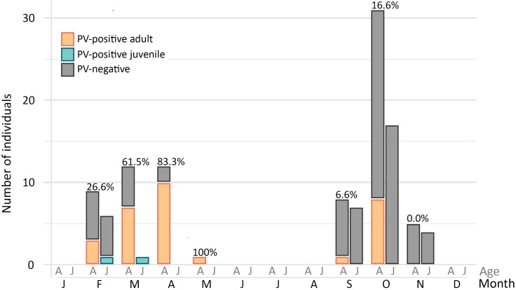
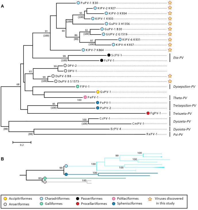
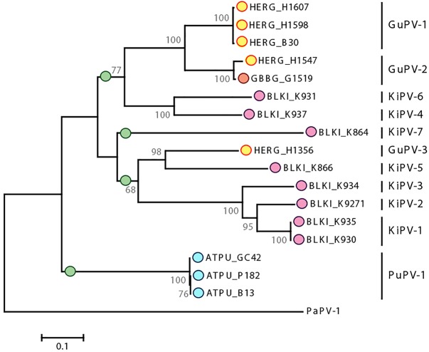
Viruses in the family Papillomaviridae have circular dsDNA genomes of approximately 5.7-8.6 kb that are packaged within non-enveloped, icosahedral capsids. The known papillomavirus (PV) representatives infect vertebrates, and there are currently more than 130 recognized PV species in more than 50 genera. We identified 12 novel avian papillomavirus (APV) types in wild birds that could represent five distinct species and two genera. Viruses were detected in paired oropharyngeal/cloacal swabs collected from six bird species, increasing the number of avian species known to harbor PVs by 40%. A new duck PV (DuPV-3) was found in mallard and American black duck (27.6% estimated prevalence) that was monophyletic with other known DuPVs. A single viral type was identified in Atlantic puffin (PuPV-1, 9.8% estimated prevalence), while a higher genetic diversity was found in other Charadriiformes. Specifically, three types [gull PV-1 (GuPV-1), -2, and -3] were identified in two gull species (estimated prevalence of 17% and 2.6% in American herring and great black-backed gull, respectively), and seven types [kittiwake PV-1 (KiPV-1) through -7] were found in black-legged kittiwake (81.3% estimated prevalence). Significantly higher DuPV-3 circulation was observed in spring compared to fall and in adults compared to juveniles. The studied host species' tendencies to be in crowded environments likely affect infection rates and their migratory behaviors could explain the high viral diversity, illustrating how host behavior can influence viral ecology and distribution. For DuPV-3, GuPV-1, PuPV-1, and KiPV-2, we obtained the complete genomic sequences, which showed the same organization as other known APVs. Phylogenetic analyses showed evidence for virus-host co-divergence at the host taxonomic levels of family, order, and inter-order, but we also observed that host-specificity constraints are relaxed among highly related hosts as we found cross-species transmission within ducks and within gulls. Furthermore, the phylogeny of viruses infecting the Charadriiformes did not match the host phylogeny and gull viruses formed distinct monophyletic clades with kittiwake viruses, possibly reflecting past host-switching events. Considering the vast PV genotype diversity in other hosts and the large number of bird species, many more APVs likely remain to be discovered.

Keywords: avian papillomavirus; molecular epidemiology; papillomavirus; viral ecology; virus discovery; virus evolution.


Figures

FIGURE 1
FIGURE 1
Genome organization of the four novel papillomaviruses identified in this study. For each virus (PuPV-1, puffin papillomavirus type 1; DuPV-3, duck papillomavirus type 3; GuPV-1, gull papillomavirus type 1, KiPV-2, kittiwake papillomavirus type 2), the size of the genome is indicated at the center of the diagram and predicted ORFs are represented by colored arrows (orange: late structural proteins; teal: core early non-structural proteins; purple: early accessory oncoproteins; light blue: avian-virus-specific putative early protein). The positions of the long control region (LCR), TATA box (TATA), and poly-adenylation signals (polyA) are also indicated.
FIGURE 2
FIGURE 2
Alignments of conserved domains identified in APV proteins. The motifs of the zinc-binding domain are indicated in the sequences of the E6 protein (A) and E7 protein (B), where the retinoblastoma tumor suppressor (pRb)-binding domain (LXCXE) is also specified. The DNA-binding domain of the E2 protein is shown in (C), while the Walker domains of the E1 helicase are show in (D). Finally, specific protein domains of the L2 protein are shown in (E). Domain designation and typical sequences of each motif are indicated below and above the alignments, respectively. Viral types identified in this study are in red.
FIGURE 3
FIGURE 3
Estimated prevalence of DuPV-3 infection over the year. The graph illustrates the number of positive (colors) and negative (gray) adult (A) and juvenile (J) ducks (American black duck, mallard, and hybrids) sampled in each month of the year. Overall viral estimated prevalence for each month for which samples were available is indicated above the bars.
FIGURE 4
FIGURE 4
Phylogenetic analysis of APVs. (A) Analysis of partial L1 nucleotide sequences (corresponding to nt 5678–6070 of FcPV-1; accession number NC_004068) of all known APVs. The tree was built with the maximum-likelihood method (Felsenstein, 1981) based on the Kimura 2 parameters model (Kimura, 1980), identified as the best-fitting model after the model test analysis in MEGA 7 (Kumar et al., 2016). A discrete Gamma distribution was used to model evolutionary rate differences among sites (+G = 0.9136) and branch lengths are proportional to genetic distances as indicated by the scale bar. The outcome of the bootstrap analysis (Felsenstein, 1985) is shown next to the nodes, while the corresponding bootstrap values obtained in a different analysis involving the whole L1 open-reading frame (Supplementary Figure S1) is shown in parenthesis. Viruses are marked by type name and the strain name is also indicated for viruses described in this study (indicated by a star), while established viral genera are shown on the right. Viruses are labeled by a colored dot that indicates the order of the birds in which they were identified, as indicated in the legend, and hosts are indicated within viral type names (Du, duck; Fc, Fringilla coelebs; Fg, Fulmarus glacialis; Fl, Francolinus leucoscepus; Gf, Gyps fulvus; Gu, gull; Ki, kittiwake; Pa, Pygoscelis adeliae; Pe, Psittacus erithacus timneh; Pu, puffin; Sc, Serinus canaria; CC, Caretta caretta; Cm, Chelonia mydas, Ec, Equus ferus caballus, Ra, Rousettus aegyptiacus). (B) Subtree including branches of viruses identified in four bird orders (Charadriiformes, Sphenisciformes, Galliformes, and Anseriformes) obtained from the phylogenetic analysis involving the complete L1 ORF (Supplementary Figure S1).
FIGURE 5
FIGURE 5
Phylogenetic analysis of APVs identified in birds within the order Charadriiformes. The tree is based on L1 sequences of all PVs identified in this study from Atlantic puffin (ATPU, blue), American herring gull (HERG, yellow), great black-backed gull (GBBG, orange), and black-legged kittiwake (BLKI, pink). The four proposed different species are indicated by green dots at the base of the species branch. The tree was built with the maximum-likelihood method (Felsenstein, 1981) based on the Tamura–Nei model (Tamura and Nei, 1993), identified as the best-fitting model after the model test analysis in MEGA 7 (Kumar et al., 2016). A discrete Gamma distribution was used to model evolutionary rate differences among sites (+G = 0.9236) and the rate variation model allowed for some sites to be evolutionarily invariable ([+I], 23.03% of sites). Branch lengths are proportional to genetic distances as indicated by the scale bar and the outcome of the bootstrap analysis (Felsenstein, 1985) is shown next to the nodes. Viral types or putative types are indicated on the right, and PaPV-1 was used as an outgroup.

Similar articles

Cited by

References

    1. Antonsson A., Erfurt C., Hazard K., Holmgren V., Simon M., Kataoka A., et al. (2003). Prevalence and type spectrum of human papillomaviruses in healthy skin samples collected in three continents. J. Gen. Virol. 84 1881–1886. 10.1099/vir.0.18836-0 - DOI - PubMed
    1. Antonsson A., McMillan N. A. J. (2006). Papillomavirus in healthy skin of Australian animals. J. Gen. Virol. 87 3195–3200. 10.1099/vir.0.82195-0 - DOI - PubMed
    1. Ayginin A. A., Pimkina E. V., Matsvay A. D., Speranskaya A. S., Safonova M. V., Blinova E. A., et al. (2018). The study of viral RNA diversity in bird samples using de novo designed multiplex genus-specific primer panels. Adv. Virol. 2018:3248285. 10.1155/2018/3248285 - DOI - PMC - PubMed
    1. Bergvall M., Melendy T., Archambault J. (2013). The E1 proteins. Virology 445 35–56. 10.1016/j.virol.2013.07.020 - DOI - PMC - PubMed
    1. Bock H., Brennicke A., Schuster W. (1994). Rps3 and rpl16 genes do not overlap in Oenothera mitochondria: GTG as a potential translation initiation codon in plant mitochondria? Plant Mol. Biol. 24 811–818. 10.1007/BF00029863 - DOI - PubMed