. 2019 Apr 12:10:713.
doi: 10.3389/fmicb.2019.00713. eCollection 2019.

Distinct and Specific Role of NlpC/P60 Endopeptidases LytA and LytB in Cell Elongation and Division of Lactobacillus plantarum

Affiliations

Distinct and Specific Role of NlpC/P60 Endopeptidases LytA and LytB in Cell Elongation and Division of Lactobacillus plantarum

Marie-Clémence Duchêne et al. Front Microbiol. .

Abstract

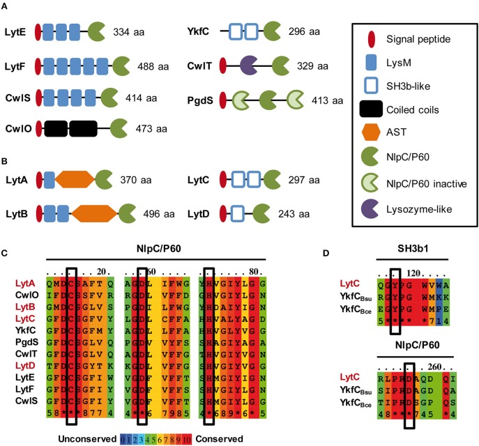
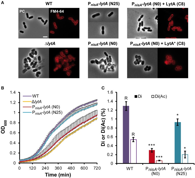
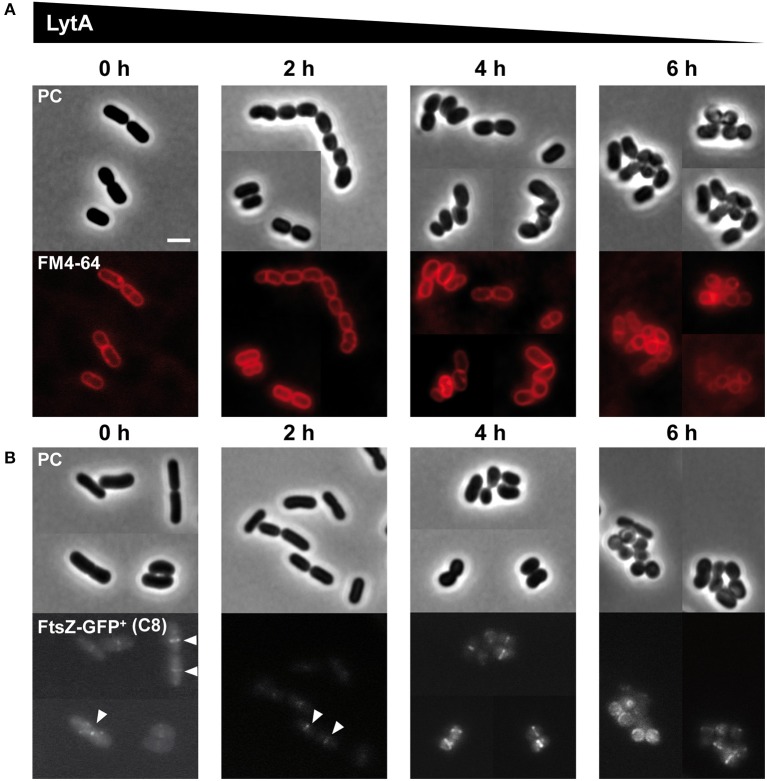
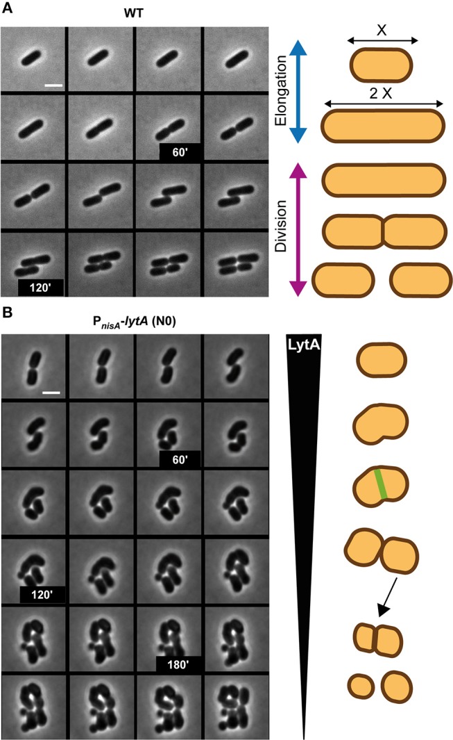
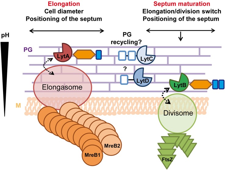
Peptidoglycan (PG) is an essential lattice of the bacterial cell wall that needs to be continuously remodeled to allow growth. This task is ensured by the concerted action of PG synthases that insert new material in the pre-existing structure and PG hydrolases (PGHs) that cleave the PG meshwork at critical sites for its processing. Contrasting with Bacillus subtilis that contains more than 35 PGHs, Lactobacillus plantarum is a non-sporulating rod-shaped bacterium that is predicted to possess a minimal set of 12 PGHs. Their role in morphogenesis and cell cycle remains mostly unexplored, except for the involvement of the glucosaminidase Acm2 in cell separation and the NlpC/P60 D, L-endopeptidase LytA in cell shape maintenance. Besides LytA, L. plantarum encodes three additional NlpC/P60 endopeptidases (i.e., LytB, LytC and LytD). The in silico analysis of these four endopeptidases suggests that they could have redundant functions based on their modular organization, forming two pairs of paralogous enzymes. In this work, we investigate the role of each Lyt endopeptidase in cell morphogenesis in order to evaluate their distinct or redundant functions, and eventually their synthetic lethality. We show that the paralogous LytC and LytD enzymes are not required for cell shape maintenance, which may indicate an accessory role such as in PG recycling. In contrast, LytA and LytB appear to be key players of the cell cycle. We show here that LytA is required for cell elongation while LytB is involved in the spatio-temporal regulation of cell division. In addition, both PGHs are involved in the proper positioning of the division site. The absence of LytA activity is responsible for the asymmetrical positioning of septa in round cells while the lack of LytB results in a lateral misplacement of division planes in rod-shaped cells. Finally, we show that the co-inactivation of LytA and LytB is synthetically affecting cell growth, which confirms the key roles played by both enzymes in PG remodeling during the cell cycle of L. plantarum. Based on the large distribution of NlpC/P60 endopeptidases in low-GC Gram-positive bacteria, these enzymes are attractive targets for the discovery of novel antimicrobial compounds.

Keywords: Lactobacillus; NlpC/P60 endopeptidase; cell cycle; cell wall; morphogenesis; muropeptidase; peptidoglycan hydrolase.


Figures

Figure 1
Figure 1
In silico analysis of NlpC/P60 endopeptidases of B. subtilis and L. plantarum. Schematic representation of LytE, LytF, CwlS, CwlO, CwlT and PgdS from B. subtilis 168 (A) and LytA, LytB, LytC, and LytD from L. plantarum WCFS1 (B) Protein size (aa) corresponds to the precursor with its exportation signal-peptide. SH3b-like domain structures were predicted by Phyre 2.0 (www.sbg.bio.ic.ac.uk/phyre2/). (C) Alignment of conserved regions of NlpC/P60 domains that contain the essential residues for catalysis (Cys, His, Asp; boxed). NlpC/P60 endopeptidases of B. subtilis and L. plantarum are indicated in black and red, respectively. (D) Conserved regions between L. plantarum LytC, B. subtilis YkfC (YkfCBsu), and B. cereus YkfC (YkfCBce) that are involved in the selection of PG stem peptides ending with L-Ala. Key residues involved in substrate selection (Tyr and Asp) are boxed. Alignments in (C,D) were obtained with PRALINE (http://www.ibi.vu.nl/programs/pralinewww/).
Figure 2
Figure 2
Effect of LytA deficiency on cell morphology, growth, and PG composition. (A) Images of L. plantarum cells obtained by phase contrast (PC) microscopy and epifluorescence microscopy for membrane labeling with FM4-64. Left panel, WT and ΔlytA mutant; middle panel, conditional PnisA-lytA mutant without (N0) or with nisin 25 ng ml−1 (N25); right panel, complementation of PnisA-lytA mutant with LytA (Pshp0064-lytA; + LytA) and a catalytic mutant of LytA (Pshp0064-lytA*; + LytA*), grown without nisin (N0) in presence of ComS (8 μM, C8). Cells were collected in exponential phase from MRS cultures (with chloramphenicol when needed) and observed on agarose pads after suspension in PBS. Similar observations were obtained from at least 3 independent experiments. The scale bar is 2 μm. (B) Growth curves of WT, ΔlytA mutant, and PnisA-lytA mutant (N0 and N25) in MRS medium. Curves were generated from triplicates (mean values + standard deviations). (C) Percentage of disaccharides-dipeptides without and with O-acetylation (Di and Di+Ac, respectively) in the PG of WT and PnisA-lytA mutant (N0 and N25) after mutanolysin digestion. Mean values of three independent extractions ± standard deviations. Significance with respect to the WT (R, reference) is based on Student's t-test. *P < 0.05 and ***P < 0.001, respectively.
Figure 3
Figure 3
Effect of progressive LytA depletion on cell morphology and division site positioning. (A) PnisA-lytA mutant cells observed during nisin depletion (0, 2, 4, 6 h) by phase contrast (PC) microscopy and FM4-64 staining. The scale bar is 2 μm. (B) PnisA-lytA mutant cells expressing an FtsZ-GFP+ fusion (Pshp0064-ftsZ-fgp+; + FtsZ-GFP+) observed during nisin depletion (0, 2, 4, 6 h) by phase contrast (PC) microscopy and epifluorescence microscopy for the localization of FtsZ-GFP+. White arrows indicate Z-rings in normal rod-shaped cells. Bacteria were cultured in MRS with erythromycin and chloramphenicol, and induced with ComS (8 μM, C8). Cultures were moderately shaken after ComS induction. The scale bar is 2 μm. For (A,B), similar observations were obtained from at least two independent experiments.
Figure 4
Figure 4
Comparison of the cell cycle of WT and LytA-depleted strain. (A) Time-lapse microscopy of the WT strain. (B) Time-lapse microscopy of the PnisA-lytA mutant strain without nisin (N0). The cell cycle of both strains is schematically represented on the right. Bacteria were grown on MRS-containing agarose pads at 30°C. The scale bar is 2 μm. For (A,B) one representative time lapse from at least two independent experiments.
Figure 5
Figure 5
Effect of LytB deficiency and its complementation on cell morphology and division site positioning. (A) Images of cells of WT, ΔlytB mutant, and ΔlytB mutant complemented with LytB (Pshp0064-lytB; + LytB) obtained by phase contrast (PC) microscopy. Yellow arrowheads show asymmetrical divisions in ΔlytB mutant cells. Bacteria were grown in MRS with erythromycin and ComS (8 μM, C8) when needed. Scale bar is 2 μm. (B) Cell length (μm) of WT, ΔlytB mutant and ΔlytB mutant + LytB without (C0) and with ComS (C8) measured in exponential growth phase. Cells were cultured in MRS with erythromycin and ComS when needed. (C) Number of septa (0 to 3; bars) related to cell length (lanes; mean values ± standard deviations) in WT and ΔlytB mutant cells. (D) Relative septum deviation (% of deviation from the median position of cells stained with FM4-64) in mono-septal cells of WT and ΔlytB mutant. For (B–D) measures were obtained from triplicates by using MicrobeJ with n > 500 cells.
Figure 6
Figure 6
Cell cycle of the LytB-deficient strain. Time-lapse microscopy of ΔlytB mutant cells showing a deregulation between elongation and division (A) and asymmetrical divisions (B). In (B) the selected cell for the scheme is indicated with a white arrow. Scheme of the cell cycle of two selected cells are displayed on the right. Bacteria were grown on MRS-containing agarose pads at 30°C. The scale bar is 2 μm.
Figure 7
Figure 7
Effect of the double LytA LytB deficiency on growth and cell morphology. (A) Growth curves of WT, PnisA-lytA mutant, ΔlytB mutant and double PnisA-lytA ΔlytB mutant. N0, non-induced; N1 and N25, nisin 1 and 25 ng ml−1, respectively. Bacteria were grown in MRS with chloramphenicol and nisin when appropriate. Curves were generated from triplicates (mean values + standard deviations). (B) Cells of the double PnisA-lytA ΔlytB mutant observed by phase contrast (PC) microscopy and FM4-64 staining under nisin depletion (2 h). Cells were collected from MRS cultures (with chloramphenicol) and observed on agarose pads after suspension in PBS. The scale bar is 2 μm. (C) PnisA-lytA ΔlytB mutant cells expressing FtsZ-GFP+ (Pshp0064-ftsZ-fgp+; + FtsZ-GFP+) observed by epifluorescence microscopy. Bacteria were cultured in MRS with erythromycin and chloramphenicol, and induced with ComS (8 μM, C8) in absence of nisin (N0). Cultures were moderately shaken after induction. The scale bar is 2 μm. (D) Cells of PnisA-lytA ΔlytB mutant complemented with LytA (Pshp0064-lytA; + LytA, left panels) and LytB (Pshp0064-lytB; + LytB, right panels) observed by phase contrast microscopy and FM4-64 staining under nisin depletion (2 h). Bacteria were cultured in MRS with erythromycin and chloramphenicol, and induced with ComS (8 μM, C8) in absence of nisin. Scale bar is 2 μm. For (B–D) similar observations were obtained from at least three independent experiments.
Figure 8
Figure 8
Roles of LytA and LytB in the cell cycle. LytA is mainly involved in cell elongation and in the control of cell diameter. Its absence (probably indirectly) also affects septum positioning. LytA potentially interacts with the elongasome and its basophilic LysM domain suggests that LytA could act in the external PG layers where the pH is higher. LytB is implicated in division (timing, septum maturation, and lateral positioning) and potentially interacts with the divisome. The presence of two acidophilic LysM domains suggests that LytB could be localized close to the cell membrane where the pH is lower. LytC is proposed to be involved in the recycling/catabolism of PG stem peptides while LytD could be an accessory endopeptidase unrelated to the cell cycle. Labeling of accessory domains of Lyt enzymes are as in Figure 1. Dotted arrows indicate potential interactions. PG, peptidoglycan; M, cytoplasmic membrane.

Similar articles

Cited by

References

    1. Anantharaman V., Aravind L. (2003). Evolutionary history, structural features and biochemical diversity of the NlpC/P60 superfamily of enzymes. Genome Biol. 4:R11. 10.1186/gb-2003-4-2-r11 - DOI - PMC - PubMed
    1. Andre G., Deghorain M., Bron P. A., van Swam I. I., Kleerebezem M., Hols P., et al. . (2011). Fluorescence and atomic force microscopy imaging of wall teichoic acids in Lactobacillus plantarum. ACS Chem. Biol. 6, 366–376. 10.1021/cb1003509 - DOI - PubMed
    1. Barrett J. F., Dolinger D. L., Schramm V. L., Shockman G. D. (1984). The mechanism of soluble peptidoglycan hydrolysis by an autolytic muramidase. A processive exodisaccharidase. J. Biol. Chem. 259, 11818–11827. - PubMed
    1. Berg K. H., Biørnstad T. J., Johnsborg O., Håvarstein L. S. (2012). Properties and biological role of streptococcal fratricins. Appl. Environ. Microbiol. 78, 3515–3522. 10.1128/AEM.00098-12 - DOI - PMC - PubMed
    1. Bernard E., Rolain T., Courtin P., Guillot A., Langella P., Hols P., et al. . (2011a). Characterization of O-acetylation of N-acetylglucosamine: a novel structural variation of bacterial peptidoglycan. J. Biol. Chem. 286, 23950–23958. 10.1074/jbc.M111.241414 - DOI - PMC - PubMed

LinkOut - more resources