. 2019 Apr 12:10:751.
doi: 10.3389/fmicb.2019.00751. eCollection 2019.

Different Hydrophobins of Fusarium graminearum Are Involved in Hyphal Growth, Attachment, Water-Air Interface Penetration and Plant Infection

Affiliations

Different Hydrophobins of Fusarium graminearum Are Involved in Hyphal Growth, Attachment, Water-Air Interface Penetration and Plant Infection

Alessandra Quarantin et al. Front Microbiol. .

Abstract

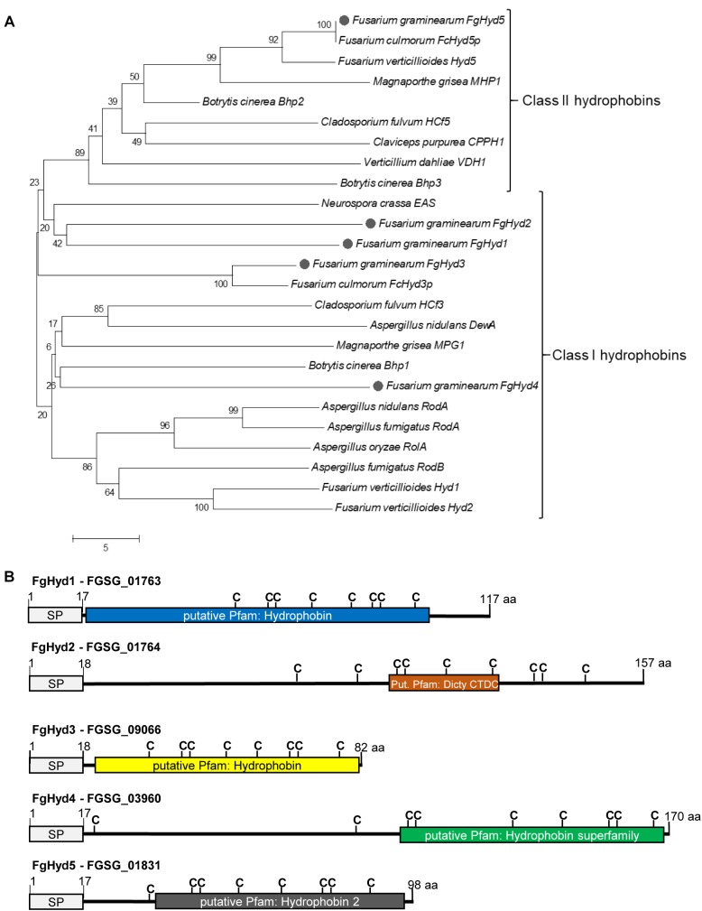
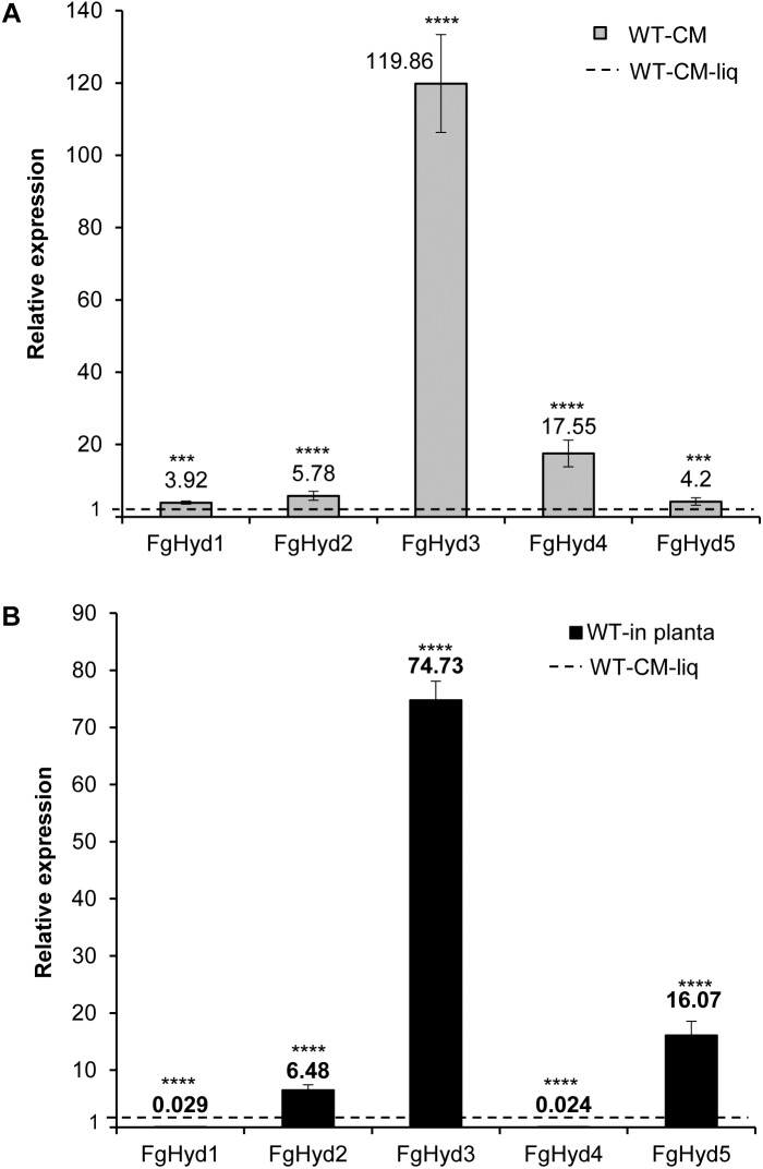
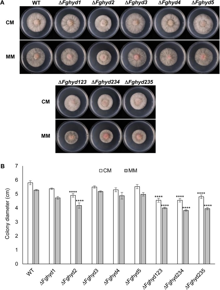
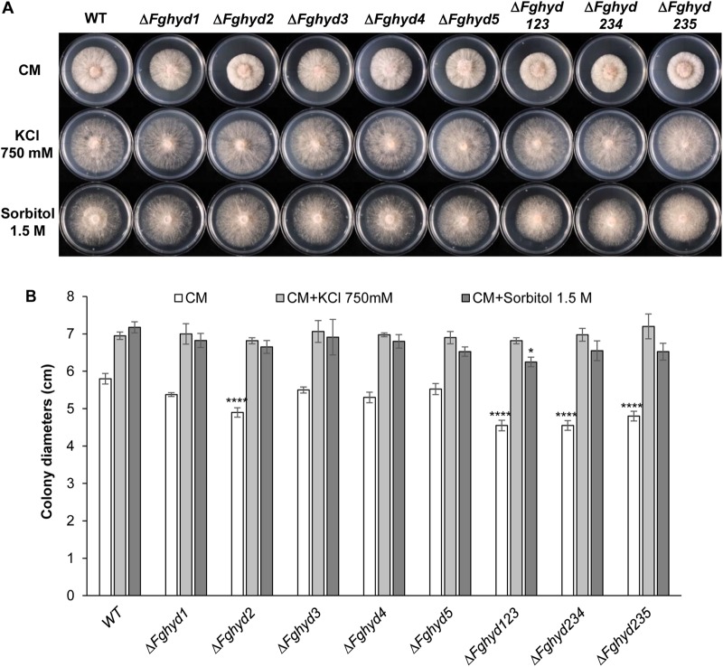
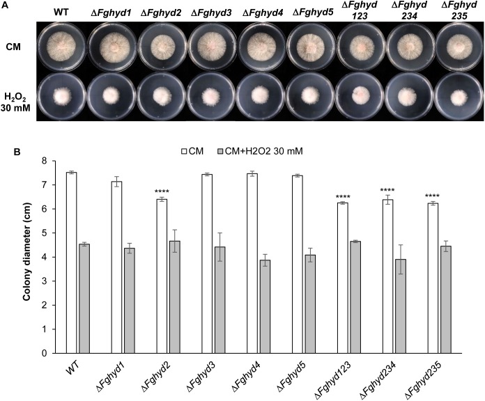
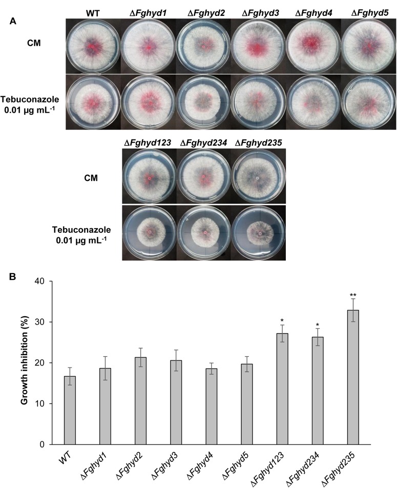
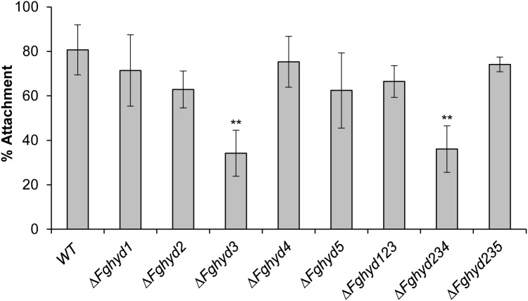
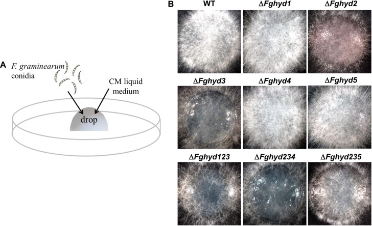
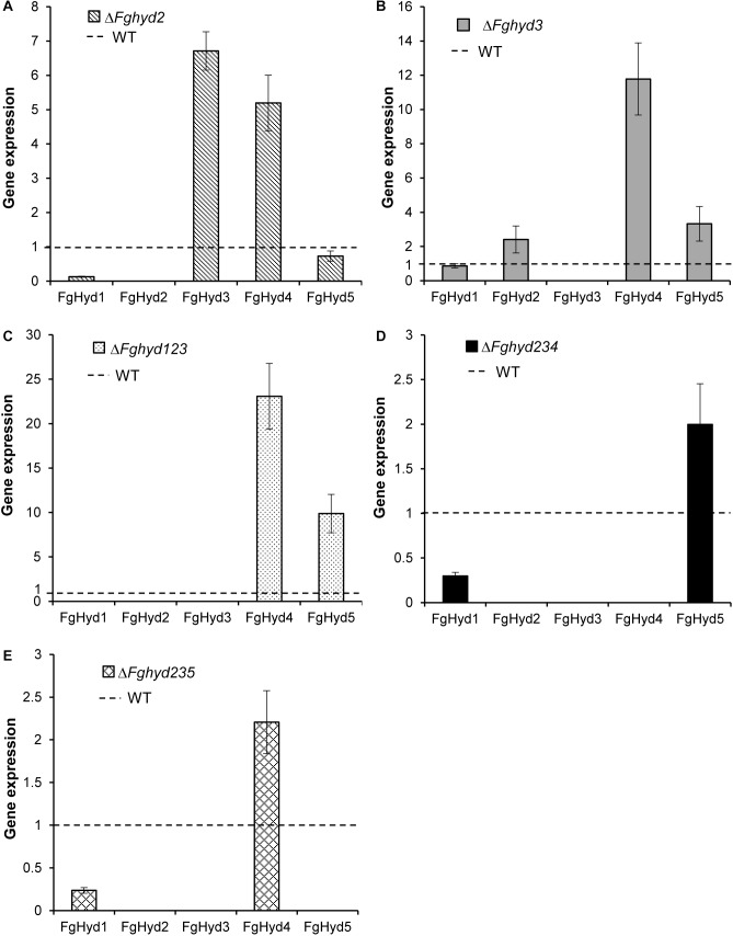
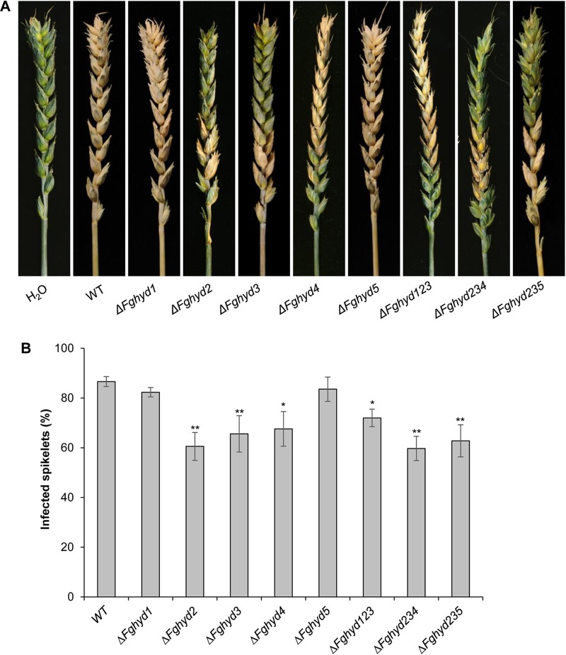
Hydrophobins (HPs) are small secreted fungal proteins possibly involved in several processes such as formation of fungal aerial structures, attachment to hydrophobic surfaces, interaction with the environment and protection against the host defense system. The genome of the necrotrophic plant pathogen Fusarium graminearum contains five genes encoding for HPs (FgHyd1-5). Single and triple FgHyd mutants were produced and characterized. A reduced growth was observed when the ΔFghyd2 and the three triple mutants including the deletion of FgHyd2 were grown in complete or minimal medium. Surprisingly, the growth of these mutants was similar to wild-type when grown under ionic, osmotic or oxidative stress conditions. All the mutant strains confirmed the ability to develop conidia and perithecia, suggesting that the FgHyds are not involved in normal development of asexual and sexual structures. A reduction in the ability of hyphae to penetrate through the water-air interface was observed for the single mutants ΔFghyd2 and ΔFghyd3 as well as for the triple mutants including the deletion of FgHyd2 and FgHyd3. Besides, ΔFghyd3 and the triple mutant ΔFghyd234 were also affected in the attachment to hydrophobic surface. Indeed, wheat infection experiments showed a reduction of symptomatic spikelets for ΔFghyd2 and ΔFghyd3 and the triple mutants only when spray inoculation was performed. This result could be ascribed to the affected ability of mutants deleted of FgHyd2 and FgHyd3 to penetrate through the water-air interface and to attach to hydrophobic surfaces such as the spike tissue. This hypothesis is strengthened by a histological analysis, performed by fluorescence microscopy, showing no defects in the morphology of infection structures produced by mutant strains. Interestingly, triple hydrophobin mutants were significantly more inhibited than wild-type by the treatment with a systemic triazole fungicide, while no defects at the cell wall level were observed.

Keywords: Fusarium graminearum; attachment; hydrophobin; virulence; water-air interface.


Figures

FIGURE 1
FIGURE 1
Fusarium graminearum hydrophobin family. Hydrophobin family of F. graminearum contains four class I (FgHyd1, FgHyd2, FgHyd3, and FgHyd4) and one class 2 (FgHyd5) hydrophobins. (A) Phylogenetic relationships of F. graminearum hydrophobins (gray circle) with hydrophobins from other ascomycetes demonstrate high homology among class 2, but low among class I hydrophobins. The Neighbor Joining model was built using MEGA 5 software. Robustness of the generated tree was determined using 1,000 bootstrap replicates. Bootstrap values are provided at the beginning of each branch and are given as a percentage. The fungal proteins used are: Cladosporium fulvum HCf3 (CAD92803); C. fulvum HCf5 (CAC27408); F. graminearum FgHyd1 (FGSG_01763); F. graminearum FgHyd2 (FGSG_01764); F. graminearum FgHyd3 (FGSG_09066); F. graminearum FgHyd4 (FGSG_03960); F. graminearum FgHyd5 (FGSG_01831); Fusarium verticillioides Hyd1 (Q6YF32); F. verticillioides Hyd2 (Q6YF31); F. verticillioides Hyd5 (Q6YD93); Botrytis cinerea Bhp1 (BC1G_15273); B. cinerea Bhp2 (BC1G_03994); B. cinerea Bhp3 (BC1G_01012); Aspergillus oryzae RolA (BAC65230.1); Verticillium dahliae VDH1 (AAY89101.1); Magnaporthe grisea MPG1 (P52751); M. grisea MHP1 (AAD18059); Fusarium culmorum FcHyd3p (ABE27987.1); F. culmorum FcHyd5p (ABE27986.1); Neurospora crassa EAS (EAA34064.1); Claviceps purpurea CPPH1 (CAD10781.1); Aspergillus nidulans RodA (AAA33321.1); A. nidulans DewA (AAC13762.1); Aspergillus fumigatus RodA (AAB60712.1) and A. fumigatus RodB (EAL91055.1). The corresponding accession numbers were obtained from the NCBI database (http://www.ncbi.nlm.nih.gov/). (B) Schematic representation of F. graminearum hydrophobin proteins displaying classical hydrophobin characteristics, signal peptide for secretion (SP, signal peptide), multiple cysteins (C, cystein) and low number of amino acids (aa, small proteins). Prediction of SP was performed using SignalP. Prediction of hydrophobin domains was performed using Motif Scan (MyHits, SIB, Switzerland). Dicty_CTDC Dictyostelium (slime mold) repeat.
FIGURE 2
FIGURE 2
FgHyd3 gene is the most highly expressed hydrophobin in F. graminearum. Expression analysis of hydrophobin genes by RT-qPCR demonstrated that Fghyd3 hydrophobin gene is the most highly expressed when F. graminearum wild type strain (WT) grows on an artificial hydrophobic surface or during infection of wheat spikes. (A) RNA extracted from mycelia of WT grown on agarized complete media (CM) with a cellophane layer for 3 days at 28°C in the dark, and WT grown in liquid CM media (CM-liq) for 3 days at 28°C with 150 rpm were used to quantify the relative expression of hydrophobin genes in axenic culture. (B) RNA extracted from wheat spikes inoculated with 200 conidia of WT (WT-in planta) for 5 days at 22°C with a photoperiod of 16/8 h light was used to quantify the relative expression of hydrophobin genes during infection. b-tubulin and EIF-5a (eukaryotic translation initiation factor 5A) were used as normalizers and expression on WT grown in liquid as calibrator (set to 1). Error bars indicate standard deviation calculated from data per triplicate (3 biological samples and 3 experimental replicates). Statistical analysis was calculated with respect to the WT using one way-Anova Bonferroni–Holm (significance: ∗∗∗∗p < 0.0001, ∗∗∗p < 0.001).
FIGURE 3
FIGURE 3
Fusarium graminearum hydrophobins deletion mutants phenotype on complete or minimal media. 5 mm agar blocks of actively growing deletion mutants or WT were placed on CM or minimal media (MM) and grown for 2 days at 28°C in the dark. Hydrophobin deletion mutants grew similarly to the WT with exception of ΔFghyd2 and triple mutants which grew slower. (A) Pictures of WT and hydrophobins deletion mutants were taken at 2 dpi. (B) Graph showing the average of colonies diameter. Error bars indicate standard deviation calculated from data representative of 2 biological experiments and 3 replicates. Statistical analysis of each treatment was calculated with respect to the WT using one way-Anova Bonferroni–Holm (significance: ∗∗∗∗p < 0.0001). The experiment was performed by using two independent knock-out mutants for each gene obtaining similar results.
FIGURE 4
FIGURE 4
Ionic and osmotic stress response of F. graminearum hydrophobin mutants on complete and minimal medium. Plates containing CM or CM supplemented with 750 mM KCl or 1.5 M sorbitol were inoculated with 5 mm plugs of actively growing mycelia of the WT strain, single or triple deletion mutants. Plates were incubated at 28°C in the dark. (A) Pictures representative of each treatment were taken at 2 dpi. (B) The susceptibility to stress was estimated by measuring colony diameters at 2 dpi. Error bars indicate standard deviation calculated from data representative of 2 biological experiments and 3 experimental replicates. Statistical analysis of each treatment was calculated with respect to the WT using one way-Anova Bonferroni–Holm (significance: p < 0.05, ∗∗∗∗p < 0.0001).
FIGURE 5
FIGURE 5
Oxidative stress response of F. graminearum hydrophobin mutants on complete and minimal medium. Plates containing CM or CM supplemented with 30 mM H2O2 were inoculated with 5 mm plugs of actively growing mycelia of the WT strain, single or triple deletion mutants. Plates were incubated at 28°C in the dark. (A) Pictures representative of each treatment were taken at 2 dpi. (B) The susceptibility to stress was estimated by measuring mycelia diameters after 2 days. Error bars indicate standard deviation calculated from data representative of 2 biological experiments and 3 experimental replicates. Statistical analysis of each treatment was calculated with respect to the WT using one way-Anova Bonferroni–Holm (significance: ∗∗∗∗p < 0.0001).
FIGURE 6
FIGURE 6
Triple hydrophobin mutants are more sensitive to the fungicide tebuconazole. While the single mutants grew similarly to the WT, the triple deletion mutants grew slower on minimal media supplemented with 0.01 μg mL-1 tebuconazole. (A) Plates with CM or CM supplemented with the fungicide tebuconazole (Folicur 250, Bayer) were inoculated with 5 μL of a conidial suspension of 1 × 10⁵ conidia mL-1 of the WT strain, single or triple hydrophobin mutants. Plates were incubated in the dark at 28°C. Pictures were taken at 3 dpi. (B) Percentage of growth inhibition was determined by measuring the radial growth at 3 dpi. Data represent the mean ± standard error (indicated by bars) of five independent experiments. Statistical analysis was calculated with respect to the WT using one way-Anova Bonferroni–Holm (significance: p < 0.05, ∗∗p < 0.01).
FIGURE 7
FIGURE 7
FgHyd3 is necessary for hydrophobic surface attachment. Six drops containing 50 μL sterile water and 1,000 conidia each were placed on the inner surface of Petri dishes and incubated at 28°C on the dark. After 24 h, fungal germlings were washed 3 times and counted. 1,000 conidia counted represent 100% of attachment. Error bars indicate standard deviation calculated from data representative of 2 biological experiments and 6 replicates each. Statistical analysis was calculated with respect to the WT using one way-Anova Bonferroni–Holm (significance: ∗∗p < 0.01). Only ΔFghyd3 and triple ΔFghyd234 mutants have a defect in attachment to hydrophobic surfaces. The experiment was performed by using two independent knock-out mutants for each gene obtaining similar results.
FIGURE 8
FIGURE 8
FgHyd2 and FgHyd3 are necessary for breaking the liquid-air interface. (A) A 50 μL drop of CM containing 500 conidia of the WT strain or hydrophobin deletion mutants were placed on the inner surface of Petri dishes and allowed to grow for 36 h at 28°C in the dark. (B) Pictures were taken with the stereo microscope (Leica- ZDFIII) at 36 h. Hyphae of single deletion mutants ΔFghyd2, ΔFghyd3 and triple deletion mutant ΔFghyd234 were not able to break the liquid-air interface. Triple deletion mutants ΔFghyd123 and ΔFghyd235 displayed less mycelia passing through the liquid-air interface than the WT. The experiment was performed by using two independent knock-out mutants for each gene obtaining similar results.
FIGURE 9
FIGURE 9
Hydrophobin genes expression in hydrophobin deletion mutants grown on artificial surface. RNAs extracted from mycelia of WT strain, ΔFghyd2, ΔFghyd3 and triple mutants grown on agarized CM with a cellophane layer were extracted and used for expression analysis using RT-qPCR. b-tubulin and EIF-5a were used as normalizers and expression on WT under similar conditions as calibrator (set to 1). (A) Lack of FgHyd2 gene increased expression of FgHyd3 and FgHyd4. (B) FgHyd2, FgHyd4, and FgHyd5 were up-regulated on the ΔFghyd3 mutant. (C) FgHyd4 and FgHyd5 were strongly expressed on the triple mutant ΔFghyd123 compared to WT. (D) Expression of FgHyd5 was moderately increased on the triple deletion mutant ΔFghyd234. (E) FgHyd4 expression was increased on the triple deletion mutant ΔFghyd235. Error bars indicate standard error calculated from data per triplicate using the relative expression analysis tool REST (Relative Expression Software Tool).
FIGURE 10
FIGURE 10
Hydrophobins of F. graminearum are necessary for virulence. (A) Wheat spikes of the susceptible cultivar Nandu were sprayed twice with 100 μL of a conidial suspension containing 500 conidia μL-1 of the WT strain, single and triple mutants. After 24 h the spikes were sprayed twice with 2 mL of water. The spikes sprayed with the single mutants ΔFgHyd2, ΔFgHyd3, and ΔFgHyd4 as well as the triple mutants developed less symptoms than the WT, while ΔFgHyd1 and ΔFgHyd5 mutants developed similar symptoms than the WT. Water was used as mock inoculation. (B) Disease symptoms were assessed at 21 dpi by counting the number of visually diseased spikelets on wheat cultivar Nandu. Infected spikelets are expressed as percentage of symptomatic spikelets on total number of spikelets of the respective head. Data represent the mean ± standard error (indicated by bars) of 10 independent infection experiments. Statistical analysis was calculated with respect to the WT using one way-Anova Bonferroni–Holm (significance: p < 0.05, ∗∗p < 0.01). The experiment was performed by using two independent knock-out mutants for each gene obtaining similar results.
FIGURE 11
FIGURE 11
Expression analysis of hydrophobin genes during infection. RNAs extracted from wheat spikes inoculated with 10 μL of a conidial suspension of 2 × 10⁴ conidia mL-1 of WT strain, ΔFghyd2, ΔFghyd3 and triple mutants were extracted at 5 dpi and used for expression analysis using RT-qPCR. b-tubulin and EIF-5a were used as normalizers and expression on WT under similar conditions as calibrator (set to 1). (A) FgHyd3 gene expression increased and expression of FgHyd1 and FgHyd4 decreased in ΔFghyd2 mutant. (B) FgHyd4 gene expression decreased on the ΔFghyd3 mutant, while other hydrophobin genes had no change in expression. (C) FgHyd4 and FgHyd5 had higher expression on the triple mutant ΔFghyd123 compared to WT. (D) Expression of FgHyd5 increased on the triple deletion mutant ΔFghyd234. (E) FgHyd4 expression was highly increased on the triple deletion mutant ΔFghyd235 compared to WT. Error bars indicate standard error calculated from data per triplicate using the relative expression analysis tool REST (Relative Expression Software Tool).

References

    1. Aimanianda V., Bayry J., Bozza S., Kniemeyer O., Perruccio K., Elluru S. R., et al. (2009). Surface hydrophobin prevents immune recognition of airborne fungal spores. Nature 460 1117–1121. 10.1038/nature08264 - DOI - PubMed
    1. Beck E., Ludwig G., Auerswald E. A., Reiss B., Schaller H. (1982). Nucleotide sequence and exact localization of the neomycin phosphotransferase gene from transposon Tn5. Gene 19 327–336. - PubMed
    1. Beckerman J. L., Ebbole D. J. (1996). MPG1, a gene encoding a fungal hydrophobin of Magnaporthe grisea, is involved in surface recognition. Mol. Plant Microbe Interact. 9 450–456. 10.1094/MPMI-9-0450 - DOI - PubMed
    1. Chen L., Zhong H. Y., Kuang J. F., Li J. G., Lu W. J., Chen J. Y. (2011). Validation of reference genes for RT-qPCR studies of gene expression in banana fruit under different experimental conditions. Planta 234 377–390. 10.1007/s00425-011-1410-3 - DOI - PubMed
    1. Christianson T. W., Sikorski R. S., Dante M., Shero J. H., Heiter P. (1992). Multifunctional yeast high-copy-number shuttle vectors. Gene 110 119–122. 10.1016/0378-1119(92)90454-W - DOI - PubMed

LinkOut - more resources