. 2019 Apr 11:10:704.
doi: 10.3389/fimmu.2019.00704. eCollection 2019.

Potent Fc Receptor Signaling by IgA Leads to Superior Killing of Cancer Cells by Neutrophils Compared to IgG

Affiliations

Potent Fc Receptor Signaling by IgA Leads to Superior Killing of Cancer Cells by Neutrophils Compared to IgG

Arianne M Brandsma et al. Front Immunol. .

Abstract

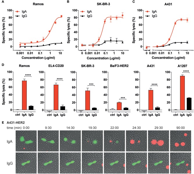
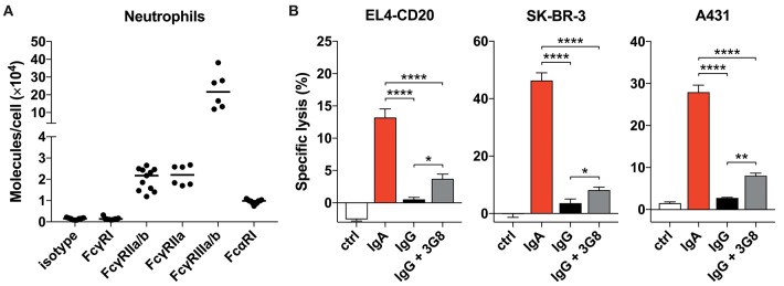
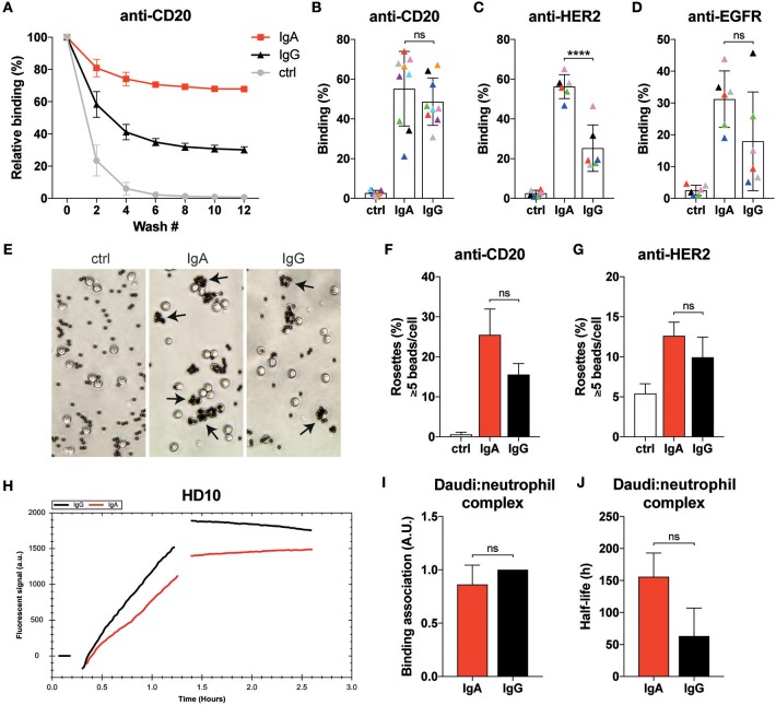
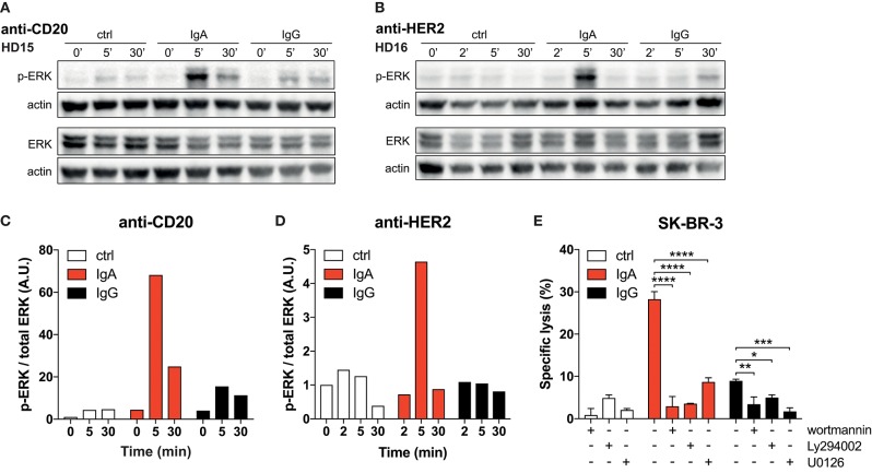
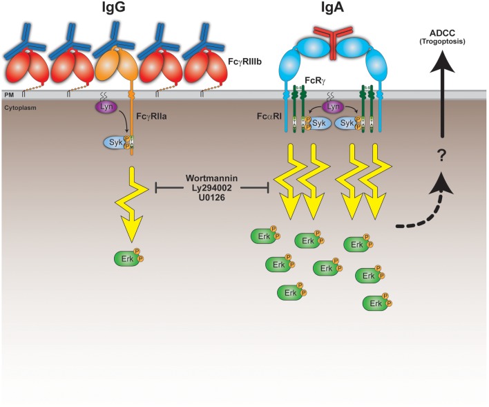
Antibody therapy of cancer is increasingly used in the clinic and has improved patient's life expectancy. Except for immune checkpoint inhibition, the mode of action of many antibodies is to recognize overexpressed or specific tumor antigens and initiate either direct F(ab')2-mediated tumor cell killing, or Fc-mediated effects such as complement-dependent cytotoxicity (CDC) and antibody-dependent cell-mediated cytotoxicity/phagocytosis (ADCC/P) after binding to activating Fc receptors. All antibodies used in the clinic are of the IgG isotype. The IgA isotype can, however, also elicit powerful anti-tumor responses through engagement of the activating Fc receptor for monomeric IgA (FcαRI). In addition to monocytes, macrophages and eosinophils as FcαRI expressing immune cells, neutrophils are especially vigorous in eliminating IgA opsonized tumor cells. However, with IgG as single agent it appears almost impossible to activate neutrophils efficiently, as we have visualized by live cell imaging of tumor cell killing. In this study, we investigated Fc receptor expression, binding and signaling to clarify why triggering of neutrophils by IgA is more efficient than by IgG. FcαRI expression on neutrophils is ~2 times and ~20 times lower than that of Fcγ receptors FcγRIIa and FcγRIIIb, but still, binding of neutrophils to IgA- or IgG-coated surfaces was similar. In addition, our data suggest that IgA-mediated binding of neutrophils is more stable compared to IgG. IgA engagement of neutrophils elicited stronger Fc receptor signaling than IgG as indicated by measuring the p-ERK signaling molecule. We propose that the higher stoichiometry of IgA to the FcαR/FcRγ-chain complex, activating four ITAMs (Immunoreceptor Tyrosine-based Activating Motifs) compared to a single ITAM for FcγRIIa, combined with a possible decoy role of the highly expressed FcγRIIIb, explains why IgA is much better than IgG at triggering tumor cell killing by neutrophils. We anticipate that harnessing the vast population of neutrophils by the use of IgA monoclonal antibodies can be a valuable addition to the growing arsenal of antibody-based therapeutics for cancer treatment.

Keywords: ADCC; CD89; Fc alpha receptor I; IgA; cancer; immunotherapy; neutrophil; signaling.


Figures

Figure 1
Figure 1
IgA mediates higher tumor cell lysis than IgG with human neutrophils. (A–D) Specific lysis of tumor cells by isolated human neutrophils (E:T = 40:1) using a 4 h 51Cr release assay. Anti-CD20 Abs were used for Ramos and EL4-CD20, anti-HER2 Abs were used for SK-BR-3 and Ba/F3-HER2, anti-EGFR Abs were used for A431 and A1207 target cells. (A–C) Specific lysis of indicated target cells using a broad titration range of IgA and IgG antibodies. (D) Antibody concentrations were identical (10 μg/mL) for IgG and IgA antibodies against all target cells except for Ramos (13.3 μg/mL) and A1207 cells (1 μg/mL). Ctrl indicates condition without antibody (or 0.001 μg/mL IgG1-anti-CD20 for Ramos cells). One representative graph is shown for n = 4–6 independent experiments with different healthy donors. p < 0.001: ***, p < 0.0001: ****, unpaired Student's t test. (E) Stills from live cell microscopy of adhered A431-HER2 cells (calcein labeled) and neutrophils in the presence of 5 μg/mL anti-HER2 IgA2 or IgG (Trastuzumab) and TO-PRO-3 (red fluorescence), time in minutes, image acquisition every 30 s for 1.5 h.
Figure 2
Figure 2
Blocking FcγRIIIb only marginally improves tumor cell lysis. (A) Quantitative expression of FcγR and FcαRI on human neutrophils as analyzed by flow cytometry (Qifikit), n = 6–11 healthy donors. (B) 3 h 51Cr release assays using EL4-CD20 with anti-CD20 IgA2 or IgG1 (rituximab), SK-BR-3 with anti-HER2 IgA1 or IgG1 and A431 with anti-EGFR IgA2 or IgG1, all at 10 ug/mL. 3G8 F(ab′)2 fragments (1 ug/mL final concentration) were added 15 min prior to start of the ADCC assays. Statistics: one-way ANOVA with Tukey's multiple comparison test, p < 0.05: *, p < 0.01: **, p < 0.0001: ****. Ctrl indicates no antibody added.
Figure 3
Figure 3
Neutrophils display similar binding characteristics to IgA- or IgG-associated surfaces. (A) Example of relative binding of a donor of which calcein labeled neutrophils were allowed to associate to anti-CD20 IgA2 or IgG1 (clone UMAB001) coated 96 well plate. Remaining calcein fluorescence was measured after repeating wash steps. Calcein fluorescence before the first wash was set 100%. Ctrl indicates that no antibody was used during coating. (B–D) As in A, but data of 6–9 different donors at wash 10 are combined per target being anti-CD20 IgA2 or IgG1 (clone UMAB001) or anti-HER2 IgA2 or IgG1. Each color indicates data from the same donor. Statistics: two-way ANOVA with Tukey's multiple comparison test, p < 0.0001: ****. (E) Examples images of neutrophils binding to albumin, anti-CD20 IgA, or IgG coated Dynabeads (ratio beads:neutrophils = 5:1). Rosettes are defined as cells binding 5 or more beads. (F,G) Quantification of rosettes of anti-CD20- or anti-HER2-coated Dynabeads with neutrophils. One representative of n = 3 individual experiments is shown. Differences were not significant according to one-way ANOVA with Tukey's multiple comparison test. (H) Binding traces for calcein labeled neutrophils binding to anti-CD20 IgA- (red) or IgG- (black) opsonized Daudi cells. One representative of n = 5 experiments is shown. (I) Binding association and (J) average half-life of the Daudi:neutrophils complex pooled from 5 different donors. Statistics: paired Student's t-test.
Figure 4
Figure 4
Strong p-ERK signal in neutrophils elicited by IgA. (A,B) Time-dependent increase and decrease of p-ERK signal in neutrophils after exposure to anti-CD20 or anti-HER2 IgA2- or IgG1-coated Dynabeads. Actin detection was performed on the same blot after destroying the p-ERK or total ERK signal. (C,D) Quantification of signaling defined as the ratio of normalized p-ERK/actin over normalized total ERK/actin. Data was normalized against the 0 min time point. (E) 4 h 51Cr release assays using SK-BR-3 with anti-HER2 IgA2 or IgG1, both at 1 ug/mL. Where indicated, pharmacological inhibitors of signaling factors were included (wortmannin at 0.5 μM, Ly294002 and U0126 both at 20 μM). Statistics: two-way ANOVA with Tukey's multiple comparisons test p < 0.05: *, p < 0.01: **, p < 0.001: ***, p < 0.0001: ****.
Figure 5
Figure 5
Model for superior IgA-mediated tumor cell killing by neutrophils. Neutrophils express the low affinity Fc receptors FcγRIIIb, FcγRIIa, and FcαRI. Both FcγRIIa and FcαRI are activating Fc receptors, because upon ligand (antibody Fc domain) binding, clustering and recruitment of kinases (e.g., Lyn), they signal through their ITAM domains and elicit cellular effector functions. The relatively high FcγRIIIb expression on neutrophils could possibly interfere with the activating capacity of FcγRIIa by competing for Fc domain binding and preventing proper formation of signaling platforms and/or organization within the lipid bilayer. This results in a poor ability of FcγRIIa to initiate sufficient signaling in neutrophils. Despite the low FcαRI expression on neutrophils, they can still engage clustered IgA Fc domains to a similar degree as IgG Fc domains bind the FcγRs. IgA can, however, bind FcαRI bivalently resulting in more stable binding and recruitment of in total 4 ITAMs. This scenario would initiate a robust ITAM signaling necessary for activating effector functions, including trogoptosis to eliminate tumor cells. This is further illustrated by the signaling inhibitors wortmannin, Ly294002 and U0126 that prevent tumor cell lysis in vitro. The question mark refers to yet unclear processes involving signaling, intracellular Ca2+, actin-myosin contraction, and immune cell-tumor cell interactions that lead to ADDC by neutrophils (trogoptosis) (23).

Similar articles

Cited by

References

    1. Scott AM, Wolchok JD, Old LJ. Antibody therapy of cancer. Nat Rev Cancer. (2012) 12:278–87. 10.1038/nrc3236 - DOI - PubMed
    1. Kimiz-Gebologlu I, Gulce-Iz S, Biray-Avci C. Monoclonal antibodies in cancer immunotherapy. Mol Biol Rep. (2018) 45:2935–40. 10.1007/s11033-018-4427-x - DOI - PubMed
    1. Overdijk MB, Verploegen S, Bleeker WK, Parren PWHI. Chapter 13 - Role of IgG Fc receptors in monoclonal antibody therapy of cancer. In: Antibody Fc, Linking Adaptive and Innate Immunity. Amsterdam: Elsevier Inc. (2014). p. 239–55. 10.1016/B978-0-12-394802-1.00013-3 - DOI
    1. Getahun A, Cambier JC. Of ITIMs, ITAMs, and ITAMis: revisiting immunoglobulin Fc receptor signaling. Immunol Rev. (2015) 268:66–73. 10.1111/imr.12336 - DOI - PMC - PubMed
    1. Leusen JHW. IgA as therapeutic antibody. Mol Immunol. (2015) 68:35–9. 10.1016/j.molimm.2015.09.005 - DOI - PubMed

Publication types