. 2019 Apr 12:10:793.
doi: 10.3389/fimmu.2019.00793. eCollection 2019.

The P2X7 Receptor Is Shed Into Circulation: Correlation With C-Reactive Protein Levels

Affiliations

The P2X7 Receptor Is Shed Into Circulation: Correlation With C-Reactive Protein Levels

Anna Lisa Giuliani et al. Front Immunol. .

Abstract

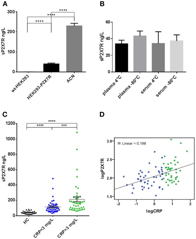
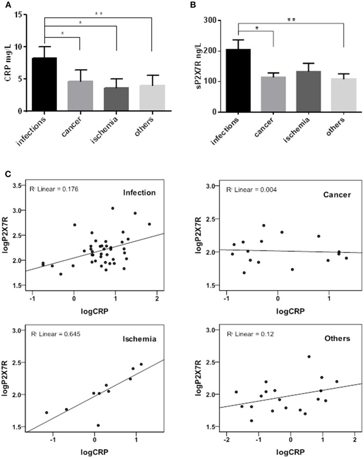
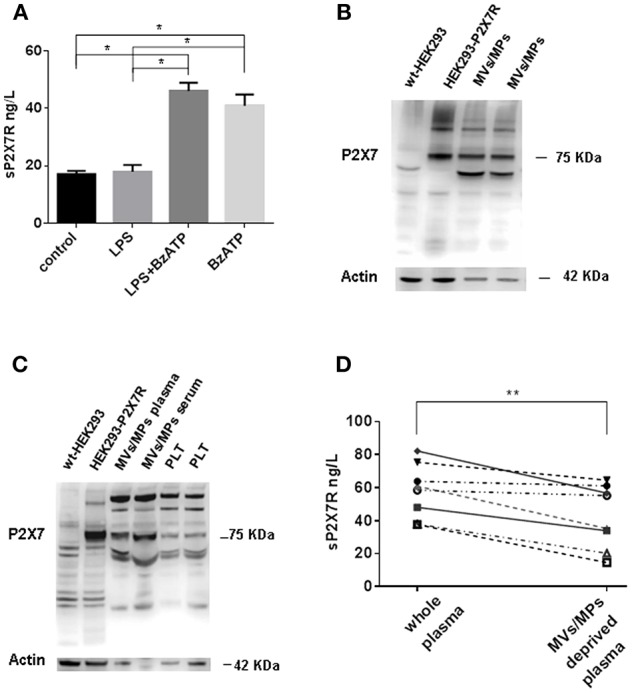
The P2X7 receptor (P2X7R) is a key pro-inflammatory plasma membrane receptor responsible for NLRP3 inflammasome activation and IL-1β release. Various inflammatory plasma membrane receptors (e.g., IL-1 type I receptor, TNF type I and II receptors, IL-2 receptor) are shed under different pathophysiological conditions. In the present study, we show that the full length P2X7R is released into circulation in patients as well as in healthy subjects. Blood levels of shed P2X7R (sP2X7R) correlate to those of the inflammatory marker C reactive protein (CRP). Blood sP2X7R ranged from 16.74 to 82.17 ng/L, mean ± SE 40.97 ± 3.82 (n = 26) in healthy subjects, from 33.1 to 484.0 ng/L, mean ± SE 114.78 ± 12.22 (n = 45) in patients with CRP <3 mg/L, and from 63.65 to 1092.3 ng/L, mean ± SE 204.2 ± 30.94 (n = 42) in patients with CRP >3 mg/L. sP2X7R in plasma was largely associated to microvesicles/microparticles. Peripheral blood monocytes from healthy subjects released sP2X7R upon stimulation with the semi-selective P2X7R agonist benzoyl ATP. These data show that the P2X7R can be released into circulation, and that its blood levels increase in various disease conditions.

Keywords: cytokines; extracellular ATP; inflammation; microvesicles; purinergic signaling.


Figures

Figure 1
Figure 1
The P2X7R was present in human plasma and serum, its concentration was higher in diseased subjects and correlated with CRP. (A) ELISA analysis for sP2X7R of lysates from 108 cells/ml of wt-HEK293, HEK293-P2X7R, and ACN neuroblastoma cells. Data are means ± SE of results from 3 separate experiments. Differences are statistically significant (One-way ANOVA, p < 0.0001; unpaired t-test between groups: ****p < 0.0001). (B) Effect of storage conditions on sP2X7R detection by ELISA in plasma and serum samples from 4 healthy subjects. Differences are not statistically significant. (C) sP2X7R levels were measured by ELISA in serum samples from 26 healthy control subjects (HC), 45 patients with CRP <3 mg/L and 42 patients with CRP >3 mg/L. Data are means ± SE. Mann Whitney test between groups: ***p < 0.001; ****p < 0.0001. (D) Linear regression analysis between log-transformed CRP and sP2X7R concentrations in serum samples from 87 diseased subjects. Color coding in (C,D): blue, CRP <3 mg/L, green. CRP >3 mg/L.
Figure 2
Figure 2
sP2X7R was differentially concentrated in different diagnosis sub-groups. CRP and sP2X7R were measured in serum samples stratified into four sub-groups according to diagnosis at admission to the hospital: infectious diseases (n = 42), cancer (n = 16), ischemia (n = 10) and others (trauma, autoimmune diseases) (n = 19). Data are means ± SE. Only significant differences are shown in the graphs. (A) Kruskal-Wallis test p = 0.0057; Mann Whitney test between groups: *p < 0.05; **p < 0.01. (B) Kruskal-Wallis test p = 0.0205; Mann Whitney test between groups: *p < 0.05; **p < 0.01. (C) Linear regression analysis between log-transformed CRP and sP2X7R in the different diagnosis sub-groups: ischemia, R2 linear = 0.645; infectious diseases, R2 linear = 0.176; cancer, R2 linear = 0.004; others, R2 linear = 0.12.
Figure 3
Figure 3
sP2X7R is shed in association with MVs/MPs by P2X7R stimulation. (A) Monocyte-derived macrophages were isolated from blood samples of 4 healthy control subjects as described in Methods and incubated in 10% FBS-supplemented RPMI under the following conditions: (1) no additions (control) for 5 h; (2) 1 μg/ml LPS for 4 h; (3) 1 μg/ml LPS for 4 h, followed by 300 μM BzATP for 1 h; (4) no additions for 4 h, followed by 300 μM BzATP for 1 h. Data are means ± SE of data from 4 separate experiments. Only significant differences are shown in the graph. Kruskal-Wallis test p = 0.0008. Mann Whitney test between groups: *p < 0.05. (B) MVs/MPs were isolated from two plasma samples by centrifugation at 2200xg followed by 14000xg. Ten μg of protein were loaded on the SDS-PAGE gel. (C) MVs/MPs were isolated from one plasma sample and from one serum sample by centrifugation at 2200xg followed by 100000xg ultracentrifugation. Platelets (PLT) were isolated from plasma by centrifugation at 2200xg. See Material and Methods for additional details. Ten μg of proteins were loaded on the SDS-PAGE gel. (D) ELISA analysis of sP2X7R content of plasma samples from 8 subjects before and after depletion of MVs/MPs by ultracentrifugation at 14000× g. Wilcoxon matched paired t-test: p = 0.0078.

Similar articles

Cited by

References

    1. Coleman LG, Jr, Maile R, Jones SW, Cairns BA, Crews FT. HMGB1/IL-1beta complexes in plasma microvesicles modulate immune responses to burn injury. PLoS ONE. (2018) 13:e0195335. 10.1371/journal.pone.0195335 - DOI - PMC - PubMed
    1. Zhang S, Dai H, Zhu L, Lin F, Hu Z, Jing R, et al. . Microvesicles packaging IL-1beta and TNF-alpha enhance lung inflammatory response to mechanical ventilation in part by induction of cofilin signaling. Int Immunopharmacol. (2018) 63:74–83. 10.1016/j.intimp.2018.07.034 - DOI - PubMed
    1. Sha Y, Markovic-Plese S. Activated IL-1RI signaling pathway induces Th17 cell differentiation via interferon regulatory factor 4 signaling in patients with relapsing-remitting multiple sclerosis. Front Immunol. (2016) 7:543. 10.3389/fimmu.2016.00543 - DOI - PMC - PubMed
    1. Moreira-Souza ACA, Almeida-da-Silva CLC, Rangel TP, Rocha GDC, Bellio M, Zamboni DS, et al. . The P2X7 receptor mediates toxoplasma gondii control in macrophages through canonical NLRP3 inflammasome activation and reactive oxygen species production. Front Immunol. (2017) 8:1257. 10.3389/fimmu.2017.01257 - DOI - PMC - PubMed
    1. Jimenez-Pacheco A, Diaz-Hernandez M, Arribas-Blazquez M, Sanz-Rodriguez A, Olivos-Ore LA, Artalejo AR, et al. . Transient P2X7 receptor antagonism produces lasting reductions in spontaneous seizures and gliosis in experimental temporal lobe epilepsy. J Neurosci. (2016) 36:5920–32. 10.1523/JNEUROSCI.4009-15.2016 - DOI - PMC - PubMed

Publication types