

Editorial
doi: 10.21037/qims.2019.03.06.
Real-time intraoperative ultrasound in brain surgery: neuronavigation and use of contrast-enhanced image fusion
Affiliations
- PMID: 31032183
- PMCID: PMC6462565
- DOI: 10.21037/qims.2019.03.06
Item in Clipboard
Editorial
Real-time intraoperative ultrasound in brain surgery: neuronavigation and use of contrast-enhanced image fusion
Quant Imaging Med Surg.
2019 Mar.
No abstract available
Conflict of interest statement
Conflicts of Interest: The authors have no conflicts of interest to declare.
Figures
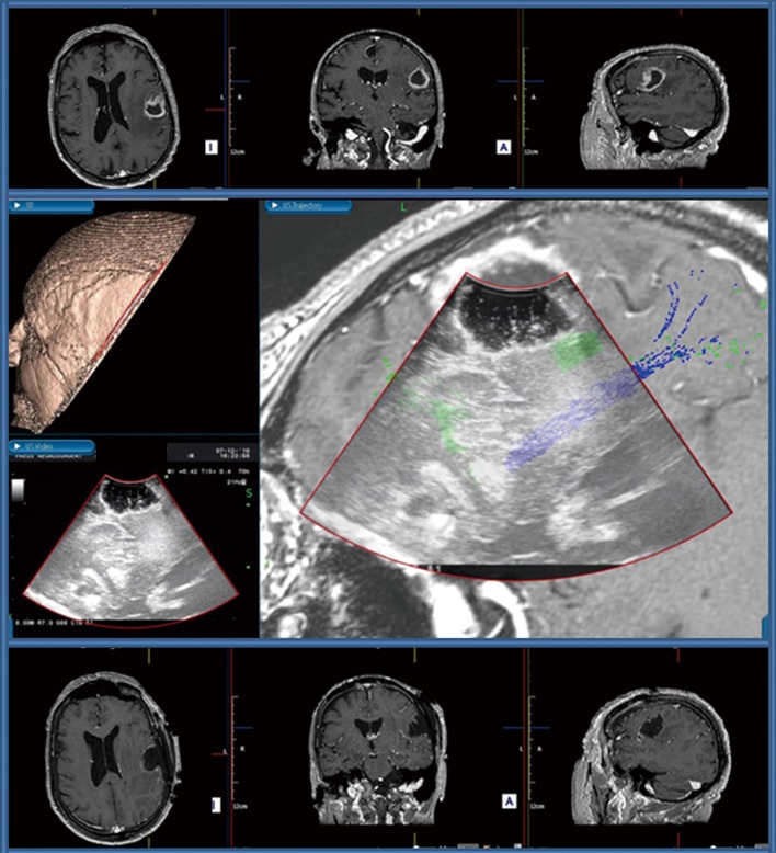
Awake craniotomy for a left-sided motor strip glioma. Upper row: Preoperative post-contrast T1WI MRI scan showing an irregular cortico-subcortical lesion in the left frontal operculum, characterised by ill-defined peripheral gadolinium uptake and central necrosis with a significant amount of surrounding vasogenic edema. Central Image: Screenshot of image fusion created for neuronavigation purposes by coupling IoUS and preoperative MRI scan. Real-time visualisation of the relation of the surgical cavity with subcortical association fibres, note the green fibres seen adjacent to the deep aspect of the surgical cavity indicating the Superior Longitudinal Fasciculus and Inferior Fronto-Occipital Fasciculus serving speech function, and the blues fibers indicating the cortico-spinal tract. The presence of these fibres was confirmed with subcortical stimulation during the procedure. Bottom row: Postoperative post-contrast T1WI MRI scan showing a complete surgical resection with a clear surgical cavity.
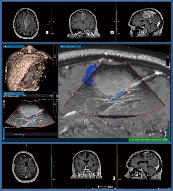
Falcine meningioma in close contact with the Anterior Cerebral Artery vessels and the motor strip. Upper row: Preoperative post-contrast T1WI MRI scan showing a mass lesion originating from the falx and causing edema in the adjacent parts of the frontal lobe, more extensive on the left where it affects the precentral region inducing displacement of the corpus callosum. Central Image: Screenshot of image fusion created for neuronavigation purposes demonstrating multimodality real-time imaging (conventional intraoperative navigation & DTI & real-time US & Doppler), note the blue fibers seen lateral to the lesion indicating the close relationship with the cortico-spinal tract, the inferior sagittal sinus adjacent to the lesion and the mass effect with inferior displacement of the cingulate gyrus and corpus callosum. Bottom row: Postoperative post-contrast T1WI MRI scan showing a complete surgical resection with re-expansion of the ventricular system, and no evidence of bleeding/ischaemic events surrounding the surgical cavity.
References
-
- Talacchi A, Turazzi S, Locatelli F, Sala F, Beltramello A, Alessandrini F, Manganotti P, Lanteri P, Gambin R, Ganau M, Tramontano V, Santini B, Gerosa M. Surgical treatment of high-grade gliomas in motor areas. The impact of different supportive technologies: a 171-patient series. J Neurooncol 2010;100:417-26. 10.1007/s11060-010-0193-x - DOI - PubMed
-
- Ma H, Wang Z, Xu K, Shao Z, Yang C, Xu P, Liu X, Hu C, Lu X, Rong Y. Three-dimensional arterial spin labeling imaging and dynamic susceptibility contrast perfusion-weighted imaging value in diagnosing glioma grade prior to surgery. Exp Ther Med 2017;13:2691-8. 10.3892/etm.2017.4370 - DOI - PMC - PubMed
Publication types
LinkOut - more resources
Full Text Sources