

Editorial
doi: 10.21037/atm.2019.01.49.
Ultrasound patterns of pulmonary edema
Affiliations
- PMID: 31032297
- PMCID: PMC6462619
- DOI: 10.21037/atm.2019.01.49
Item in Clipboard
Editorial
Ultrasound patterns of pulmonary edema
Ann Transl Med.
2019 Mar.
No abstract available
Conflict of interest statement
Conflicts of Interest: The authors have no conflicts of interest to declare.
Figures
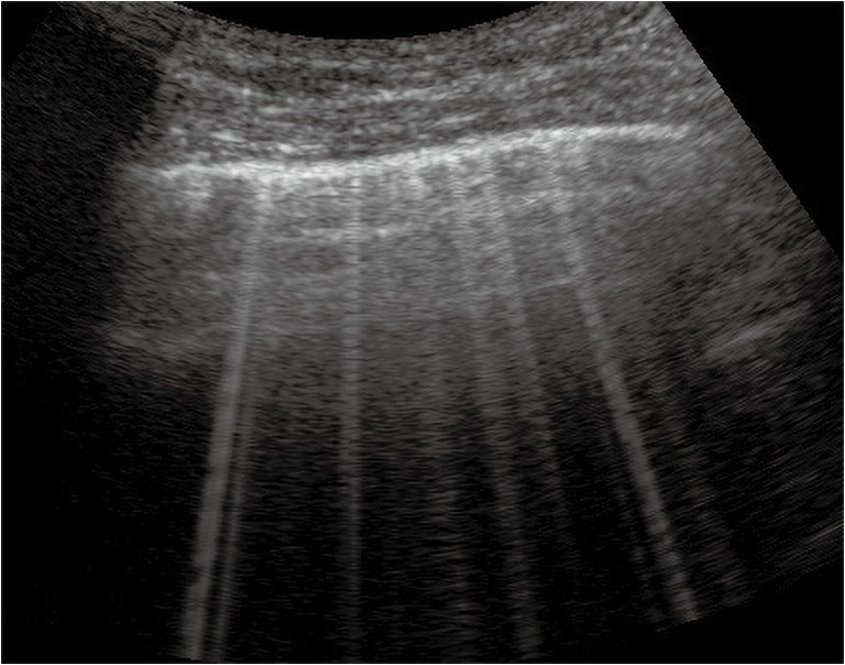
Septal pattern of early CPE. The pleural line is regular. B-lines are separated, laser like artefacts, spreading from the pleural line to the bottom of the screen and they show an internal sequence of alternating horizontal bands (convex probe, fundamental imaging, 6 MHz). CPE, cardiogenic pulmonary edema.
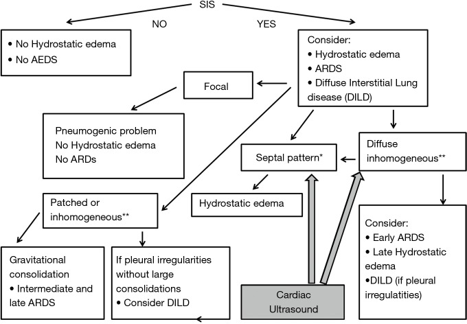
Algorithmic approach for the ultrasound diagnosis of ARDS. The presence of SIS is mandatory for the identification of a parenchymal subpleural involvement. Focal SIS is indicative of a pneumogenic pathology. Septal pattern is indicative of increased hydrostatic extravascular lung water. Inhomogeneous SIS and gravitational consolidations are suggestive of established ARDS. A chronic evolution of the disease, SIS and the presence of coarse pleural irregularities indicates pulmonary fibrosis. Cardiac ultrasound can be resolutive when the differential diagnosis between late CPE and early ARDS is necessary, and mixed lung involvement (hydrostatic an fibrogenic) is possible. *, presence of separated B-lines that appear as discrete laser-like vertical hyperechoic artifacts, arising with a narrow base from the pleural line, extending to the bottom of the screen without fading, and moving synchronously with lung sliding. Typical septal B-lines show a sequence of alternating white and black (or gray) horizontal bands. **, this term refers to the inhomogeneity of the individual B-lines and to the spatial variability of the B-lines arrangement. ARDS, acute respiratory distress syndrome; SIS, sonographic interstitial syndrome; CPE, cardiogenic pulmonary edema.
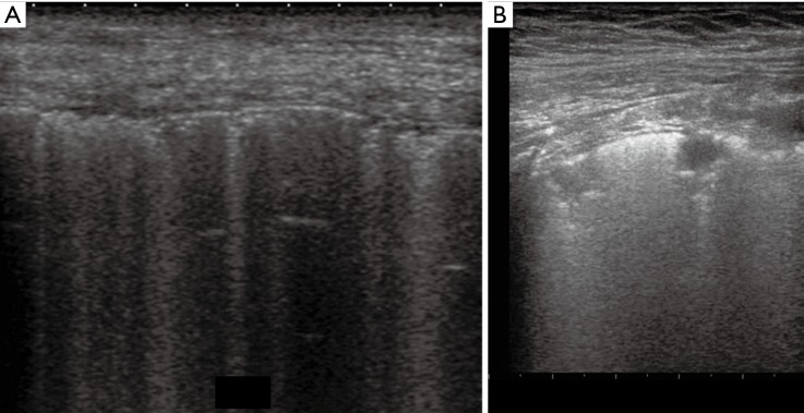
Early ARDS (A) and ARDS with consolidations (B). (A) Diffuse inhomogeneous SIS with spared areas; (B) white lung with small consolidations coexist. Note pleural line irregularities. ARDS, acute respiratory distress syndrome; SIS, sonographic interstitial syndrome.
Comment on
-
Diagnostic value of cardiopulmonary ultrasound in elderly patients with acute respiratory distress syndrome.BMC Pulm Med. 2018 Aug 13;18(1):136. doi: 10.1186/s12890-018-0666-9. BMC Pulm Med. 2018. PMID: 30103730 Free PMC article.
References
-
- Ranieri VM, Rubenfeld GD, Thompson BT, et al. ARDS Definition Task Force Acute respiratory distress syndrome: the Berlin Definition. JAMA 2012;307:2526-33. - PubMed
Publication types
LinkOut - more resources
Full Text Sources