

Surface Immobilization of TiO2 Nanotubes with Bone Morphogenetic Protein-2 Synergistically Enhances Initial Preosteoblast Adhesion and Osseointegration
- PMID: 31032352
- PMCID: PMC6457305
- DOI: 10.1155/2019/5697250
Surface Immobilization of TiO2 Nanotubes with Bone Morphogenetic Protein-2 Synergistically Enhances Initial Preosteoblast Adhesion and Osseointegration
Abstract
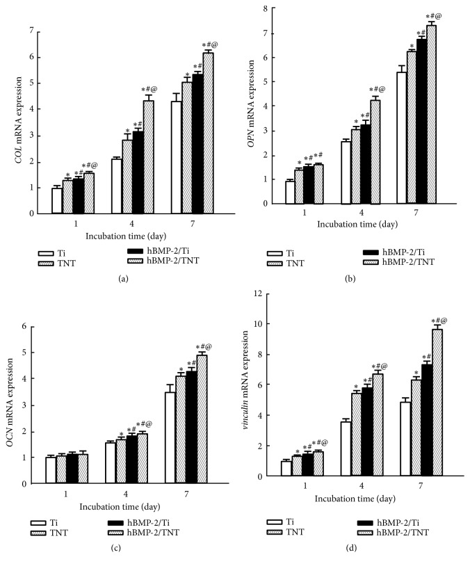
Although titanium (Ti) alloys have been widely used as implant materials, the bioinertness of pristine Ti impairs their bioactivity and early osseointegration. In the present work, we prepared TiO2 nanotubes (TNT) layer on the titanium (Ti) surface by anodic oxidation. The anodized surface was functionalized with human bone morphogenetic protein-2 coating to form the hBMP-2/TNT surface. The release behavior of hBMP-2 on the hBMP-2/TNT surface displayed a controlled and sustained pattern, compared to that on the hBMP-2/Ti surface, which showed a rapid release. In vitro cellular activity tests demonstrated that both TNT and hBMP-2/Ti surfaces, particularly the hBMP-2/TNT surface, enhanced adhesion, proliferation, and differentiation of osteoblast cells. Increased cell adhesion, improved cytoskeleton organization, and immunofluorescence staining of vinculin were observed on the modified surfaces. The TNT, hBMP-2/Ti, and hBMP-2/TNT surfaces, especially the hBMP-2/TNT surface, further displayed an upregulated gene expression of adhesion and osteogenic markers vinculin, collagen type 1, osteopontin, and osteocalcin, compared to the pristine Ti surface. In vivo experiments using a rat model demonstrated that the TNT and hBMP-2/Ti surfaces, in particular the hBMP-2/TNT surface, improved osseointegration and showed a superior bone bonding ability compared to Ti. Our study revealed a synergistic role played by TiO2 nanotubes nanotopography and hBMP-2 in promoting initial osteoblast adhesion, proliferation, differentiation, and osseointegration, thus suggesting a promising method for better modifying the implant surface.
Figures


Similar articles
-
Improved osteoblast adhesion and osseointegration on TiO2 nanotubes surface with hydroxyapatite coating.Dent Mater J. 2019 Mar 31;38(2):278-286. doi: 10.4012/dmj.2018-118. Epub 2018 Dec 11. Dent Mater J. 2019. PMID: 30541994
-
Electrically polarized TiO2 nanotubes on Ti implants to enhance early-stage osseointegration.Acta Biomater. 2019 Sep 15;96:686-693. doi: 10.1016/j.actbio.2019.07.028. Epub 2019 Jul 19. Acta Biomater. 2019. PMID: 31326668 Free PMC article.
-
Enhanced Osseointegration of Titanium Implants by Surface Modification with Silicon-doped Titania Nanotubes.Int J Nanomedicine. 2020 Nov 3;15:8583-8594. doi: 10.2147/IJN.S270311. eCollection 2020. Int J Nanomedicine. 2020. PMID: 33173295 Free PMC article.
-
Effects of titanium nanotubes on the osseointegration, cell differentiation, mineralisation and antibacterial properties of orthopaedic implant surfaces.Bone Joint J. 2018 Jan;100-B(1 Supple A):9-16. doi: 10.1302/0301-620X.100B1.BJJ-2017-0551.R1. Bone Joint J. 2018. PMID: 29292334 Free PMC article. Review.
-
Cell response of anodized nanotubes on titanium and titanium alloys.J Biomed Mater Res A. 2013 Sep;101(9):2726-39. doi: 10.1002/jbm.a.34575. Epub 2013 Feb 21. J Biomed Mater Res A. 2013. PMID: 23436766 Review.
Cited by
-
Growth Factor Immobilization Strategies for Musculoskeletal Disorders.Curr Osteoporos Rep. 2022 Feb;20(1):13-25. doi: 10.1007/s11914-022-00718-x. Epub 2022 Feb 4. Curr Osteoporos Rep. 2022. PMID: 35118607 Free PMC article. Review.
-
Biomaterials Designed to Modulate Reactive Oxygen Species for Enhanced Bone Regeneration in Diabetic Conditions.J Funct Biomater. 2024 Aug 8;15(8):220. doi: 10.3390/jfb15080220. J Funct Biomater. 2024. PMID: 39194658 Free PMC article. Review.
-
Photothermal-Controlled Release of IL-4 in IL-4/PDA-Immobilized Black Titanium Dioxide (TiO2) Nanotubes Surface to Enhance Osseointegration: An In Vivo Study.Materials (Basel). 2022 Aug 29;15(17):5962. doi: 10.3390/ma15175962. Materials (Basel). 2022. PMID: 36079344 Free PMC article.
-
Effects of rmBMP-7 on Osteoblastic Cells Grown on a Nanostructured Titanium Surface.Biomimetics (Basel). 2022 Sep 16;7(3):136. doi: 10.3390/biomimetics7030136. Biomimetics (Basel). 2022. PMID: 36134940 Free PMC article.
-
Fluorine-incorporated TiO2 nanotopography enhances adhesion and differentiation through ERK/CREB pathway.J Biomed Mater Res A. 2021 Aug;109(8):1406-1417. doi: 10.1002/jbm.a.37132. Epub 2020 Dec 16. J Biomed Mater Res A. 2021. PMID: 33253478 Free PMC article.
References
MeSH terms
Substances
LinkOut - more resources
Full Text Sources
Research Materials
