. 2019 Apr 24;5(4):eaav1110.
doi: 10.1126/sciadv.aav1110. eCollection 2019 Apr.

An aerobic eukaryotic parasite with functional mitochondria that likely lacks a mitochondrial genome

Affiliations

An aerobic eukaryotic parasite with functional mitochondria that likely lacks a mitochondrial genome

Uwe John et al. Sci Adv. .

Abstract

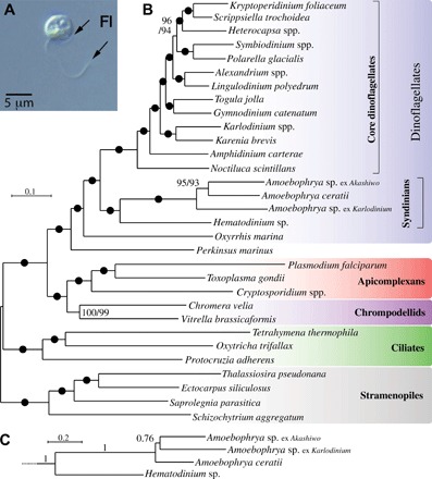
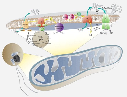
Dinoflagellates are microbial eukaryotes that have exceptionally large nuclear genomes; however, their organelle genomes are small and fragmented and contain fewer genes than those of other eukaryotes. The genus Amoebophrya (Syndiniales) comprises endoparasites with high genetic diversity that can infect other dinoflagellates, such as those forming harmful algal blooms (e.g., Alexandrium). We sequenced the genome (~100 Mb) of Amoebophrya ceratii to investigate the early evolution of genomic characters in dinoflagellates. The A. ceratii genome encodes almost all essential biosynthetic pathways for self-sustaining cellular metabolism, suggesting a limited dependency on its host. Although dinoflagellates are thought to have descended from a photosynthetic ancestor, A. ceratii appears to have completely lost its plastid and nearly all genes of plastid origin. Functional mitochondria persist in all life stages of A. ceratii, but we found no evidence for the presence of a mitochondrial genome. Instead, all mitochondrial proteins appear to be lost or encoded in the A. ceratii nucleus.


Figures

Fig. 1
Fig. 1. Multiprotein phylogeny of Amoebophrya isolated from three separate hosts, 15 other dinoflagellates, and 13 related eukaryotes.
(A) Free-living stage of the parasite Amoebophrya. Fl, flagellum. (B) The best maximum likelihood tree (IQ-TREE) under the LG + G4 + I + F model with ultrafast/nonparametric bootstrap supports at branches (black circles denote 100/100 support). (C) Relationships among Amoebophrya isolates in a PhyloBayes GTR + CAT + G4 inference with posterior probabilities at branches; the rest of the tree is identical to (B) and is fully supported at all branches.
Fig. 2
Fig. 2. Shikimate (g6770) and tryptophan (g13589) synthesis pathway multidomain genes.
(A) Individual domains of the shikimate pathway are illustrated by colored boxes, and domains of the tryptophan pathway are represented with differently shaded gray boxes. (B) Schematic view of the biosynthetic pathway for tryptophan in A. ceratii. Circles represent intermediates that can be synthesized in A. ceratii, and arrows indicate the respective enzymatic activities. Arrows without circles indicate missing pathway components in A. ceratii. The colors for the shikimate enzymatic activities are as in (A). For simplicity, all tryptophan pathway steps are depicted in gray.
Fig. 3
Fig. 3. Investigation of mitochondria in A. ceratii cells.
(A) Electron microscopy transmission image of A. ceratii dinospore showing the fine structure of the mitochondrion (Mi), nucleus (Nc), and flagella (Fl). Confocal microscopy images showing (B) SYTO-13–stained DNA of the nucleus (Nc), (C) mitochondria stained with MitoTracker, (D) an image of a free-swimming biflagellate dinospore cell, and (E) overlay of images.
Fig. 4
Fig. 4. CoxI fragment alignment.
Scaffold fragment, gene model, gDNA PCR amplicon sequence, and cDNA sequence with and without intron sequence. The predicted coxI domain is marked with a shaded background.
Fig. 5
Fig. 5. Model of mitochondrial functions in A. ceratii based on the genome gene content.
The C. velia model from (11) was taken as a template. Mitochondrial complex I has been replaced by an alternative NADH dehydrogenase (DH), which reduced the NADH from the tricarboxylic acid (TCA) cycle. Both alternative NADH dehydrogenase and succinate dehydrogenase (complex II) channel electrons through the carrier ubiquinone (Q) to the alternative oxidase (yellow arrows). Electrons may also be passed by other sources, such as d-lactate:cytochrome c oxidoreductase (d-LDH) and galacto-1,4-lactone:cytochrome c oxidoreductase (G-1,4-LDH) to cytochrome c (yellow arrows), which passes them on to complex IV (cytochrome c oxidase). Stippled yellow arrows indicate alternative pathways of electron flow as proposed in Chromera (11).

References

    1. Bachvaroff T. R., Gornik S. G., Concepcion G. T., Waller R. F., Mendez G. S., Lippmeier J. C., Delwiche C. F., Dinoflagellate phylogeny revisited: Using ribosomal proteins to resolve deep branching dinoflagellate clades. Mol. Phylogenet. Evol. 70, 314–322 (2014). - PMC - PubMed
    1. Keeling P. J., Burger G., Durnford D. G., Lang B. F., Lee R. W., Pearlman R. E., Roger A. J., Gray M. W., The tree of eukaryotes. Trends Ecol. Evol. 20, 670–676 (2005). - PubMed
    1. Lin S., Genomic understanding of dinoflagellates. Res. Microbiol. 162, 551–569 (2011). - PubMed
    1. Gornik S. G., Ford K. L., Mulhern T. D., Bacic A., McFadden G. I., Waller R. F., Loss of nucleosomal DNA condensation coincides with appearance of a novel nuclear protein in dinoflagellates. Curr. Biol. 22, 2303–2312 (2012). - PubMed
    1. LaJeunesse T. C., Lambert G., Andersen R. A., Coffroth M. A., Galbraith D. W., Symbiodinium (Pyrrhophyta) genome sizes (DNA content) are smallest among dinoflagellates. J. Phycol. 41, 880–886 (2005).

Publication types

Substances

LinkOut - more resources