Review
. 2019 Mar;5(1):142-154.
doi: 10.21037/jss.2019.03.01.

Acute post-operative airway complications following anterior cervical spine surgery and the role for cricothyrotomy

Affiliations
Review

Acute post-operative airway complications following anterior cervical spine surgery and the role for cricothyrotomy

Monika P Debkowska et al. J Spine Surg. 2019 Mar.

Abstract

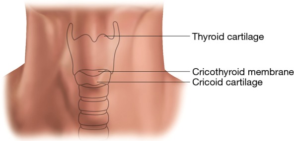
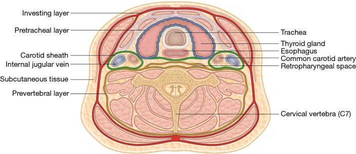
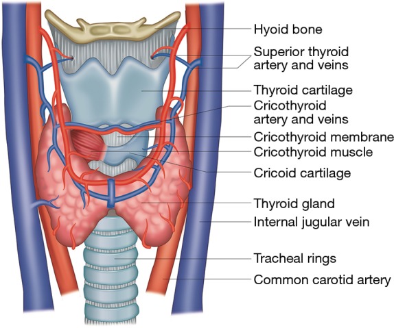
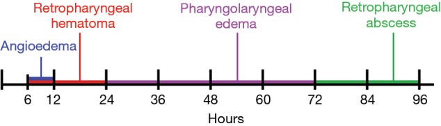
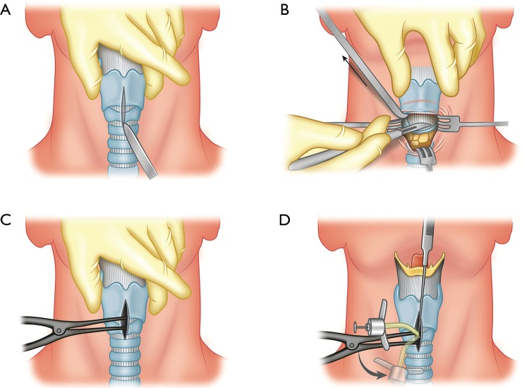
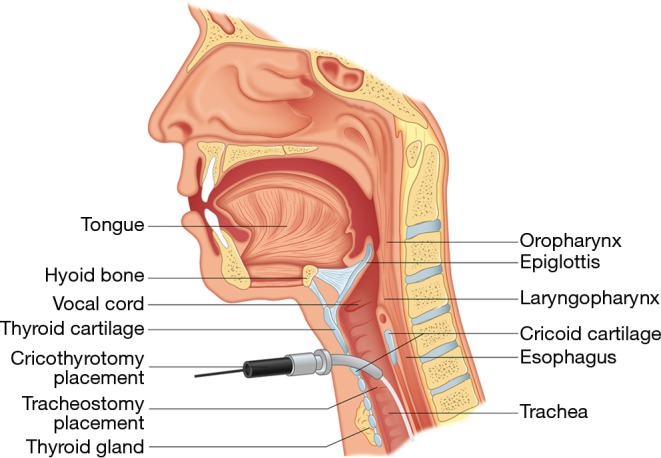
Anterior cervical spine surgery (ACSS) is a common procedure, but not without its own risks and complications. Complications that can cause airway compromise occur infrequently, but can rapidly lead to respiratory arrest, leading to severe morbidity or death. Knowing emergent post-operative airway management including surgical airway placement is critical. We aim to review the different etiologies of post-operative airway compromise following ACSS, the predictable timeline in which they occur, and the most appropriate treatment and management for each. We place special emphasis on the timing and proper surgical technique for an emergent cricothyrotomy. Angioedema is seen the earliest as a cause of post-operative airway compromise, typically within 6-12 hours. Retropharyngeal hematomas can be seen between 6-24 hours, most commonly within 12 hours. Pharyngolaryngeal edema is seen within 24-72 hours. After 72 hours, retropharyngeal abscess is the most likely etiology. Several studies have utilized delayed extubation protocols following ACSS based on patient risk factors and found reduced postoperative airway complications and reintubation rates. The administration of perioperative corticosteroids continues to be controversial with high-level studies recommending both for and against their use. Animal studies showed that after cardiac arrest, the brain can recover if oxygenation is restored within 5 minutes, but this time is likely shorter with asphyxia prior to cardiac arrest. Experience and training are essential to reduce the time for successful cricothyrotomy placement. Physicians must be prepared to diagnose and treat acute postoperative airway complications following ACSS to prevent anoxic brain injury or death. If emergent intubation cannot be accomplished on the first attempt, physicians should not delay placement of a surgical airway such as cricothyrotomy.

Keywords: Anterior cervical spine surgery (ACSS); cricothyrotomy; pharyngolaryngeal edema; post-operative airway compromise; resuscitation; retropharyngeal hematoma.


Conflict of interest statement

Conflicts of Interest: The authors have no conflicts of interest to declare.

Figures

Figure 1
Figure 1
Neck topography showing the major landmarks used for a cricothyrotomy.
Figure 2
Figure 2
Cross sectional drawing of the cervical spine at C7 showing the three deep cervical fascial layers: the investing layer, pretracheal layer, and prevertebral layer. The retropharyngeal space is posterior to the esophagus.
Figure 3
Figure 3
The internal cervical neck anatomy showing the cricothyroid membrane between the thyroid and cricoid cartilage. The membrane is incised during a cricothyrotomy. The cricothyroid artery and vein are at the superior margin of the cricothyroid membrane and may be injured during cricothyrotomy.
Figure 4
Figure 4
Etiologies of post-operative airway compromise following anterior cervical spine surgery occur in a predictable time sequence.
Figure 5
Figure 5
Prevertebral soft tissue swelling (PSTS). Lateral radiographs of a 56-year-old male who underwent anterior cervical fusion of C3–C7. The pre-operative image (A) demonstrates prevertebral soft tissue thickness of 4 and 14 mm at the C2 and C6 levels respectively. The post-operative day 6 image (B) shows increased PSTS, measuring 27 and 34 mm at the C2 and C6 levels respectively, causing mild narrowing of the upper airway.
Figure 6
Figure 6
Cervical abscess. (A) Immediate post-operative lateral radiograph of a 63-year-old male who underwent ACDF of C4–C6, demonstrating no significant PSTS; (B) axial view of a CT with IV contrast of the same patient on post-operative day 7 demonstrating a peripherally enhancing prevertebral fluid collection found to be an abscess; (C) sagittal view shows the abscess anteriorly displacing and mildly narrowing the airway. ACDF, anterior cervical discectomy and fusion; IV, intravenous.
Figure 7
Figure 7
Retropharyngeal hematoma. Axial and sagittal non-enhanced CT images of a 73-year-old female who underwent ACDF of C3–C4, post-operative day 7. A prevertebral fluid collection is seen, representing a hematoma with mild compression and narrowing of the adjacent airway. Of note, the patient had a prior fusion of C4–C6 with hardware removal. ACDF, anterior cervical discectomy and fusion.
Figure 8
Figure 8
The essential tools for a cricothyrotomy kit include antiseptic skin cleaning preparation such as ChloraPrep, scalpel, hemostat, a small cuffed endotracheal or tracheostomy tube, and a 10-mL syringe.
Figure 9
Figure 9
Steps of performing a cricothyrotomy. (A) Position the neck extended, exposing the anterior trachea. Palpate laryngeal landmarks. Make a midline skin incision from the thyroid notch to the cricoid cartilage and deep to the cricothyroid membrane; (B) make a transverse incision through the cricothyroid membrane; (C) insert the hemostat and dilate the opening; (D) insert the cuffed endotracheal tube or tracheostomy tube into the opening and inflate the cuff.
Figure 10
Figure 10
A sagittal drawing of the upper respiratory tract showing the location of a cricothyrotomy and tracheostomy as compared to the surrounding vital structures.

Similar articles

Cited by

References

    1. Rhee J, Voyadzis JM. Anterior Cervical Diskectomy and Fusion. In: Surgical Anatomy and Techniques to the Spine. 2014:131-8.
    1. Helgeson MD, Albert TJ. Anterior surgical approach to the cervical spine. In: Shen FH, Samartzis D, Fessler RG, editors. Textbook of the Cervical Spine. 1st edition. Philadelphia: Saunders Elsevier; 2014. p. 33-8.
    1. Wilson AS, Samartzis D, Shen FH. Anterior Cervical Diskectomy and Fusion. In: Textbook of the Cervical Spine. 2015:285-93.
    1. Daniels AH, Riew KD, Yoo JU, et al. Adverse events associated with anterior cervical spine surgery. J Am Acad Orthop Surg 2008;16:729-38. 10.5435/00124635-200812000-00005 - DOI - PubMed
    1. Lied B, Sundseth J, Helseth E. Immediate (0-6 h), early (6-72 h) and late (>72 h) complications after anterior cervical discectomy with fusion for cervical disc degeneration; discharge six hours after operation is feasible. Acta Neurochir 2008;150:111-8. 10.1007/s00701-007-1472-y - DOI - PubMed