. 2019 Apr 29;129(6):2237-2250.
doi: 10.1172/JCI123135.

microRNA-21-5p dysregulation in exosomes derived from heart failure patients impairs regenerative potential

Affiliations

microRNA-21-5p dysregulation in exosomes derived from heart failure patients impairs regenerative potential

Li Qiao et al. J Clin Invest. .

Abstract

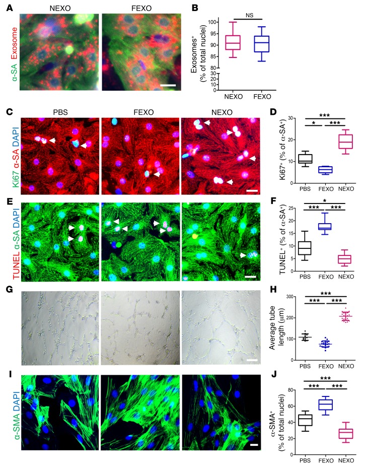
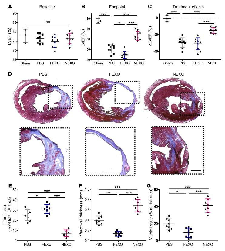
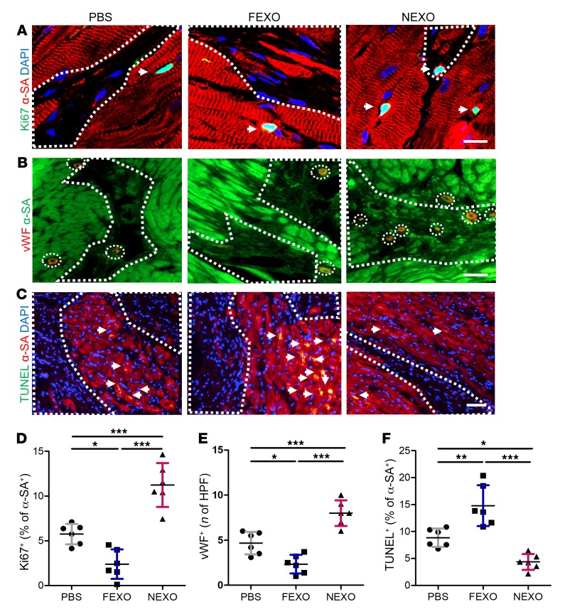
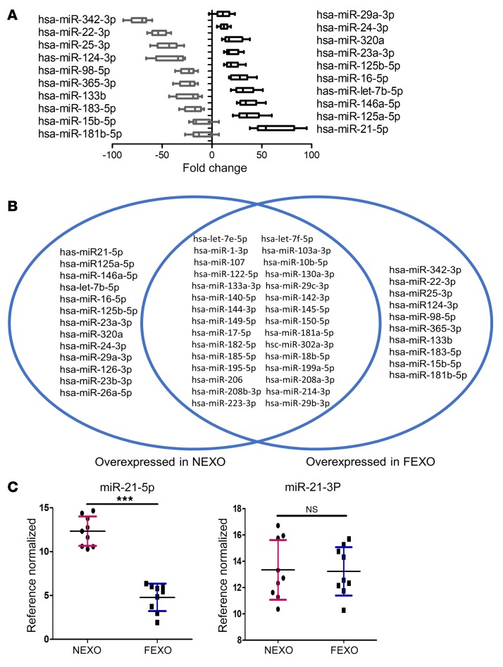
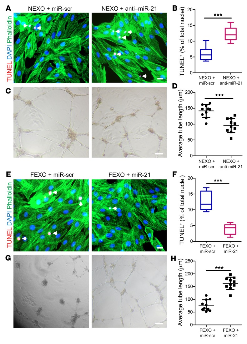
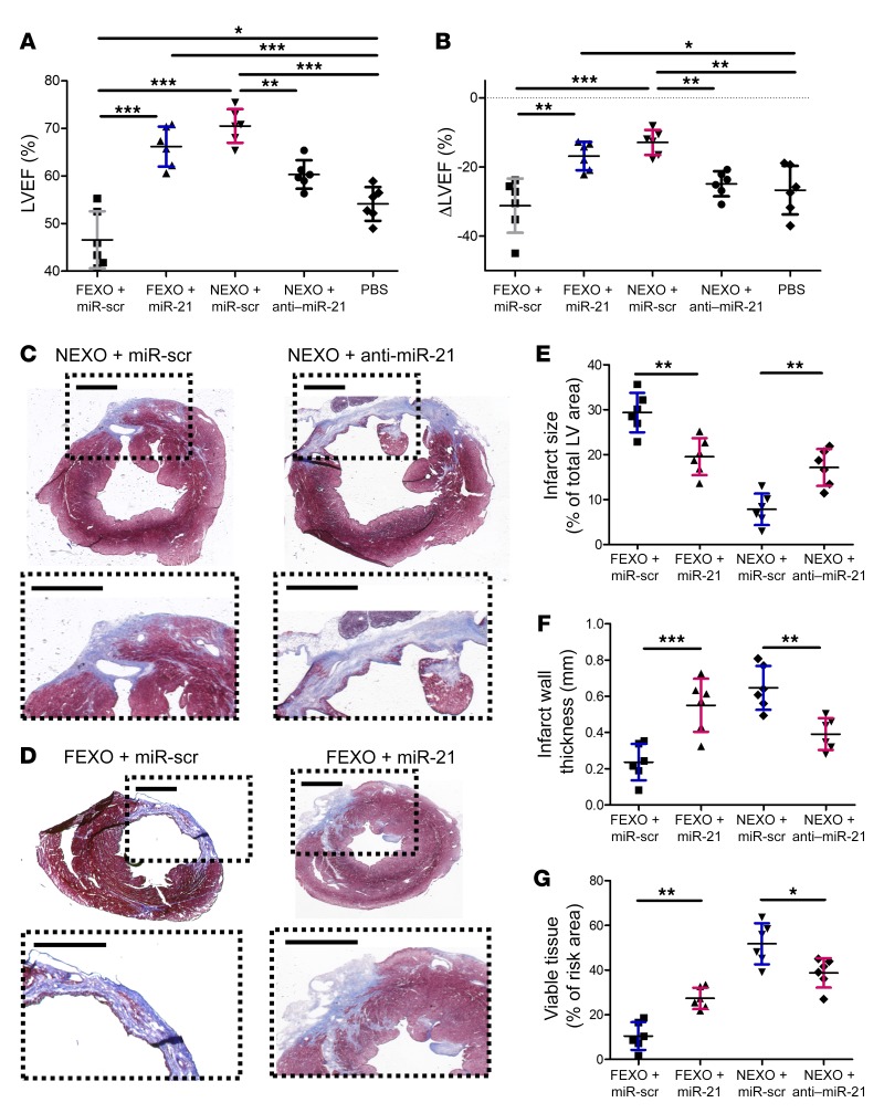
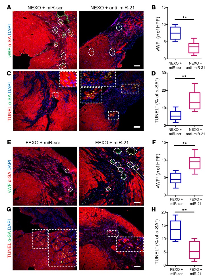
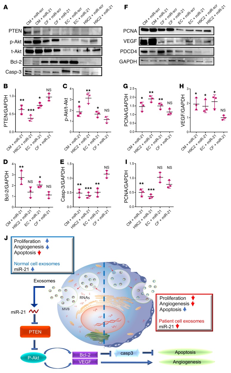
Exosomes, as functional paracrine units of therapeutic cells, can partially reproduce the reparative properties of their parental cells. The constitution of exosomes, as well as their biological activity, largely depends on the cells that secrete them. We isolated exosomes from explant-derived cardiac stromal cells from patients with heart failure (FEXO) or from normal donor hearts (NEXO) and compared their regenerative activities in vitro and in vivo. Patients in the FEXO group exhibited an impaired ability to promote endothelial tube formation and cardiomyocyte proliferation in vitro. Intramyocardial injection of NEXO resulted in structural and functional improvements in a murine model of acute myocardial infarction. In contrast, FEXO therapy exacerbated cardiac function and left ventricular remodeling. microRNA array and PCR analysis revealed dysregulation of miR-21-5p in FEXO. Restoring miR-21-5p expression rescued FEXO's reparative function, whereas blunting miR-21-5p expression in NEXO diminished its therapeutic benefits. Further mechanistic studies revealed that miR-21-5p augmented Akt kinase activity through the inhibition of phosphatase and tensin homolog. Taken together, the heart failure pathological condition altered the miR cargos of cardiac-derived exosomes and impaired their regenerative activities. miR-21-5p contributes to exosome-mediated heart repair by enhancing angiogenesis and cardiomyocyte survival through the phosphatase and tensin homolog/Akt pathway.

Keywords: Cardiology; Heart failure; Human stem cells; Mouse models; Stem cells.


Conflict of interest statement

Conflict of interest: KC is an equity holder of Xollent Biotech Inc.

Figures

Figure 1
Figure 1. Effects of exosome treatment on cardiomyocytes, endothelial cells, and cardiac fibroblasts in vitro.
(A) Representative fluorescent micrographs showing uptake of DiI-labeled NEXO and FEXO by NRCMs. Endocytosed exosomes (red) can be seen within the cytoplasm of cardiomyocytes (green). Scale bar: 10 μm. (B) Quantitation of exosomes uptake (n = 10). Two-tailed t test. (C) NRCM proliferation in response to NEXO, FEXO, or PBS treatment. White arrows indicate Ki-67+/α-SA+ cells. Scale bar: 20 μm. (D) Quantitation of proliferating cardiomyocytes (n = 6). (E) Apoptotic NRCMs in response to NEXO, FEXO, or PBS treatment. White arrows indicate TUNEL+/α-SA+ cells. Scale bar: 20 μm. (F) Quantitation of apoptotic cardiomyocytes (n = 6). (G) Measurement of tube formation in HUVECs co-cultured with NEXO, FEXO, or PBS. Scale bar: 100 μm. (H) Quantitation of average HUVEC tube length (n = 20). (I) Neonatal rat fibroblasts underwent phenotypic transition to myofibroblasts in response to NEXO, FEXO, or PBS treatment. Scale bar: 20 μm. (J) Quantitation of myofibroblasts (n = 12). *P < 0.05, ***P < 0.001. NS, no significance. (D, F, H, and J) One-way ANOVA with Bonferroni post hoc correction. All values are mean ± SD. FEXO, exosomes derived from the cardiac cells of patients with heart failure. NEXO, exosomes derived from the cardiac cells of normal heart donors.
Figure 2
Figure 2. Effects of NEXO and FEXO treatment in a mouse model of acute MI.
(A and B) LVEF was measured by echocardiography at baseline (1 day before MI) (A) and endpoint (3 weeks after MI) (B). (C) Treatment effects (changes in LVEF at 3 weeks relative to baseline) in each group (AC, n = 9 animals per treatment group, n = 3 animals for the sham group). (D) Representative Masson’s trichrome staining of myocardial section 3 weeks after treatment with NEXO, FEXO, or PBS. Scale bar: 0.5 mm. (EG) Quantitative analyses of infarct size, infarct wall thickness, and viable tissue from Masson’s trichrome-stained heart sections (n = 8 animals per treatment group). *P < 0.05, ***P < 0.001. One-way ANOVA with Bonferroni post hoc correction. All values are mean ± SD. FEXO, exosomes derived from the cardiac cells of patients with heart failure. NEXO, exosomes derived from the cardiac cells of normal heart donors.
Figure 3
Figure 3. Mechanisms of exosome-mediated cardiac repair.
(A) Representative images of post-MI heart sections stained with Ki-67 (green), α-SA (red), and DAPI. White boundaries show infarct area, and white arrows indicate Ki-67+ cells in the peri-infarct zone. Scale bar: 10 μm (B) Heart sections stained with vWF (red), α-SA (green) in response to NEXO, FEXO, or PBS treatment. White arrows indicate capillary structures in the peri-infarct zone. Scale bar: 50 μm. (C) Heart sections stained with TUNEL (green), α-SA (red), and DAPI (blue). White arrows indicate apoptotic cardiomyocytes in the peri-infarct zone. Scale bar: 50 μm. (D) Quantification of cycling cardiomyocytes (Ki-67+/α-SA+). (E) Quantification of capillary density (vWF+). (F) Quantification of cardiomyocyte apoptosis (TUNEL+/α-SA+). (DF) n = 6 animals per group, 3 heart sections for each animal. *P < 0.05, **P < 0.01, ***P < 0.001. One-way ANOVA with Bonferroni post hoc correction. All values are mean ± SD. FEXO, exosomes derived from the cardiac cells of patients with heart failure. NEXO, exosomes derived from the cardiac cells of normal heart donors.
Figure 4
Figure 4. Dysregulation of miR-21-5p in heart failure exosomes.
(A) miRNA array showing fold changes of miRNA abundance in NEXO or FEXO (n = 3 biological replicates, 3 technical replicates for each biological replicate). (B) Venn diagram showing the variable miRNA profile between NEXO and FEXO. (C) RT-PCR analysis validated that miR-21-5p was highly enriched in NEXO (n = 3 biological replicates, 3 technical replicates for each biological replicate). ***P < 0.001. NS, no significance. Two-tailed t test. All values are mean ± SD. FEXO, exosomes derived from the cardiac cells of patients with heart failure. NEXO, exosomes derived from the cardiac cells of normal heart donors.
Figure 5
Figure 5. Manipulation of miR-21-5p in exosomes modulates their proangiogenic and antiapoptosis activities in vitro.
(A) Antiapoptotic effects were diminished after miR-21-5p knockdown in NEXOs. White arrows indicate TUNEL+ cells. Scale bar: 50 μm. (B) Quantitation of apoptotic cells (n = 6). (C) The proangiogenic effects of NEXO were diminished by miR-21-5p knockdown. Scale bar: 50 μm. (D) Quantitation of average tube length (n = 10). (E) Enhancing miR-21-5p expression in FEXO partly rescued its ability to promote cardiomyocyte proliferation. Scale bar: 50 μm. (F) Quantitation of apoptotic cells (n = 6). (G) Tube formation assay showing enhanced proangiogenic activity of FEXO with restored miR-21-5p expression. Scale bar: 50 μm. (H) Quantitation of average tube length (n = 10). ***P < 0.001. Two-tailed t test. All values are mean ± SD. FEXO + miR-scr, exosomes derived from the cardiac cells of patients with heart failure transfected with scrambled miR oligo. FEXO + miR-21, exosomes derived from the cardiac cells of patients with heart failure transfected with miR-21-5p oligo. NEXO + miR-scr, exosomes derived from the cardiac cells of the normal hearts transfected with scrambled miR oligo. NEXO + anti–miR-21, exosomes derived from the cardiac cells of the normal hearts transfected with anti–miR-21-5p oligo. FEXO, exosomes derived from the cardiac cells of patients with heart failure. NEXO, exosomes derived from the cardiac cells of normal heart donors.
Figure 6
Figure 6. Manipulation of miR21 in exosomes modulates their therapeutic potency.
miR-21-5p–deficient exosomes were produced by transfecting healthy cardiac cells with anti–miR-21-5p oligo (NEXO + anti–miR-21). miR-21-5p–restored exosomes were engineered by transfecting heart failure cardiac cells with miR-21-5p oligo (FEXO + miR-21), followed by media conditioning and exosome isolation, as previously described. Scrambled miR oligo was used as a control (NEXO/FEXO + miR-scr). (A) LVEF was measured by echocardiography 3 weeks after treatment. (B) Treatment effects (changes in LVEF at 3 weeks relative to baseline) were calculated for each group. (C and D) Representative Masson’s trichrome staining of myocardial section 3 weeks after treatments. Scale bar: 1 mm. (EG) Quantitative analysis of infarct size, infarct wall thickness, and viable tissue from Masson’s trichrome-stained images. n = 6 animals per group. *P < 0.05, **P < 0.01, ***P < 0.001. One-way ANOVA with Bonferroni post hoc correction. All values are mean ± SD.
Figure 7
Figure 7. Manipulation of miR21 in exosomes modulates their proangiogenic and antiapoptotic activities in vivo.
(A) Representative images of post-MI heart sections stained with vWF (green), α-SA (red), and DAPI (blue). White circles indicate capillaries in the peri-infarct zone. Scale bar: 100 μm. (B) Quantitation of capillary density (vWF+). (C) Heart sections stained with TUNEL (green), α-SA (red), and DAPI (blue). White squares indicate apoptotic cardiomyocytes in the peri-infarct zone. Scale bar: 50 μm. (D) Quantitation of cardiomyocyte apoptosis (TUNEL+/a-SA+). (E) Representative images of post-MI heart sections stained with vWF (green), α-SA (red), and DAPI (blue). White circles indicate capillaries in the peri-infarct zone. Scale bar: 50 μm. (F) Quantitation of capillary density (vWF+). (G) Heart sections stained with TUNEL (green), α-SA (red), and DAPI (blue). White squares indicate apoptotic cardiomyocytes in the peri-infarct zone. Scale bar: 50 μm. (H) Quantitation of cardiomyocyte apoptosis (TUNEL+/α-SA+). (B, D, F, and H) n = 6 animals for each group and 3 heart sections for each animal. **P < 0.01. Two-tailed t test. All values are mean ± SD. FEXO + miR-scr, exosomes derived from the cardiac cells of patients with heart failure transfected with scrambled miR oligo. FEXO + miR-21, exosomes derived from the cardiac cells of patients with heart failure transfected with miR-21-5p oligo. NEXO + miR-scr, exosomes derived from the cardiac cells of the normal hearts transfected with scrambled miR oligo. NEXO + anti–miR-21, exosomes derived from the cardiac cells of the normal hearts transfected with anti–miR-21-5p oligo.
Figure 8
Figure 8. miR-21-5p targets the PTEN pathway in post-MI pathology.
(A) Representative Western blot images showing the expression of various PTEN/Akt pathway components. (BE) Quantitation of the levels of PTEN, p-Akt, t-Akt, Bcl-2, and caspase-3 (n = 3). (F) Representative Western blot images showing the expression of PCNA (proliferation marker), VEGF, and PDCD4 (miR-21 target). (G and H) Quantitation of the levels of PCNA, VEGF, and PDCD4 (n = 3). (BI) All miR-21 groups were normalized to the relevant scrambled controls. *P < 0.05 compared to scrambled control. **P < 0.01 compared to scrambled control. ***P < 0.001 compared to scrambled control. NS, no significance compared to scrambled control. Two-tailed t test. All values are mean ± SD. (J) Schematic showing the working model of our study. CM/H9C2/EC/CF +miR-scr, HCs/H9C2 cells/HUVECs/human cardiac fibroblasts transfected with scrambled miR. CM/H9C2/EC/CF + miR-21, HCs/H9C2 cells/HUVECs/human cardiac fibroblasts transfected with miR-21-5p mimic.

References

    1. Luo L, et al. Fabrication of synthetic mesenchymal stem cells for the treatment of acute myocardial infarction in mice. Circ Res. 2017;120(11):1768–1775. doi: 10.1161/CIRCRESAHA.116.310374. - DOI - PMC - PubMed
    1. Tang J, et al. Targeted repair of heart injury by stem cells fused with platelet nanovesicles. Nat Biomed Eng. 2018;2:17–26. doi: 10.1038/s41551-017-0182-x. - DOI - PMC - PubMed
    1. Tang J, et al. Heart repair using nanogel-encapsulated human cardiac stem cells in mice and pigs with myocardial infarction. ACS Nano. 2017;11(10):9738–9749. doi: 10.1021/acsnano.7b01008. - DOI - PMC - PubMed
    1. Tang J, et al. Therapeutic microparticles functionalized with biomimetic cardiac stem cell membranes and secretome. Nat Commun. 2017;8:13724. - PMC - PubMed
    1. Kreke M, Smith RR, Marbán L, Marbán E. Cardiospheres and cardiosphere-derived cells as therapeutic agents following myocardial infarction. Expert Rev Cardiovasc Ther. 2012;10(9):1185–1194. doi: 10.1586/erc.12.102. - DOI - PubMed

Publication types

MeSH terms