Evidence of biomechanical and collagen heterogeneity in uterine fibroids
- PMID: 31034494
- PMCID: PMC6488189
- DOI: 10.1371/journal.pone.0215646
Evidence of biomechanical and collagen heterogeneity in uterine fibroids
Abstract
Objective: Uterine fibroids (leiomyomas) are common benign tumors of the myometrium but their molecular pathobiology remains elusive. These stiff and often large tumors contain abundant extracellular matrix (ECM), including large amounts of collagen, and can lead to significant morbidities. After observing structural multiformities of uterine fibroids, we aimed to explore this heterogeneity by focusing on collagen and tissue stiffness.
Methods: For 19 fibroids, ranging in size from 3 to 11 centimeters, from eight women we documented gross appearance and evaluated collagen content by Masson trichrome staining. Collagen types were determined in additional samples by serial extraction and gel electrophoresis. Biomechanical stiffness was evaluated by rheometry.
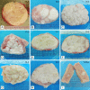
Results: Fibroid slices displayed different gross morphology and some fibroids had characteristics of two or more patterns: classical whorled (n = 8); nodular (n = 9); interweaving trabecular (n = 9); other (n = 1). All examined fibroids contained at least 37% collagen. Tested samples included type I, III, and V collagen of different proportions. Fibroid stiffness was not correlated with the overall collagen content (correlation coefficient 0.22). Neither stiffness nor collagen content was correlated with fibroid size. Stiffness among fibroids ranged from 3028 to 14180 Pa (CV 36.7%; p<0.001, one-way ANOVA). Stiffness within individual fibroids was also not uniform and variability ranged from CV 1.6 to 42.9%.
Conclusions: The observed heterogeneity in structure, collagen content, and stiffness highlights that fibroid regions differ in architectural status. These differences might be associated with variations in local pressure, biomechanical signaling, and altered growth. We conclude the design of all fibroid studies should account for such heterogeneity because samples from different regions have different characteristics. Our understanding of fibroid pathophysiology will greatly increase through the investigation of the complexity of the chemical and biochemical signaling in fibroid development, the correlation of collagen content and mechanical properties in uterine fibroids, and the mechanical forces involved in fibroid development as affected by the various components of the ECM.
Conflict of interest statement
We have the following interests: BioSpecifics Technologies Corporation (BTC) provided partial funding for the work presented here. None of the authors are employed by this company. None of the authors own stock in the company. Two and a half years ago PCL presented findings of a study, now published [Jayes, 2016] to the Board of Directors of the company. PCL is listed on a patent (Phyllis Leppert, Thomas Wegman, Darlene Taylor: Treatment method and product for uterine fibroids using purified collagenase US9744138B2). The proceeds, if any, of this patent are assigned to BTC and a portion of the profit assigned to Duke University. Our submitted manuscript is unrelated to the patent. Our manuscript does not involve the use of collagenase as a treatment. Both PCL and FLJ are involved in a phase I clinical trial of collagenase for fibroid treatment being conducted by James Segars, PI at Johns Hopkins University. There are no further patents, products in development or marketed products to declare. This does not alter our adherence to all the PLOS ONE policies on sharing data and materials.
Figures


References
-
- Baird DD, Dunson DB, Hill MC, Cousins D, Schectman JM. High cumulative incidence of uterine leiomyoma in black and white women: ultrasound evidence. Am J Obstet Gynecol. 2003;188(1):100–7. Epub 2003/01/28. - PubMed
-
- Stewart EA, Friedman AJ, Peck K, Nowak RA. Relative overexpression of collagen type I and collagen type III messenger ribonucleic acids by uterine leiomyomas during the proliferative phase of the menstrual cycle. J Clin Endocrinol Metab. 1994;79(3):900–6. Epub 1994/09/01. 10.1210/jcem.79.3.8077380 . - DOI - PubMed
-
- Cramer SF, Patel A. The frequency of uterine leiomyomas. Am J Clin Pathol. 1990;94(4):435–8. Epub 1990/10/01. . - PubMed
-
- Konishi I, Fujii S, Ban C, Okuda Y, Okamura H, Tojo S. Ultrastructural study of minute uterine leiomyomas. Int J Gynecol Pathol. 1983;2(2):113–20. Epub 1983/01/01. . - PubMed
Publication types
MeSH terms
Substances
LinkOut - more resources
Full Text Sources
Other Literature Sources
Medical

