. 2019 Apr 29;14(4):e0216055.
doi: 10.1371/journal.pone.0216055. eCollection 2019.

Cancer cell-induced neutrophil extracellular traps promote both hypercoagulability and cancer progression

Affiliations

Cancer cell-induced neutrophil extracellular traps promote both hypercoagulability and cancer progression

Hye Soo Jung et al. PLoS One. .

Abstract

Introduction: Neutrophils can generate extracellular net-like structures by releasing their DNA-histone complexes and antimicrobial peptides, which is called neutrophil extracellular traps (NETs). Various stimuli can induce NET formation. In particular, neutrophils and NET formation are abundant in tumor tissue. This study investigated how cancer cells induce NET formation and whether this NET formation promotes plasma thrombin generation and cancer progression.

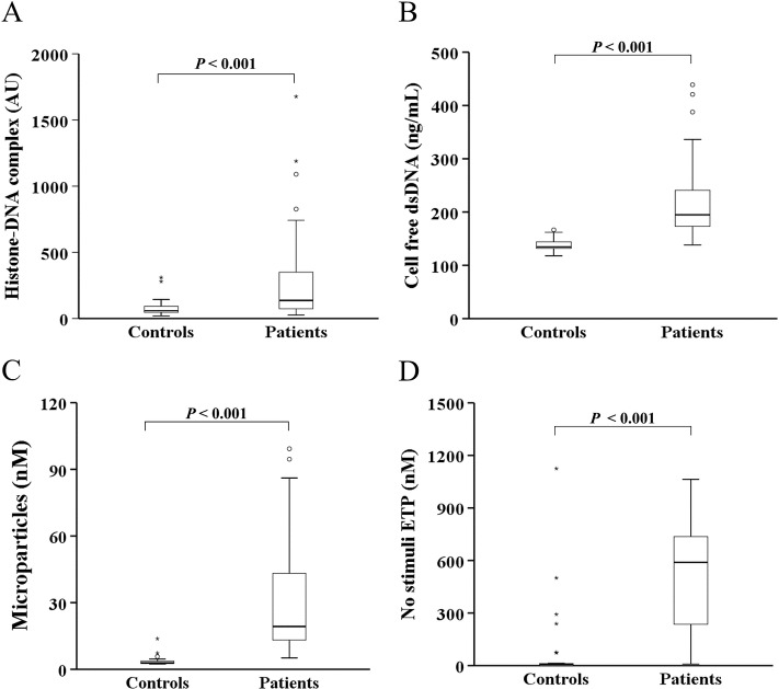
Methods: Induction of NET formation by a pancreatic cancer cell line (AsPC-1) was assessed by measuring the histone-DNA complex level. The endogenous thrombin potential (ETP) was measured by thrombin generation assay. In vitro migration, invasion, and tubule formation assays were performed. The circulating levels of NET markers and hypercoagulability markers were assessed in 62 patients with pancreatobiliary malignancy and 30 healthy controls.

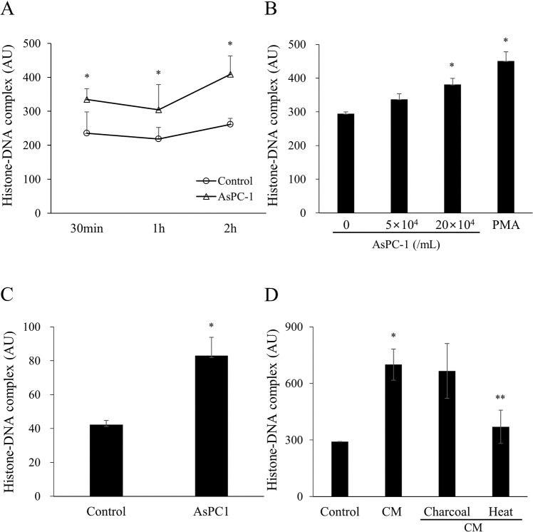
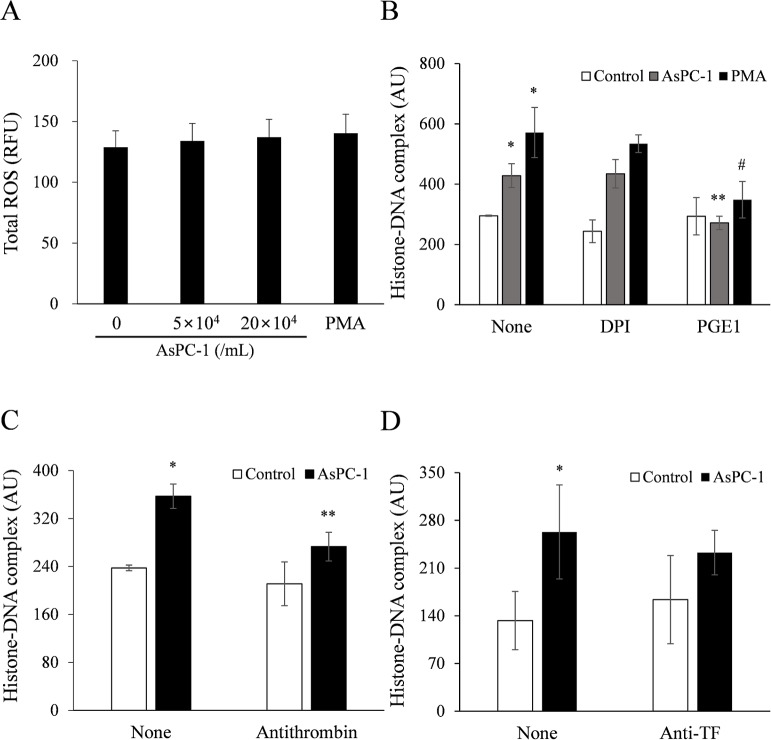
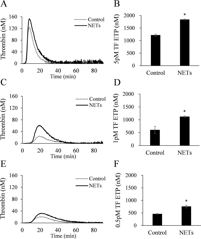
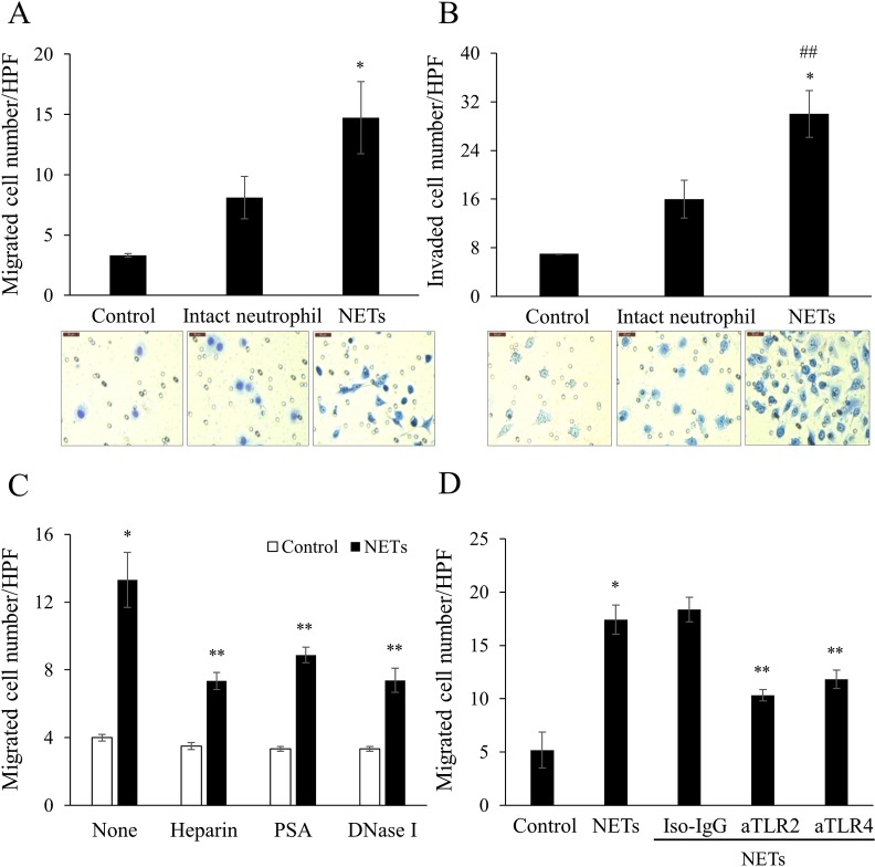
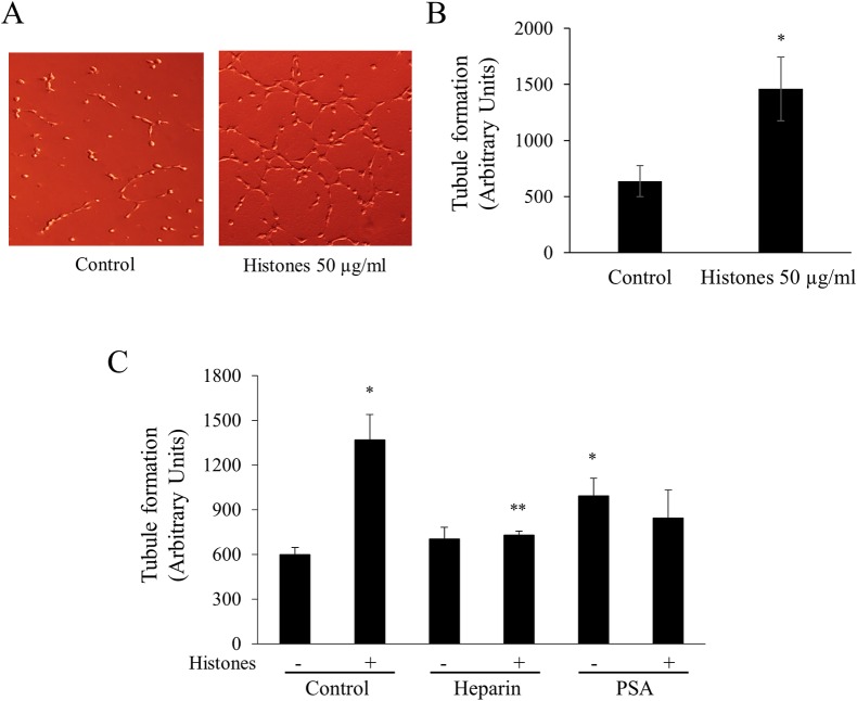
Results: AsPC-1 significantly induced NET formation in a dose-dependent manner. Conditioned medium (CM) from AsPC-1 also induced NETs. Interestingly, NET-formation was abolished by heat-inactivated CM, but not by lipid-extracted CM, suggesting an important role of protein components. A reactive oxygen species inhibitor did not inhibit cancer cell-induced NET formation, but prostaglandin E1 (PGE1, cyclic adenosine monophosphate inducer) and antithrombin did. NETs significantly increased ETP of normal plasma. Of note, NETs promoted cancer cell migration and invasion as well as angiogenesis, which were inhibited by histone-binding agents (heparin, polysialic acid), a DNA-degrading enzyme, and Toll-like receptor neutralizing antibodies. In patients with pancreatobiliary malignancy, elevated NET markers correlated well with hypercoagulability makers.

Conclusion: Our findings indicate that cancer cell-induced NET formation enhances both hypercoagulability and cancer progression and suggest that inhibitors of NET formation such as PGE1 and antithrombin can be potential therapeutics to reduce both hypercoagulability and cancer progression.


Conflict of interest statement

The authors have declared that no competing interests exist.

Figures

Fig 1
Fig 1. Pancreatic cancer cells AsPC-1 induce neutrophil extracellular traps.
(A) AsPC-1 (5 × 10⁴ cells/mL) were incubated with whole blood for 30 min, 1 h, or 2 h at 37°C. Control represents whole blood without AsPC-1. The histone–DNA complex levels were measured in the supernatants. A significant difference was observed among the groups in histone-DNA complex level (NET formation) during incubation time. Statistical analysis was performed by two-way ANOVA followed by Bonferroni’s post-hoc test. *P < 0.05 versus control group. (B) AsPC-1 (5 × 10⁴ or 20 × 104 cells/mL) were incubated with whole blood for 2 h. Phorbol 12-myristate-13-acetate (PMA, 25 nM) was used as a positive control of NET formation. Statistical analysis was performed by one-way ANOVA followed by Bonferroni’s post-hoc test. *P < 0.05 versus whole blood without AsPC-1. (C) AsPC-1 cells were incubated with isolated neutrophils for 2 h and the histone–DNA complex level was measured in the supernatants. (D) Conditioned medium (CM) harvested from AsPC-1 culture was incubated with whole blood for 2h and the histone-DNA complex level was measured in the supernatants. Charcoal was added to CM to remove lipids and heat treatment was used to degrade proteins. Data are expressed as mean ± SEM of 3 experiments. *P < 0.05 versus control; **P < 0.05 versus CM. Abbreviations: AU, arbitrary units.
Fig 2
Fig 2. AsPC-1–induced NET formation is not ROS-dependent but cyclic AMP and thrombin-dependent.
(A) AsPC-1 or PMA (positive control) was incubated with whole blood for 2 h at 37°C, and total ROS activity was measured in the supernatants. (B) Whole blood was pretreated with diphenyleneiodonium (DPI, 20 μM; ROS inhibitor) or prostaglandin E1 (PGE1, 1 μg/mL; cyclic AMP inducer) for 10 min at room temperature and then incubated with PMA or AsPC-1 for 2 h at 37°C. The histone–DNA complex level was measured in the supernatants. *P < 0.05 versus whole blood without AsPC-1 or PMA-treatment (control); **P < 0.05 versus whole blood with AsPC-1-treatment DPI or PGE1 untreated; #P < 0.1 versus whole blood with PMA-treatment DPI or PGE1 untreated. (C, D) Whole blood was treated with (C) antithrombin (5 IU/mL) or (D) monoclonal antibody against human tissue factor (anti-TF, 30 μg/mL) for 10 min at 37°C and the histone–DNA complex level was measured in the supernatants. Data are expressed as mean ± SEM of 3 experiments. *P < 0.05 versus whole blood without AsPC-1-treatment (control); **P < 0.05 versus whole blood with AsPC-1-treatment antithrombin untreated. Abbreviations: RFU, relative fluorescence units; AU, arbitrary units.
Fig 3
Fig 3. NETs increase thrombin generation in normal plasma.
NETs were prepared from isolated neutrophils treated with PMA and added (6.4 mg/dL final protein level) to normal plasma. Endogenous thrombin potential (ETP) was measured by thrombin generation assay after stimulation with (5, 1, or 0.5 pM TF. All data are presented as mean ± SEM from 4 different experiments. *P < 0.05 versus control.
Fig 4
Fig 4. NETs promote migration and invasion of pancreatic cancer cells.
Intact isolated neutrophils (3.3 × 10⁵ cells/mL) and NETs (300 mg/dL final protein concentration) were used as chemoattractants. They were added to the lower chambers of (A) an uncoated transwell and (B) rehydrated Matrigel–coated transwell. AsPC-1 (2 × 10⁵ cells/mL) were added to upper chambers. After 22 h incubation, the (A) migrated and (B) invaded cells were stained with Diff-Quik kit and imaged under an optical microscope (×400). Averaged data of six randomly selected high-power fields (HPF) are shown. Scale bar: 50 μm. Statistical analysis was performed by one-way ANOVA followed by Bonferroni’s post-hoc test. *P < 0.05 versus control; ##P < 0.05 versus intact neutrophil. (C) NETs were pre-treated with heparin (200 IU/mL), polysialic acid (PSA, 62.5 μg/mL), or DNase I (50 IU/mL) and then the migration assay was performed. (D) AsPC-1 cells were pre-treated with mouse IgG2a,K antibody (Iso-IgG, 50 μg/mL), anti-Toll-like receptor-2 (aTLR2, 50 μg/mL), or anti-TLR4 (aTLR4, 50 μg/mL) and then the migration assay was performed. The numbers of migrated or invaded AsPC-1 cells are shown as mean ± SEM of 3 experiments. *P < 0.05 versus control; #P < 0.1 versus control; **P < 0.05 versus NETs.
Fig 5
Fig 5. NETs promote endothelial cell angiogenesis.
(A) Endothelial cells EA.hy926 were incubated with or without histones (50 μg/mL) for 4 h in a Matrigel–coated well and tubule images were taken under an optical microscope (×100). (B) EA.hy926 (5 × 10⁴ cells/well) were incubated with various concentrations of histones (none, 10, 20, 50, 100 μg/mL) for 4 h in Matrigel-coated wells. The tubule formation length was calculated by Image J software in four randomly selected fields. (C) As inhibitors of tubule formation, heparin (100 IU/mL) and PSA (62.5 μg/mL) were pre-incubated with or without histones for 1 h. EA.hy926 cells were then added for 4 h. *P < 0.05 versus control; **P < 0.05 versus histone-treated.
Fig 6
Fig 6. The circulating levels of NET and hypercoagulability markers are increased in patients with pancreatobiliary malignancy.
The levels of (A) histone–DNA complex, (B) cell-free dsDNA, (C) microparticles, and (D) ETP in the absence of stimuli (no stimuli ETP) were measured in patients with pancreatobiliary malignancy (n = 62) and healthy controls (n = 30). Abbreviations: AU, arbitrary units.

References

    1. Erpenbeck L, Schon MP. Neutrophil extracellular traps: protagonists of cancer progression? Oncogene. 2017;36(18):2483–90. Epub 2016/12/13. 10.1038/onc.2016.406 . - DOI - PubMed
    1. Brinkmann V, Reichard U, Goosmann C, Fauler B, Uhlemann Y, Weiss DS, et al. Neutrophil extracellular traps kill bacteria. Science. 2004;303(5663):1532–5. 10.1126/science.1092385 . - DOI - PubMed
    1. Nauseef WM, Borregaard N. Neutrophils at work. Nat Immunol. 2014;15(7):602–11. Epub 2014/06/19. 10.1038/ni.2921 . - DOI - PubMed
    1. Fuchs TA, Abed U, Goosmann C, Hurwitz R, Schulze I, Wahn V, et al. Novel cell death program leads to neutrophil extracellular traps. The Journal of cell biology. 2007;176(2):231–41. 10.1083/jcb.200606027 - DOI - PMC - PubMed
    1. Fuchs TA, Brill A, Duerschmied D, Schatzberg D, Monestier M, Myers DD Jr., et al. Extracellular DNA traps promote thrombosis. Proc Natl Acad Sci U S A. 2010;107(36):15880–5. Epub 2010/08/28. 10.1073/pnas.1005743107 - DOI - PMC - PubMed

Publication types