. 2019 Apr 29;14(4):e0216134.
doi: 10.1371/journal.pone.0216134. eCollection 2019.

The conservation and functionality of the oxygen-sensing enzyme Factor Inhibiting HIF (FIH) in non-vertebrates

Affiliations

The conservation and functionality of the oxygen-sensing enzyme Factor Inhibiting HIF (FIH) in non-vertebrates

Rachel J Hampton-Smith et al. PLoS One. .

Abstract

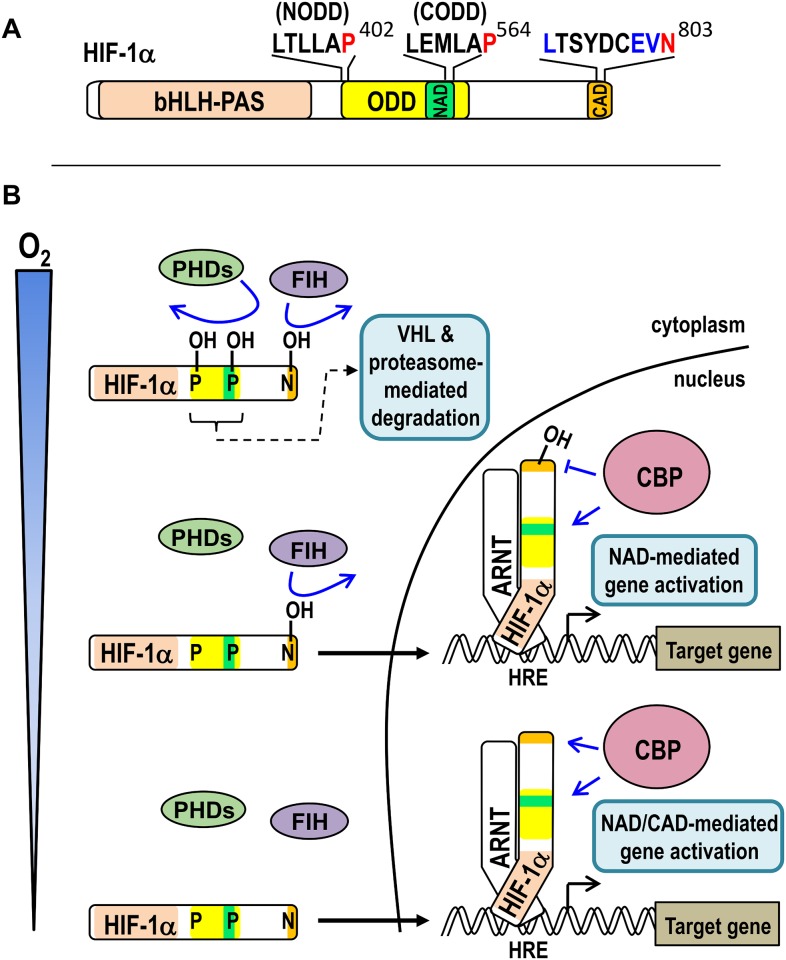
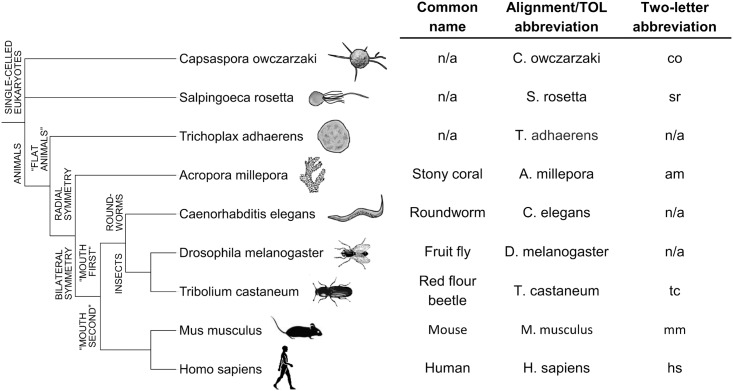
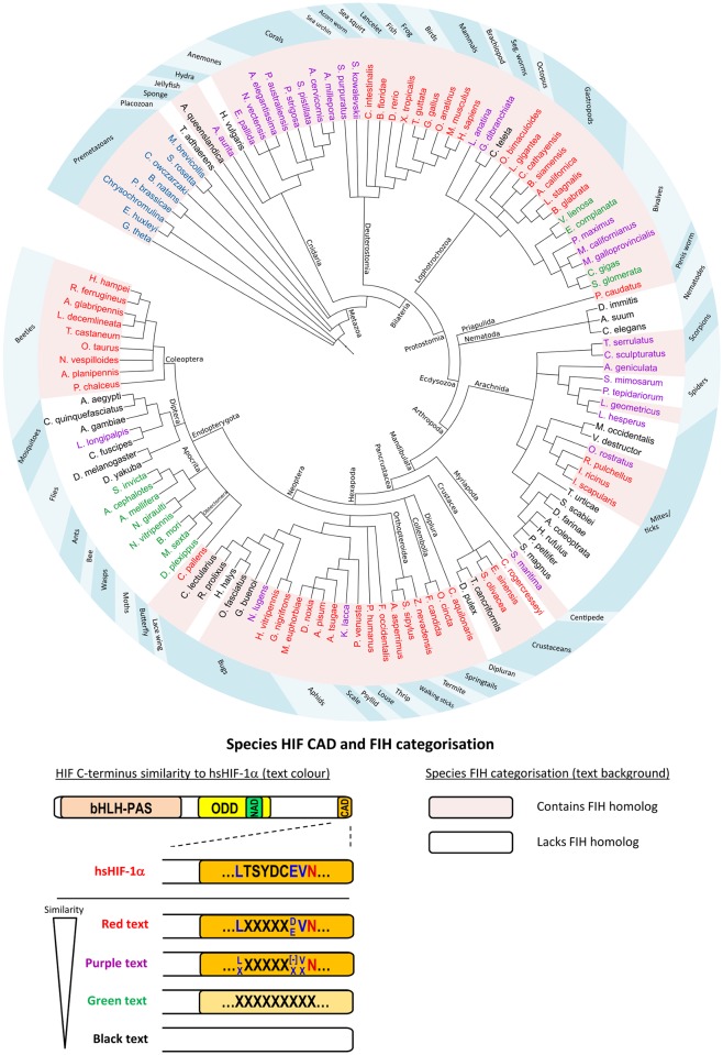
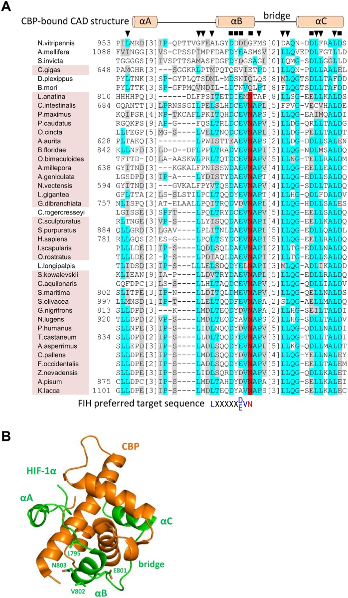
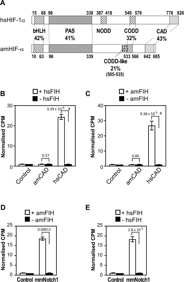
The asparaginyl hydroxylase, Factor Inhibiting HIF (FIH), is a cellular dioxygenase. Originally identified as oxygen sensor in the cellular response to hypoxia, where FIH acts as a repressor of the hypoxia inducible transcription factor alpha (HIF-α) proteins through asparaginyl hydroxylation, FIH also hydroxylates many proteins that contain ankyrin repeat domains (ARDs). Given FIH's promiscuity and the unclear functional effects of ARD hydroxylation, the biological relevance of HIF-α and ARD hydroxylation remains uncertain. Here, we have employed evolutionary and enzymatic analyses of FIH, and both HIF-α and ARD-containing substrates, in a broad range of metazoa to better understand their conservation and functional importance. Utilising Tribolium castaneum and Acropora millepora, we provide evidence that FIH from both species are able to hydroxylate HIF-α proteins, supporting conservation of this function beyond vertebrates. We further demonstrate that T. castaneum and A. millepora FIH homologs can also hydroxylate specific ARD proteins. Significantly, FIH is also conserved in several species with inefficiently-targeted or absent HIF, supporting the hypothesis of important HIF-independent functions for FIH. Overall, these data show that while oxygen-dependent HIF-α hydroxylation by FIH is highly conserved in many species, HIF-independent roles for FIH have evolved in others.


Conflict of interest statement

The authors have declared that no competing interests exist.

Figures

Fig 1
Fig 1. Activation of HIF-1 signalling by hypoxia.
(A) Schematic of human HIF-1α (hsHIF-1α) showing the regions involved in DNA-binding and dimerisation with ARNT (bHLH-PAS), oxygen-dependent degradation (ODD), and coactivator binding (the N- and C-terminal transactivation domains, NAD and CAD, respectively). The asparaginyl residue (Asn803) hydroxylated by FIH is shown in red with residues constituting the remainder of the “FIH preferred target sequence” shown in blue above the CAD. The PHD-targeted prolyl residues which are central to the N- and C-terminal ODDs (Pro402 (NODD) and Pro564 (CODD), respectively) are similarly indicated above the ODD. (B) Schematic showing the consequences of different oxygen levels (from “adequate” or normoxic at the top of the schematic to severely hypoxic at the bottom) on FIH/PHD enzyme and hsHIF-1α activity. When adequate oxygen is present, the PHDs and FIH are both active, resulting in hydroxylation of their target residues in HIF-1α (coloured as in part A). Prolyl hydroxylation results in efficient VHL-mediated ubiquitination and rapid proteasomal degradation of HIF-1α, thus ensuring minimal HIF-1 target gene activation. At intermediate levels of oxygen, the PHDs are inactive, resulting in HIF-1α stabilisation, translocation to the nucleus, and partnering with ARNT on hypoxia response elements (HREs). Ongoing FIH-mediated hydroxylation at this oxygen tension, however, precludes CBP binding to the CAD, thus only the NAD recruits CBP for target gene activation. Under more severe hypoxia, both PHDs and FIH are inactive, thus both the NAD and CAD of HRE-bound HIF-1α can recruit CBP for target gene activation.
Fig 2
Fig 2. Evolutionary relationships and nomenclature of key eukaryotic species.
Species which have previously undergone detailed molecular analysis of their HIF pathway components, including Caenorhabditis elegans [8], Drosophila melanogaster [34] and Trichoplax adhaerens [23], are shown in a cladogram with biochemically characterised species in the current work. An adjacent table shows species name abbreviations used in Fig 3 and sequence alignment figures, as well as two-letter abbreviations utilised for concise reference to various protein homologs (e.g. Tribolium castaneum FIH is abbreviated to “tcFIH”). A more comprehensive list of species name abbreviations can be found in S1 Table. n/a = not applicable.
Fig 3
Fig 3. Conservation of HIF-α CAD and FIH across the Eukaryota.
BLAST and hidden markov models were used to identify HIF-α and FIH homologs in a wide variety of Eukaryotes. Evolutionary relationships between representative analysed species are indicated by a cladogram. An explanation of species name background shading and text colour is provided in a schematic below the cladogram. Background shading of species names is used to indicate FIH status, with pink and white shading representing FIH-containing and FIH-lacking species, respectively. Amongst Metazoa, the colour of the text for the name of each species (red, purple, green or black) refers to its HIF-α characteristics, specifically, its level of C-terminal similarity to that of hsHIF-1α CAD. The features which define the four C-terminus types are represented pictorially using an orange-shaded CAD and text within the CAD to indicate the high similarity of CBP or FIH-binding residues, respectively. For comparison, the hsHIF-1α CAD is shown in the schematic coloured orange, with its FIH target sequence shown within the CAD using the same colouring as Fig 1. Red coloured species in the cladogram have strong CBP-binding residue conservation, and also contain hsFIH’s preferred target sequence. Purple species also have strong CBP-binding residue conservation, but at least 1 or more of the residues in FIH’s preferred target sequence (excluding the Asn) are not conserved (indicated by “X”s), which may render the protein an inefficient FIH substrate [47, 48]. “[–]”indicates an acidic residue (Asp or Glu). Green species have only moderate CBP-binding residue conservation (indicated by pale orange colouring of the CAD) and no FIH target sequence, while black species have no recognisable CAD. Premetazoans with blue text in the cladogram lack a HIF-α homolog. Cladogram branches are labelled with taxonomic classifications. The blue ring outside the cladogram indicates species common names. Cladogram tree generated using phyloT and displayed using Interactive Tree of Life [49].
Fig 4
Fig 4. Conservation of CBP and FIH binding residues in HIF-α CAD homologs.
(A) Known and putative HIF-α CAD sequences from representative species were aligned using MUSCLE [50]. Identical and similar residues are indicated by cyan and grey highlights, respectively, while likely FIH target Asn residues are highlighted in red. To facilitate viewing of as large a CAD region as possible, low similarity regions of the alignment were deleted and replaced with bracketed numbers which indicate how many residues in each sequence are not shown. Amino acid numbers within the HIF-α proteins (where available) are shown to the left of the sequences. To compare functional regions of hsHIF-1α CAD with the aligned homologs, CBP and FIH-interacting residues are also depicted. Specifically, the secondary structure of hsHIF-1α CAD when bound to CBP [45] as well as residues predicted to be involved in polar (black squares) or hydrophobic (black triangles) interactions with CBP are shown above the alignment. The FIH preferred target sequence is shown below the alignment, coloured as for Fig 1. Conservation of FIH in a species is indicated by pink background shading of the species’ name, and correlates strongly with the level of sequence similarity to hsHIF-1α CAD. (B) The secondary structure depicted above the alignment in part A is shown in the context of the NMR structure of CBP (orange) bound to hsHIF-1α CAD (green) [45]. The different regions of hsHIF-1α CAD that interact with CBP, including helices αA-αC and the αB-αC bridge, and the FIH target sequence residue sidechains, Leu795, Glu801, Val802 and Asn803 are labelled with green text. Structure image generated using Pymol [Schrodinger, 2015 #1226] and PBD structure 1L8C [45].
Fig 5
Fig 5. Alignment of putative premetazoan FIH homologs with human FIH.
The sequences of FIH homologs C-terminal to the first 2-OG-binding residue (Tyr145 in hsFIH) were aligned using Clustal Omega [51]. Residues strongly or partially conserved are shown with cyan and grey highlights, respectively. Residues involved in iron coordination (red), 2-OG binding (dark blue), and target asparagine positioning (pink) are indicated. The secondary structure of hsFIH is depicted above the alignment, with yellow arrows indicating the beta strands which make up the double-stranded β-helix (DSBH), and dark brown helices denoting those involved in dimerisation. Amino acid numbers are shown to the left of the alignment. Alignment shading performed using the BoxShade Server. Species name abbreviations and sequence IDs can be found in S1 Table.
Fig 6
Fig 6. T. castaneum PHD mediates degradation of T. castaneum HIF-α.
(A) Comparative domain structure of hsHIF-1α and tcHIF-α showing the percent identity of amino acid sequence in conserved regions, including the basic helix loop helix (bHLH), Per ARNT Sim homology domain (PAS), N- and C-terminal oxygen-dependent degradation domains (NODD and CODD) and C-terminal activation domain (CAD). Amino acid numbers at the start and end of each domain are shown. (B) The NODD and CODD amino acid sequences from tcHIF-α are shown aligned with the equivalent domains from hsHIF-1α. Conserved and similar residues are indicated by dark and light grey highlights, respectively, and the hydroxylated prolines (Pro533 in the NODD, Pro635 in the CODD) are in black. Amino acid numbers are to the left of each sequence. (C) The ability of tcPHD to facilitate degradation of tcHIF-α was assessed in mammalian cells. pEF-IRES-myc-6His-Puro6 plasmids, either empty (“-“) or encoding wild-type (wt) tcHIF-α, tcHIF-α single proline to alanine mutants P533A or P635A, or tcHIF-α double mutant P533A/P635A were transiently transfected into HEK293T cells along with pcDNA3.1 encoding a V5 epitope tag and wild-type tcPHD (“w”), catalytic mutant tcPHD H321A (“m”) or non-specific control Aryl Hydrocarbon Receptor aa 84–287 (“c”). Cells were then incubated for 8 hrs in normoxia. Levels of tcHIF-α and tcPHD protein in cell extracts were subsequently analysed by western blotting for myc and V5 epitope tags, respectively. α-tubulin served as a loading control.
Fig 7
Fig 7. T. castaneum FIH is a putative asparaginyl hydroxylase that hydroxylates HIF-α substrates.
(A) Thioredoxin-6 Histidine-tagged-tcHIF-α (790–879) (Trx-6H-tcCAD), Trx-6H-tcNotch (1747–1989) (Trx-6H-tcNotch ARD) and maltose binding protein-tagged tcFIH (MBP-tcFIH) were expressed in E. coli, purified by Ni2+ affinity or amylose agarose chromatography, and analysed by Coomassie-stained SDS-PAGE. (B) In vitro hydroxylation reactions were set up containing 60 μM tcHIF-α CAD (tcHIF-α residues 790–879), hsHIF-1α CAD (hsHIF-1α residues 736–826) and their corresponding Asn mutants, and either 1 μM tcFIH (white bars, analysed in triplicate) or buffer (black bars, analysed in duplicate). Reactions were incubated at 37°C degrees for 30 min, and then the counts per minute (CPM) of [14C]CO2 released during the reaction analysed by scintillation counting. Data were normalised to CPM observed for the hsHIF-1α CAD N803A + FIH sample. Bars are mean +/- SEM of combined data from 3 independent experiments. (C) As for part B, but using hsFIH enzyme. (D) Schematic representation of the reporter gene assay used to test transactivation capacity of tcHIF-α CAD. A firefly luciferase reporter gene downstream of a Gal4 response element (GRE) is transfected into mammalian cells. The GRE facilitates recruitment of yeast Gal4-DNA-binding domain (GalDBD)-tagged HIF-α CAD proximal to the luciferase gene promoter, with the activity of the CAD driving transcription. FIH-dependent HIF-1α CAD hydroxylation represses activity of the CAD, reducing transcription of luciferase. (E) Reporter gene assay testing tcHIF-α CAD transactivation potential, and the ability of hsFIH and tcFIH to repress CAD-mediated firefly luciferase production. FIH-/- mouse embryonic fibroblasts (MEFs) were transiently transfected in triplicate with pGal-O-tcHIF-α CAD (GalDBD-tcCAD), pGal-O-tcHIF-α CAD N856A (GalDBD-tcCAD N856A) or empty vector (GalDBD), together with firefly and control renilla luciferase reporter constructs. Each well was also transfected with hsFIH or tcFIH, their catalytically inactive mutants (H199A and H185A, respectively), or empty pcDNA3.1 vector (“Control”). Relative luciferase units (RLU) were calculated from the ratio of firefly to renilla luminescence. Data were normalised to the average RLU from the GalDBD-tcCAD samples (with the exception of the sample co-expressed with hsFIH due to its small magnitude). Bars are mean +/- SEM of combined data from 3 independent experiments. (F) As for part E, except using GalDBD-hsHIF-1α CAD and GalDBD-hsHIF-1α CAD N803A in place of GalDBD-tcHIF-α CAD and GalDBD-tcHIF-α CAD N856A. Statistical analysis for parts B, C, E and F was carried out on non-normalised, log-transformed data using a 2-tailed paired t-test, with p values indicated above the bars. p values < 0.05 are considered significant. # indicates comparison also significant using the conservative Bonferroni-adjusted significance value of 0.0125 (for parts A and B) and 0.00625 (for parts E and F) for multiple comparisons.
Fig 8
Fig 8. Hydroxylation of ARD-containing substrates by T. castaneum FIH.
(A) 1 μM MBP-tagged tcFIH was tested in triplicate in the presence of 40 μM Trx-6H-tagged mmNotch1 (1862–2104) (mmNotch1 ARD) or Trx-6H-tcNotch (1747–1989) (tcNotch ARD) and their Asn to Ala mutants mmNotch1 ARD NN1945/2012AA (mmNotch1 ARD dbl mut) and tcNotch ARD NN1830/1897AA (tcNotch ARD dbl mut) by in vitro hydroxylation assay (white bars). As a control, each substrate was also tested in duplicate with buffer in place of enzyme (black bars). Data from each experiment were normalised to CPM (counts per minute of [14C]CO2 released) for the mmNotch1 ARD dbl mut + FIH sample. Bars are mean +/- SEM of combined data from 3 independent experiments. (B) As for (A), but using hsFIH. (C) Alignment of sequences within tcNotch predicted to contain target Asns with their equivalent regions in mmNotch1. Conserved and similar residues are indicated by dark and light grey highlights, respectively, and the (putative) target Asns are in black. Amino acid numbers are to the left of each sequence. Statistical analysis for parts A and B was carried out on non-normalised, log-transformed data using a 2-tailed paired t-test, with p values indicated above the bars. p values < 0.05 are considered significant. # indicates comparison also significant using the conservative Bonferroni-adjusted significance value of 0.0125 (for parts A and B) for multiple comparisons.
Fig 9
Fig 9. Hydroxylation of CAD and ARD substrates by A. millepora FIH.
(A) Comparative domain structure of hsHIF-1α and A. millepora HIF-α (amHIF-α) showing the percent identity of conserved regions, including the basic helix loop helix (bHLH), Per ARNT Sim homology domain (PAS), C-terminal oxygen-dependent degradation domain (CODD) and C-terminal activation domain (CAD). Unlike human and T. castaneum HIF-α, amHIF-α does not appear to contain a NODD, but instead contains a second sequence with low similarity to the human CODD (“CODD-like”) just N-terminal of a more robustly conserved CODD sequence. Amino acid numbers at the start and end of each domain are shown. (B) MBP-tagged hsFIH (1 μM) was tested in triplicate by in vitro hydroxylation assay with 25 μM Trx-6H-tagged hsHIF-α CAD and amHIF-α CAD substrates (hsHIF-1α (736–826) and amHIF-α (604–693), respectively), or buffer alone control. (C) As for part B, except using 1 μM MBP-tagged A. millepora FIH (amFIH). (D) MBP-tagged amFIH (at 1 μM) was tested in triplicate with mouse Trx-6H-Notch1 (1862–2104) (mmNotch1, 25 μM) or buffer alone (control) by in vitro hydroxylation assay. (E) As for D, but using hsFIH. For parts B-E, data from each experiment were normalised to CPM (counts per minute of [14C]CO2 released during the reaction) observed for the control minus FIH sample. Bars are mean +/- SEM of combined data from 3 independent experiments. Statistical analysis was carried out on non-normalised data using a 2-tailed paired t-test, with p values indicated above the bars. p values < 0.05 are considered significant. For parts B and C, # indicates comparison also significant using the conservative Bonferroni-adjusted significance value of 0.025 for multiple comparisons.

Similar articles

Cited by

References

    1. Prabhakar NR, Semenza GL. Oxygen Sensing and Homeostasis. Physiology (Bethesda). 2015;30(5):340–8. Epub 2015/09/04. 10.1152/physiol.00022.2015 . - DOI - PMC - PubMed
    1. Hewitson KS, McNeill LA, Riordan MV, Tian YM, Bullock AN, Welford RW, et al. Hypoxia-inducible factor (HIF) asparagine hydroxylase is identical to factor inhibiting HIF (FIH) and is related to the cupin structural family. J Biol Chem. 2002;277(29):26351–5. 10.1074/jbc.C200273200 . - DOI - PubMed
    1. Lando D, Peet DJ, Gorman JJ, Whelan DA, Whitelaw ML, Bruick RK. FIH-1 is an asparaginyl hydroxylase enzyme that regulates the transcriptional activity of hypoxia-inducible factor. Genes Dev. 2002;16(12):1466–71. 10.1101/gad.991402 . - DOI - PMC - PubMed
    1. Lando D, Peet DJ, Whelan DA, Gorman JJ, Whitelaw ML. Asparagine hydroxylation of the HIF transactivation domain a hypoxic switch. Science. 2002;295(5556):858–61. 10.1126/science.1068592 . - DOI - PubMed
    1. Mahon PC, Hirota K, Semenza GL. FIH-1: a novel protein that interacts with HIF-1alpha and VHL to mediate repression of HIF-1 transcriptional activity. Genes Dev. 2001;15(20):2675–86. 10.1101/gad.924501 . - DOI - PMC - PubMed

Publication types

Substances