. 2019 Jun 20;47(11):5465-5479.
doi: 10.1093/nar/gkz247.

Site-specific replacement of phosphorothioate with alkyl phosphonate linkages enhances the therapeutic profile of gapmer ASOs by modulating interactions with cellular proteins

Affiliations

Site-specific replacement of phosphorothioate with alkyl phosphonate linkages enhances the therapeutic profile of gapmer ASOs by modulating interactions with cellular proteins

Michael T Migawa et al. Nucleic Acids Res. .

Abstract

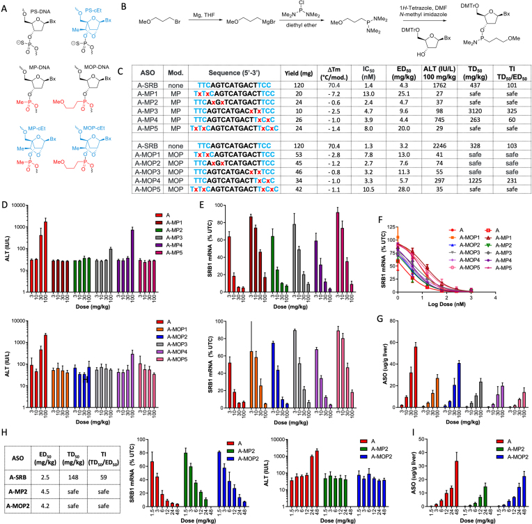
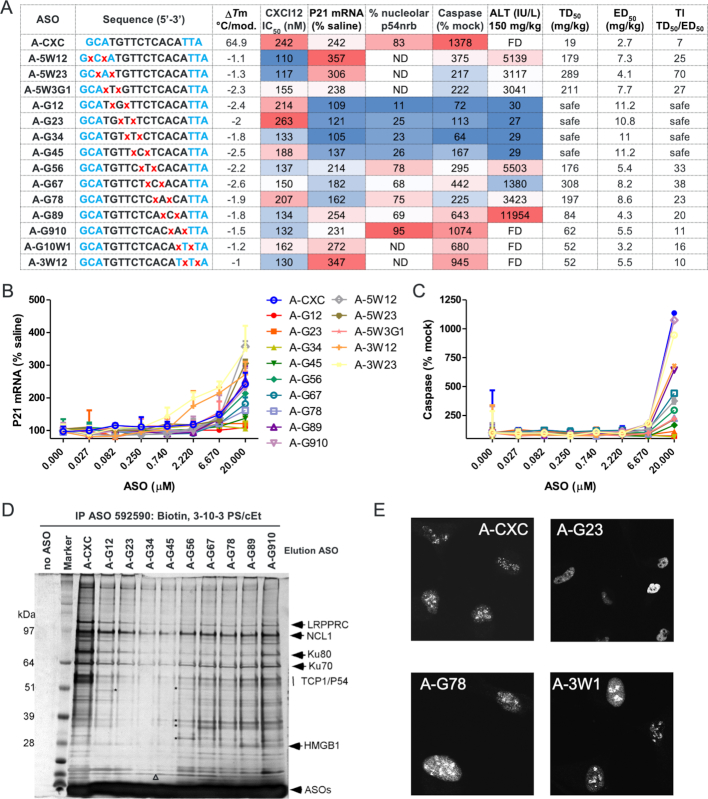
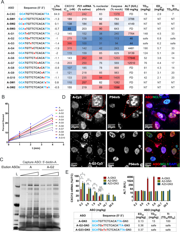
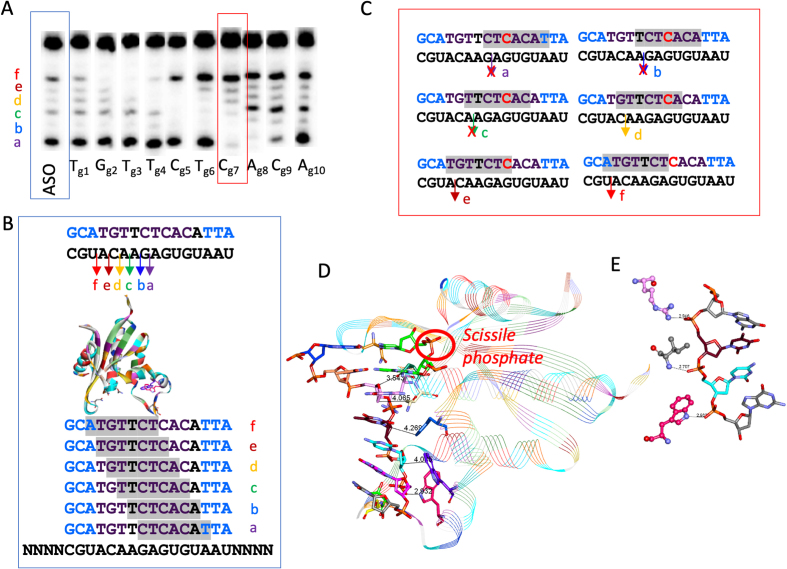
Phosphorothioate-modified antisense oligonucleotides (PS-ASOs) interact with a host of plasma, cell-surface and intracellular proteins which govern their therapeutic properties. Given the importance of PS backbone for interaction with proteins, we systematically replaced anionic PS-linkages in toxic ASOs with charge-neutral alkylphosphonate linkages. Site-specific incorporation of alkyl phosphonates altered the RNaseH1 cleavage patterns but overall rates of cleavage and activity versus the on-target gene in cells and in mice were only minimally affected. However, replacing even one PS-linkage at position 2 or 3 from the 5'-side of the DNA-gap with alkylphosphonates reduced or eliminated toxicity of several hepatotoxic gapmer ASOs. The reduction in toxicity was accompanied by the absence of nucleolar mislocalization of paraspeckle protein P54nrb, ablation of P21 mRNA elevation and caspase activation in cells, and hepatotoxicity in mice. The generality of these observations was further demonstrated for several ASOs versus multiple gene targets. Our results add to the types of structural modifications that can be used in the gap-region to enhance ASO safety and provide insights into understanding the biochemistry of PS ASO protein interactions.


Figures

Figure 1.
Figure 1.
Replacing PS with neutral alkylphosphonate linkages near 5′-end of the DNA gap improves therapeutic profile of toxic gapmer ASOs. (A) Structures of MP and MOP-modified DNA and cEt nucleotides. (B) Synthesis of MOP-modified nucleoside phosphoramidites. (C) Sequence, ASO design (blue letters – cEt, black – PS DNA, red – MOP linkage), Tm versus complementary RNA, activity in primary mouse hepatocytes after ASO delivery by free uptake, potency for reducing SRB1 mRNA by 50% in mouse liver (ED50), plasma ALT (IU/L) in mice 72 hours after injection of 100 mg/kg ASO, ASO dose (mg/kg) required to produce half-maximal elevations in plasma ALT (TD50), Therapeutic index (TI) was calculated as the ratio of TD50 divided by the ED50. ASOs were deemed safe if no elevation in plasma ALT were observed at the highest dose tested. (D) Plasma ALT (IU/L) levels following treatment with MP- or MOP-ASOs. (E) SRB1 mRNA reduction in liver in mice following treatment with MP- or MOP-ASOs. Balb-c mice were injected subcutaneously with 3, 10, 30 and 100 mg/kg of ASO and sacrificed 72 h after injection. Livers were harvested and changes in SRB1 mRNA relative to saline-treated control animals were quantified by qRT-PCR. (F) SRB1 mRNA reduction in primary mouse hepatocytes treated with MP- or MOP-ASOs by free uptake. (G) Liver accumulation of MOP-ASOs from dose-response experiment described in Figure 1E. ASO tissue levels were determined utilizing an HELISA assay. (H) Confirmation dose response to determine ED50 and plasma ALT (IU/L) levels for ASOs A, A-MP2 and A-MOP2. Balb-c mice were injected subcutaneously with 1.5, 3, 6, 12, 24 and 48 mg/kg of ASO and sacrificed 72 h after injection. Livers were harvested and changes in SRB1 mRNA relative to saline-treated control animals were quantified by qRT-PCR. (I) Liver accumulation (ug/g) for ASOs A, A-MP2 and A-MOP2 from confirmatory dose–response study as determined using HELISA. All errors are ±std.dev.
Figure 2.
Figure 2.
Site-specific introduction of two MOP-linkages near the 5′-end of the DNA gap reduces toxicity of a toxic gapmer ASO in cells and in mice. (A) Sequence, ASO design and chemistry (blue letters – cEt, black – PS DNA, red – MOP linkage); Tm for complementary RNA; Inhibition concentration (IC50) for reducing CXCl12 mRNA by 50% in 3T3L1 cells; P21 mRNA elevation in 3T3L1 cells; and caspase activation in Hepa1-6 cells following ASO (20 μM) delivery by electroporation; plasma ALT (IU/L) in mice 72 hours after injection of 150 mg/kg ASO; ASO dose (mg/kg) required to produce half-maximal elevations in plasma ALT (TD50); and effective dose (mg/kg) for reducing CXCl12 mRNA in mouse liver by 50% (ED50). Columns were formatted using the conditional formatting option in MS excel using the red-white-blue color scale. Blue indicates reduced toxicity and improved activity while red indicates enhanced toxicity and reduced activity respectively. Therapeutic index (TI) was calculated as the ratio of TD50 divided by the ED50. ASOs were deemed safe if no elevation in plasma ALT were observed at the highest dose tested. (B) qRT-PCR quantification of P21 mRNA levels in 3T3L1 cells treated with ASOs at indicated concentrations by electroporation. Errors are ±std.dev. (C) Caspase activity levels in Hepa1-6 cells treated by electroporation with different ASOs at indicated concentrations for 18–24 h. Errors are ±std.dev. (D) PS-ASO-binding proteins were isolated from HeLa lysate with 5′-biotinylated PS-ASOs and were eluted by competition using unconjugated MOP-ASOs. Silver staining of eluted proteins resolved on SDS-PAGE. L, size marker. (E) Toxic ASOs A-CXC, A-3W1 and A-G78 causes mislocalization of P54nrb to the nucleolus after transfection into HeLa cells (200 nM for 2 h) while the safe MOP ASO A-G23 does not. FD = found dead.
Figure 3.
Figure 3.
Replacing PS with a single MOP-linkage at gap positions 2 and 3 reduces toxicity without impacting the potency of a toxic gapmer ASO in cells and in mice. (A) Sequence, ASO design and chemistry (blue letters – cEt, black – PS DNA, red – MOP linkage); Tm for complementary RNA; Inhibition concentration (IC50) for reducing CXCl12 mRNA by 50% in 3T3L1 cells; P21 mRNA elevation in 3T3L1 cells; and caspase activation in Hepa1-6 cells following ASO (20 μM) delivery by electroporation; plasma ALT (IU/l) in mice 72 h after injection of 150 mg/kg ASO; ASO dose (mg/kg) required to produce half-maximal elevations in plasma ALT (TD50); and ED50 (mg/kg) for reducing CXCl12 mRNA in mouse liver. Columns were formatted using the conditional formatting option in MS excel using the red-white-blue color scale. Blue indicates reduced toxicity and improved activity while red indicates enhanced toxicity and reduced activity respectively. Therapeutic index (TI) was calculated as the ratio of TD50 divided by the ED50. ASOs were deemed safe if no elevation in plasma ALT were observed at the highest dose tested. (B) Initial rates for cleavage of MOP-ASO/RNA duplexes by human recombinant RNaseH1. (C) PS-ASO-binding proteins were isolated from HeLa lysate with 5′-biotinylated PS-ASOs and were eluted by competition using unconjugated PS-ASOs at three different elution concentrations. Silver staining of eluted proteins resolved on SDS-PAGE. L, size marker. (D) ASO A-Cy3 causes mislocalization of P54nrb to the nucleolus after transfection into Hela cells (200 nM for 2 hours) while the MOP ASO A2-Cy3 does not. (E) ED50 for reducing CXCl12 mRNA in the liver in mice using GalNAc conjugates of select MOP ASOs. Balb-c mice were injected subcutaneously with a single dose of 0.2, 0.6, 1.8 and 5.4 mg/kg of A-GN3 or with 0.2, 0.6, 1.8, 5.4, 15 and 50 mg/kg of A2-GN3 or A23-GN3. Mice were sacrificed after 72 h and plasma ALT levels were recorded. Livers were homogenized and reduction of CXCl12 mRNA was measured by qRT-PCR. Errors are ±std.dev. P-values were calculated based on unpaired t-test using Prism. *P < 0.05; **P < 0.01; NS, not significant. FD = found dead. NT = not tested.
Figure 4.
Figure 4.
MOP at gap position 2 reduces toxicity of ASOs targeting diverse gene targets. (A) Balb-c mice were injected subcutaneously with the parent GalNAc (GN3) gapmer ASOs or their MOP-counterparts at 50 mg/kg. Mice were sacrificed after 72 h and plasma ALT (IU/l) levels were measured. (B) Structure of GN3-conjugated ASOs. Therapeutic indices of parent and MOP GN3-ASOs targeting (C) mouse HDAC2 mRNA, (D) FBO1a mRNA (E) Dynamin2 mRNA. Balb-c mice were injected subcutaneously with increasing doses of the ASOs as indicated. Mice were sacrificed 72 h after injection and plasma ALT levels were measured to determine the TD50. Livers were homogenized, and reduction of the targeted mRNA were quantified by qRT-PCR and ED50 (mg/kg) was determined. All errors are ±std.dev.
Figure 5.
Figure 5.
Structural rationale for the changes in RNaseH1 cleavage patterns for MOP-ASO/RNA duplexes (A) Human RNaseH1 cleaves the parent ASO A-CXC/RNA duplex at six distinct sites (a–f) and site-specific incorporation of MOP produced distinct changes in RNaseH1 cleavage patterns. (B) The distinct but overlapping 7-nucleotide footprints of the catalytic domain of human RNaseH1 for cleavage sites a–f. (C) Insertion of MOP at C7 in the gap ablates cleavage sites a, b and c indicating that MOP is not tolerated at positions 3, 4 and 5 of the seven-nucleotide footprint. (D) Structure of the catalytic domain of human RNaseH1 with the scissile site on the RNA (only two nucleotides flanking the scissile site are shown for clarity). The catalytic domain makes three critical contacts with the backbone phosphates at positions 3, 4 and 5 of the footprint. (E) Insertion of neutral linkages at positions 3, 4 and 5 of the footprint ablates RNaseH1 cleavage.

References

    1. Crooke S.T., Wang S., Vickers T.A., Shen W., Liang X-h.. Cellular uptake and trafficking of antisense oligonucleotides. Nat. Biotech. 2017; 35:230–237. - PubMed
    1. Seth P.P., Tanowitz M., Bennett C.F.. Selective tissue targeting of synthetic nucleic acid drugs. J.Clin.Investig. 2019; 129:915–925. - PMC - PubMed
    1. Liang X-h., Sun H., Shen W., Crooke S.T.. Identification and characterization of intracellular proteins that bind oligonucleotides with phosphorothioate linkages. Nucleic Acids Res. 2015; 43:2927–2945. - PMC - PubMed
    1. Liang X.-H., Shen W., Sun H., Kinberger G.A., Prakash T.P., Nichols J.G., Crooke S.T.. Hsp90 protein interacts with phosphorothioate oligonucleotides containing hydrophobic 2′-modifications and enhances antisense activity. Nucleic Acids Res. 2016; 44:3892–3907. - PMC - PubMed
    1. Shen W., Liang X-h., Crooke S.T.. Phosphorothioate oligonucleotides can displace NEAT1 RNA and form nuclear paraspeckle-like structures. Nucleic Acids Res. 2014; 42:8648–8662. - PMC - PubMed

Publication types

MeSH terms