. 2019 Aug;33(8):8865-8877.
doi: 10.1096/fj.201802418R. Epub 2019 Apr 29.

Severe acute respiratory syndrome coronavirus ORF3a protein activates the NLRP3 inflammasome by promoting TRAF3-dependent ubiquitination of ASC

Affiliations

Severe acute respiratory syndrome coronavirus ORF3a protein activates the NLRP3 inflammasome by promoting TRAF3-dependent ubiquitination of ASC

Kam-Leung Siu et al. FASEB J. 2019 Aug.

Abstract

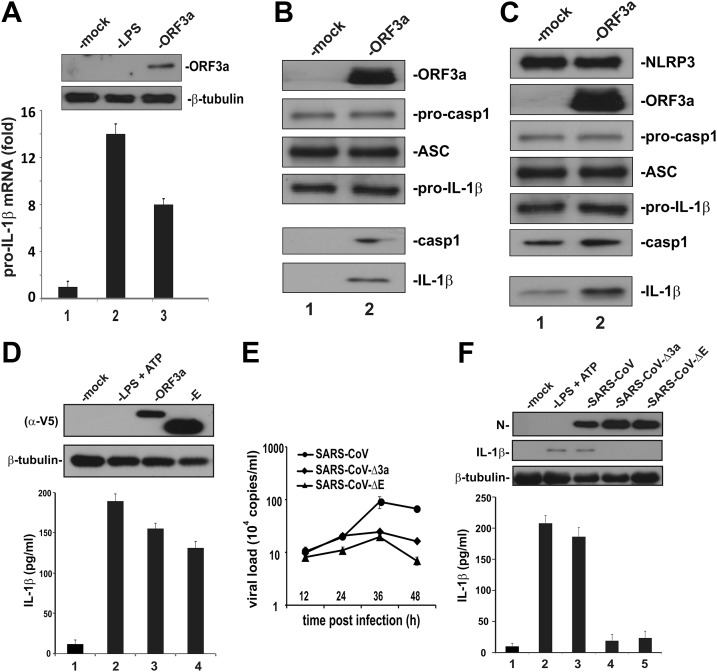
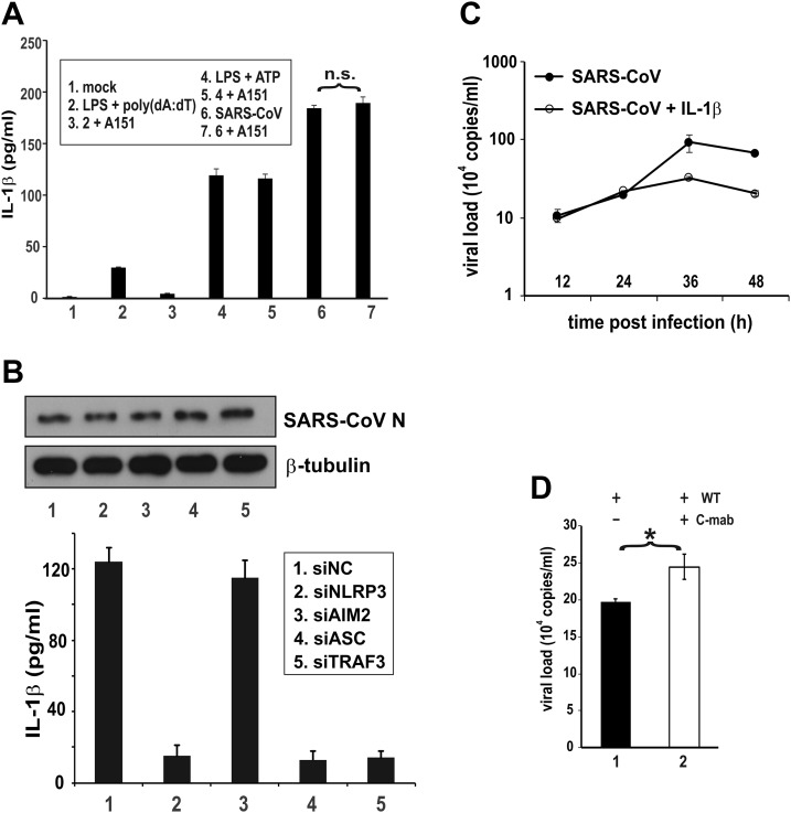
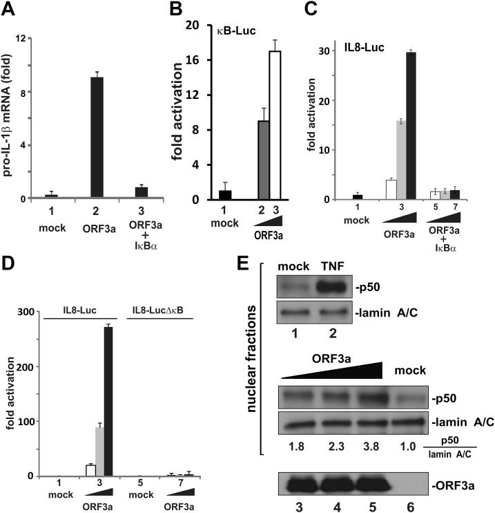
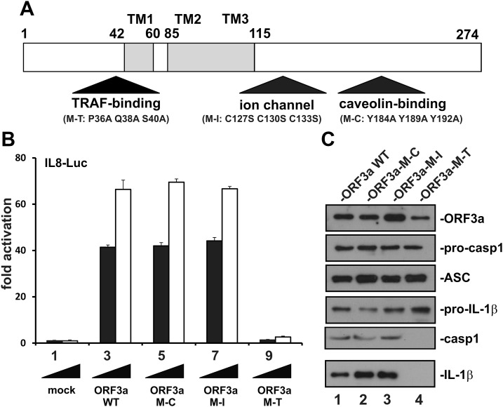
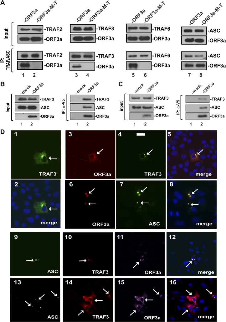
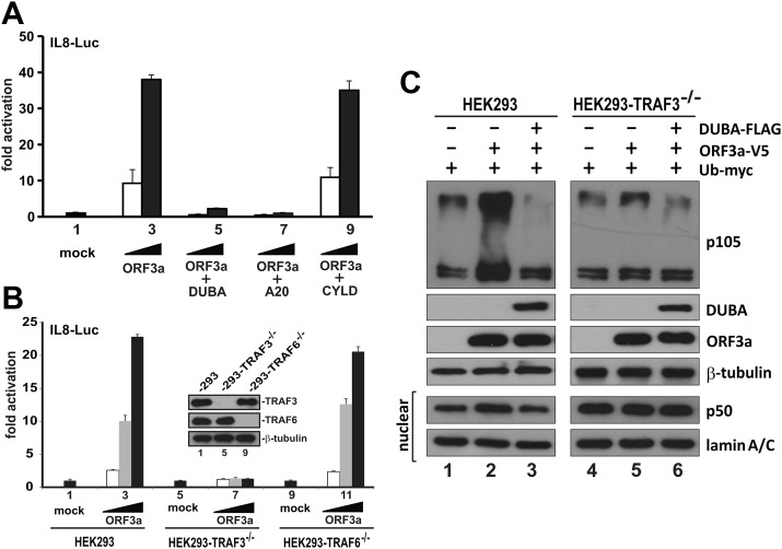
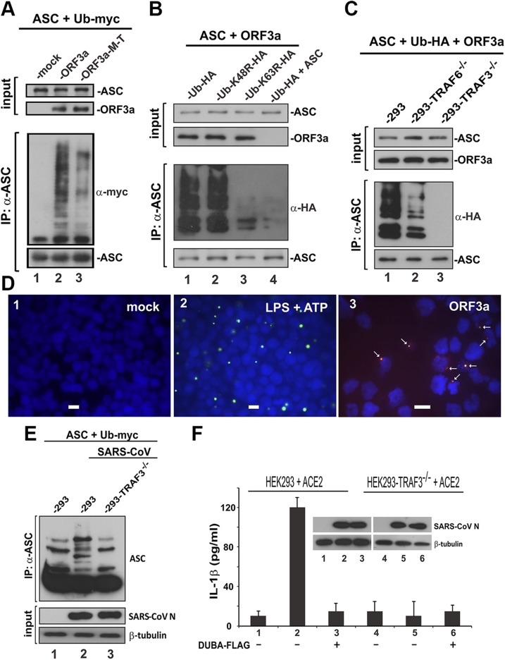
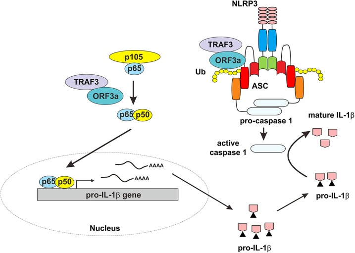
Severe acute respiratory syndrome coronavirus (SARS-CoV) is capable of inducing a storm of proinflammatory cytokines. In this study, we show that the SARS-CoV open reading frame 3a (ORF3a) accessory protein activates the NLRP3 inflammasome by promoting TNF receptor-associated factor 3 (TRAF3)-mediated ubiquitination of apoptosis-associated speck-like protein containing a caspase recruitment domain (ASC). SARS-CoV and its ORF3a protein were found to be potent activators of pro-IL-1β gene transcription and protein maturation, the 2 signals required for activation of the NLRP3 inflammasome. ORF3a induced pro-IL-1β transcription through activation of NF-κB, which was mediated by TRAF3-dependent ubiquitination and processing of p105. ORF3a-induced elevation of IL-1β secretion was independent of its ion channel activity or absent in melanoma 2 but required NLRP3, ASC, and TRAF3. ORF3a interacted with TRAF3 and ASC, colocalized with them in discrete punctate structures in the cytoplasm, and facilitated ASC speck formation. TRAF3-dependent K63-linked ubiquitination of ASC was more pronounced in SARS-CoV-infected cells or when ORF3a was expressed. Taken together, our findings reveal a new mechanism by which SARS-CoV ORF3a protein activates NF-κB and the NLRP3 inflammasome by promoting TRAF3-dependent ubiquitination of p105 and ASC.-Siu, K.-L., Yuen, K.-S., Castaño-Rodriguez, C., Ye, Z.-W., Yeung, M.-L., Fung, S.-Y., Yuan, S., Chan, C.-P., Yuen, K.-Y., Enjuanes, L., Jin, D.-Y. Severe acute respiratory syndrome coronavirus ORF3a protein activates the NLRP3 inflammasome by promoting TRAF3-dependent ubiquitination of ASC.

Keywords: SARS coronavirus.


Conflict of interest statement

The authors thank Lilong Jia (Department of Microbiology, The University of Hong Kong) for assistance with the infection study. This work was supported by the Hong Kong Health and Medical Research Fund (13121032, 14130822, and HKM-15-M01), Hong Kong Research Grants Council (T11-707/15-R, C7011-15R, and 17124415), Government of Spain (BIO2013-42869-R), U.S. National Institutes of Health (NIH) National Institute of Allergy and Infectious Diseases (2P01AI060699-06A1), and European Commission (Innovative Medicines Initiative Grant Agreement 115760). C.C.-R. received a fellowship from Fundacion La Caixa. K.-L.S., K.-S.Y., and C.C.-R. are co-first authors. The authors declare no conflicts of interest.

Figures

Figure 1
Figure 1
Activation of IL-1β production by SARS-CoV and its ORF3a protein. A) Induction of pro–IL-1β transcript by ORF3a. THP-1 cells were either mock-transfected with an empty vector or transfected with an ORF3a expression vector for 24 h. Cells in the control group were treated with 10 ng/ml of LPS for 8 h. The IL-1β transcript was analyzed by qRT-PCR. The level of IL-1β mRNA was relative to that of HGPRT mRNA. Bars represent the means of 3 biologic replicates, and error bars indicate sd. B, C) Induction of IL-1β secretion by ORF3a in a HEK293 reconstitution system. HEK293 cells were transfected with expression plasmids for procaspase 1, ASC and pro–IL-1β. Cells were mock-transfected with an empty vector or cotransfected with ORF3a plasmid alone (B) or NLRP3 + ORF3a plasmids (C) for 24 h. Cell lysates and conditioned medium was collected. Secreted IL-1β in the conditioned medium was immunoprecipitated with anti-FLAG antibody. Cell lysates (upper) and precipitates (lower) were probed by Western blotting. Pro–IL-1β expression was driven by a cytomegalovirus promoter and was therefore nonresponsive to ORF3a. Procasp1, procaspase 1; casp1, caspase 1 (p20). D) Induction of IL-1β secretion by SARS-CoV ORF3a and E proteins in THP-1 cells. THP-1 cells were transfected with ORF3a and E-expression plasmids for 24 h. Cell lysates were collected and probed by Western blotting. Secreted IL-1β in the conditioned medium was analyzed by ELISA. As a positive control, cells were also treated with 10 ng/ml LPS and 5 mM ATP. Both ORF3a and E proteins were V5 tagged and therefore detected with anti-V5 (alpha-V5). E) Virus growth kinetics. THP-1 cells were inoculated with SARS-CoV, SARS-CoV-Δ3a, or SARS-CoV-ΔE at an MOI of 0.01. Samples were harvested at the indicated time points, and viral load was determined by qRT-PCR. Data points represent means of triplicates, and error bars indicate sd. F) Induction of IL-1β secretion by SARS-CoV in THP-1 cells. THP-1 cells were infected with SARS-CoV, SARS-CoV-Δ3a, or SARS-CoV-ΔE (MOI = 0.01) for 24 h. Cell lysates were collected and probed by Western blotting. Secreted IL-1β in the conditioned medium was analyzed by ELISA.
Figure 2
Figure 2
SARS-CoV–induced activation of IL-1β production requires NLRP3, ASC, and TRAF3. A) Sensitivity of SARS-CoV–induced IL-1β secretion to AIM2 inhibitor. THP-1 cells were treated, transfected, or infected with the indicated stimuli for 24 h; cells in groups 2 and 3 were transfected with 1 μg/ml poly(dA:dT) for 3 h. Cells in groups 3, 5, and 7 were also treated with 1 μM A151. Secreted IL-1β in the conditioned medium was measured by ELISA. The difference between the indicated groups was not significant (n.s.) by Student’s t test. B) Sensitivity of SARS-CoV–induced IL-1β secretion to NLRP3, AIM2, ASC, and TRAF3 knockdown. THP-1 cells were transfected with the indicated siRNA for 48 h and then infected with SARS-CoV for 24 h. Secreted IL-1β in the conditioned medium was measured by ELISA. siNC: negative control siRNA. C) Effect of IL-1β on SARS-CoV replication. Huh7 cells were infected with SARS-CoV at an MOI of 0.01. Cells were mock-treated or treated with 10 ng/ml recombinant IL-1β. Samples were harvested at the indicated time points for qRT-PCR analysis of viral load. D) Effect of anti–IL-1β on SARS-CoV replication. Huh7 cells were infected with SARS-CoV WT at an MOI of 0.01 for 24 h. Cells were mock-treated or treated with 10 ng/ml Canakinumab (C-mab). Viral load was measured by qRT-PCR. The differences between the indicated groups were statistically significant Student’s t test. *P < 0.05.
Figure 3
Figure 3
Activation of NF-κB by ORF3a. A) Activation of pro–IL-1β gene transcription by ORF3a requires NF-κB. Coexpression of IκB-α superrepressor (i.e., S32A S36A mutant) with ORF3a in THP-1 cells erased ORF3a-induced elevation of the pro–IL-1β transcript. The mean ± sd from 3 biologic replicates is presented. B) Activation of κB-Luc by ORF3a in HEK293 cells. C) Coexpression of the IκB-α superrepressor with ORF3a in HEK293 cells blunted ORF3a-induced activation of the IL-8 promoter. Dual luciferase assay was performed. Normalized results represent 3 biologic replicates, and error bars indicate sd. D) Deletion of the κB element from the IL-8 promoter prevented its activation by ORF3a in HEK293 cells. E) ORF3a facilitated p50 processing in HEK293 cells. Cells were transfected with increasing doses of ORF3a plasmid. Cells in the control group were treated with 30 ng/ml of TNF-α. Cell fractionation was performed, and nuclear fractions were collected. Proteins were detected by Western blotting. Quantitative analysis of the relative amount of p50 normalized to that of lamin A/C was obtained by densitometry, and the ratios are provided below the gel panel. ORF3a in the cytoplasm was also probed.
Figure 4
Figure 4
Definition of a TRAF binding domain in ORF3a required for activation of NF-κB and IL-1β secretion. A) Functional domains in ORF3a. Point mutants (M-T, M-I, and M-C) in which the TRAF-binding, ion channel and caveolin binding domains are individually disrupted are indicated. TM, transmembrane domain. B) Requirement of TRAF binding domain for ORF3a-induced activation of IL-8 promoter. Increasing doses of ORF3a (WT and mutants) were expressed for 24 h in HEK293 cells. Dual-luciferase reporter assay was performed. Bars represent the means of 3 biologic replicates, and error bars indicate sd. C) Requirement of the TRAF binding domain for ORF3a-induced secretion of IL-1β in HEK293 reconstitution system. Pro–IL-1β expression was driven by a cytomegalovirus promoter and therefore nonresponsive to ORF3a. Procasp1, procaspase 1; casp1, caspase 1 (p20).
Figure 5
Figure 5
Association of ORF3a with TRAF3 and ASC. A) The TRAF binding domain is required for the association of ORF3a with TRAF2, 3, and 6 and ASC. The indicated proteins were expressed in HEK293 cells for 24 h. Cell lysates were collected for immunoprecipitation (IP). TRAF proteins were precipitated with mouse anti-FLAG. ASC was pulled down with mouse anti-ASC. Input lysates and precipitates were analyzed by Western blotting. TRAF proteins were detected with rabbit anti-FLAG. ASC was probed with rabbit anti-ASC. B) Reciprocal immunoprecipitation and Western blotting. ORF3a was expressed in HEK293 cells for 24 h. V5-tagged ORF3a was immunoprecipitated with anti-V5. TRAF3 was probed with mouse anti-TRAF3. ASC was detected with rabbit anti-ASC. C) Association of ORF3a with endogenous TRAF3 and ASC. THP-1 cells were transfected with ORF3a plasmid. Immunoprecipitation of ORF3a was performed with anti-V5 (a-V5). Precipitates were probed for endogenous TRAF3 and ASC as above. D) Colocalization of ORF3a with TRAF3 and ASC. The indicated proteins were ectopically expressed in A549 cells for 24 h. Cells were fixed and stained for the overexpressed proteins using anti-tag antibodies. TRAF3 was stained with anti-FLAG. ORF3a was probed with anti-V5. ASC was detected with anti-T7. Fluorescent signals of different colors were merged with nuclear morphology in blue revealed by DAPI staining. Transfected cells are highlighted by arrows. Scale bar, 20 µm.
Figure 6
Figure 6
ORF3a-induced activation of NF-κB requires TRAF3. A) Effect of TRAF deubiquitinases on ORF3a-induced activation of IL-8 promoter in HEK293 cells. B) Loss of ORF3a activity on IL-8 promoter in TRAF3−/− HEK293 cells. Expression of TRAF3 and TRAF6 proteins was verified by Western blotting (inset). Similar results were also obtained from 2 other clones of TRAF3−/− and TRAF6−/− HEK293 cells. C) TRAF3-dependent enhancement of p105 ubiquitination and processing by ORF3a. HEK293 and TRAF3−/− HEK293 cells were transfected with the indicated combinations of expression plasmids for DUBA, ORF3a, and ubiquitin (Ub). Cells were harvested for nuclear fractionation (lower 2 panels) and Western blot analysis 24 h post-transfection.
Figure 7
Figure 7
Promotion of TRAF3-dependent ubiquitination of ASC by ORF3a. A) ORF3a promoted ASC polyubiquitination. The indicated proteins were expressed in HEK293 cells for 24 h. Cell lysates were collected, and ASC was immunoprecipitated (IP); a-ASC, anti-ASC; a-myc, anti-myc. B) ORF3a promoted K63-linked polyubiquitination of ASC; a-HA, anti-HA. C) ORF3a-facilitated polyubiquitination of ASC was not seen in TRAF3−/− HEK293 cells. D) ORF3a augmented ASC speck formation. THP-1 cells were transfected with ORF3a expression plasmid for 24 h. Cells in the positive control group (panel 2) were treated with 10 ng/ml LPS and 5 mM ATP. Cells were spun down onto the slide by centrifuging at 1000 rpm and then fixed with ice-cold 50% methanol/50% acetone. Cells were stained for ORF3a with anti-V5 (red) and for endogenous ASC (green). Nuclear morphology was revealed by DAPI (blue). Fluorescent signals were merged, and colocalization of ORF3a and ASC appears in yellow. Scale bars, 20 μM. E) ASC ubiquitination in infected cells. HEK293 and TRAF3−/− HEK293 cells were transfected with expression plasmids for ubiquitin (Ub), SARS-CoV receptor ACE2, and ASC for 24 h. Cells were infected with SARS-CoV for 1 h and harvested for the immunoprecipitation of ASC at 24 h postinfection. F) IL-1β secretion from infected cells. HEK293 and TRAF3−/− HEK293 cells were transfected with ACE2 expression plasmid. DUBA was also expressed in cells in groups 3 and 6. Cells were infected with SARS-CoV for 1 h at 24 h post-transfection. Then, cell lysates and conditioned media were collected at 24 h post infection for the Western blot analysis and ELISA, respectively.
Figure 8
Figure 8
A working model for ORF3a-induced activation of the NLRP3 inflammasome. On one hand, ORF3a interacts with TRAF3 to activate NF-κB, resulting in transcription of the pro–IL-1β gene. On the other hand, ORF3a interacts with TRAF3 to promote ASC ubiquitination, leading to activation of caspase 1 and IL-1β maturation. ORF3a sufficiently activates both signals required for NLRP3 inflammasome activation. Ub, ubiquitin.

References

    1. Chan J. F., Lau S. K., To K. K., Cheng V. C., Woo P. C., Yuen K. Y. (2015) Middle East respiratory syndrome coronavirus: another zoonotic betacoronavirus causing SARS-like disease. Clin. Microbiol. Rev. 28, 465–522 - PMC - PubMed
    1. Tang B. S., Chan K. H., Cheng V. C., Woo P. C., Lau S. K., Lam C. C., Chan T. L., Wu A. K., Hung I. F., Leung S. Y., Yuen K. Y. (2005) Comparative host gene transcription by microarray analysis early after infection of the Huh7 cell line by severe acute respiratory syndrome coronavirus and human coronavirus 229E. J. Virol. 79, 6180–6193 - PMC - PubMed
    1. De Lang A., Baas T., Teal T., Leijten L. M., Rain B., Osterhaus A. D., Haagmans B. L., Katze M. G. (2007) Functional genomics highlights differential induction of antiviral pathways in the lungs of SARS-CoV-infected macaques. PLoS Pathog. 3, e112 - PMC - PubMed
    1. Broz P., Dixit V. M. (2016) Inflammasomes: mechanism of assembly, regulation and signalling. Nat. Rev. Immunol. 16, 407–420 - PubMed
    1. Agostini L., Martinon F., Burns K., McDermott M. F., Hawkins P. N., Tschopp J. (2004) NALP3 forms an IL-1β-processing inflammasome with increased activity in Muckle-Wells autoinflammatory disorder. Immunity 20, 319–325 - PubMed

Publication types

MeSH terms

Substances