. 2019 Aug;33(8):8925-8934.
doi: 10.1096/fj.201802754R. Epub 2019 Apr 29.

The mitochondrial metabolic function of DJ-1 is modulated by 14-3-3β

Affiliations

The mitochondrial metabolic function of DJ-1 is modulated by 14-3-3β

Maria Weinert et al. FASEB J. 2019 Aug.

Abstract

Mitochondrial metabolic plasticity is a key adaptive mechanism in response to changes in cellular metabolic demand. Changes in mitochondrial metabolic efficiency have been linked to pathophysiological conditions, including cancer, neurodegeneration, and obesity. The ubiquitously expressed DJ-1 (Parkinsonism-associated deglycase) is known as a Parkinson's disease gene and an oncogene. The pleiotropic functions of DJ-1 include reactive oxygen species scavenging, RNA binding, chaperone activity, endocytosis, and modulation of major signaling pathways involved in cell survival and metabolism. Nevertheless, how these functions are linked to the role of DJ-1 in mitochondrial plasticity is not fully understood. In this study, we describe an interaction between DJ-1 and 14-3-3β that regulates the localization of DJ-1, in a hypoxia-dependent manner, either to the cytosol or to mitochondria. This interaction acts as a modulator of mitochondrial metabolic efficiency and a switch between glycolysis and oxidative phosphorylation. Modulation of this novel molecular mechanism of mitochondrial metabolic efficiency is potentially involved in the neuroprotective function of DJ-1 as well as its role in proliferation of cancer cells.-Weinert, M., Millet, A., Jonas, E. A., Alavian, K. N. The mitochondrial metabolic function of DJ-1 is modulated by 14-3-3β.

Keywords: metabolic plasticity; mitochondrial efficiency; neuroprotective oncogenes.


Conflict of interest statement

This study was funded by the Imperial College London funds provided to K.N.A. The authors declare no conflicts of interest.

Figures

Figure 1
Figure 1
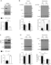
DJ-1 interacts with 14-3-3β in a phosphorylation-dependent manner. A, B) 14-3-3β is present in Flag IP from HEK cells overexpressing myc-Flag–tagged DJ-1 (DJ-1mycFlag) (A) and in IP of endogenous DJ-1 from rat brain (B). C) 14-3-3β co-IP after treatment of HEK lysates containing DJ-1mycFlag with AP (n = 3, unpaired Student’s t test). D, dimer. Data are expressed as means ± sem. *P = 0.0164.
Figure 2
Figure 2
Binding to 14-3-3β and mitochondrial translocation of DJ-1 is inversely correlated after hypoxic stress. A) HEK cells overexpressing DJ-1mycFlag were treated for 24 h with hypoxia (Hyp, 1% O2) prior to Flag IP and quantification of 14-3-3β co-IP relative to DJ-1mycFlag (n = 3, unpaired Student’s t test). *P = 0.0244. B) Endogenous DJ-1 in cytosolic and mitochondrial fractions from HEK cells after 24 h hypoxia. Ctl, control; d, dimer; m, monomer. Data are represented as means ± sem; n = 3, unpaired Student’s t test. Cytosol: P = 0.1028; mitochondria: *P = 0.0172.
Figure 3
Figure 3
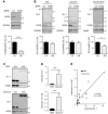
14-3-3β inhibits mitochondrial accumulation of DJ-1 in proliferating cells. A) 14-3-3β knockdown in HEK cells stably expressing scrambled (Scr) or 14-3-3β–targeting shRNA (sh14-3-3β) (n = 6, unpaired Student’s t test). ****P < 0.0001. B) DJ-1 levels in cytosolic and mitochondrial fractions of stable Scr and sh14-3-3β HEK cells after 24 h normoxia (Nor) or hypoxia (Hyp) (n = 3, 1-way ANOVA with Sidak’s multiple comparisons test). *P = 0.0365, **P = 0.005. C) 14-3-3β levels in HEK cells stably expressing pcDNA3 (Ctl) or pcDNA3-14-3-3β (14-3-3βOE) (n = 3, unpaired Student’s t test). **P = 0.0056. D) DJ-1 levels in cytosolic and mitochondrial (monomer and dimer) fractions of stable Ctl and 14-3-3βOE HEK cells after 24 h normoxia or hypoxia (n = 3, 1-way ANOVA with Sidak’s multiple comparisons test). α-tub, α-tubulin; Ctl, control; d, dimer; en, endogenous; m, monomer; tg, transgene; Tom40, translocase of outer mitochondrial membrane 40 homolog. Data are expressed as means ± sem. **P = 0.0015, ***P = 0.0001.
Figure 4
Figure 4
Mitochondrial levels of 14-3-3β and DJ-1 are high in oxidative neurons compared with proliferative HEK cells. A) Western blot and quantification of 14-3-3β in Cas9–transduced primary rat cortical neurons coexpressing nontargeting (Ctl) and 14-3-3β–targeting guide RNA (gRNA) (14-3-3β) (n = 4, unpaired Student’s t test). ***P = 0.0004. B) Total cytosolic (cyto) and mitochondrial (mito) DJ-1 levels in rat cortical neurons transduced as in A (n = 4, unpaired Student’s t test). *P = 0.0191. C) Western blot of cytosolic (in duplicate) and mitochondrial (in duplicate) levels of DJ-1 and 14-3-3β in HEK cells (top) and primary rat cortical neurons (bottom). D) Quantification of the mitochondrial over cytosolic (mito/cyto) ratio of DJ-1 (top) and 14-3-3β (bottom) in HEK cells and neurons (n = 7 for HEK, n = 10 for neurons; Mann-Whitney U test). Top: ***P = 0.0007, bottom: ***P = 0.0001. E) Combined correlation analysis of mito/cyto ratios of DJ-1 and 14-3-3β from HEK cells and neurons shows a significant positive correlation (n = 7 for HEK, n = 10 for neurons; Pearson correlation: r = 0.876, R2 = 0.767). P = 0.0001. Ctl, control; d, dimer; GAPDH, glyceraldehyde-3-phosphate dehydrogenase; Hsp60, heat shock protein 60; KD, knockdown; m, monomer; p, posttranslationally modified. Data are expressed as means ± sem.
Figure 5
Figure 5
Targeting of endogenous DJ-1 to mitochondria shifts metabolism toward oxidative phosphorylation. HEK cells stably expressing scrambled (Scr) shRNA or shRNA against DJ-1 (shDJ-1) or 14-3-3β (sh14-3-3β) were tested for relative ΔΨm (A, B) and proliferation (C, D). A, B) Automated quantification of peak fluorescence intensity per mitochondrion (A) and representative confocal images (B) of TMRE fluorescence (from 3 independent experiments; shDJ-1: n = 30, Scr: n = 23, sh14-3-3β: n = 25; 1-way ANOVA with Dunnett’s multiple comparisons test against Scr). Magnification value (B, right panel), 4.9x. Scale bar, 20 μM. *P = 0.0125, **P = 0.0012. C, D) Proliferation after 72 h (C) and after 48 h with 2.5 nM oligomycin (D) (D: n = 22, G: n = 32, 1-way ANOVA with Dunnett’s multiple comparisons test). A.u., arbitrary units; untr, untreated (0.025% ethanol). Data are expressed as means ± sem. **P = 0.0015, ****P < 0.0001.

References

    1. Deng H., Wang P., Jankovic J. (2018) The genetics of Parkinson disease. Ageing Res. Rev. 42, 72–85 - PubMed
    1. Zhang L., Shimoji M., Thomas B., Moore D. J., Yu S. W., Marupudi N. I., Torp R., Torgner I. A., Ottersen O. P., Dawson T. M., Dawson V. L. (2005) Mitochondrial localization of the Parkinson’s disease related protein DJ-1: implications for pathogenesis. Hum. Mol. Genet. 14, 2063–2073 - PubMed
    1. Bonifati V., Rizzu P., Squitieri F., Krieger E., Vanacore N., van Swieten J. C., Brice A., van Duijn C. M., Oostra B., Meco G., Heutink P. (2003) DJ-1(PARK7), a novel gene for autosomal recessive, early onset parkinsonism. Neurol. Sci. 24, 159–160 - PubMed
    1. Hayashi T., Ishimori C., Takahashi-Niki K., Taira T., Kim Y. C., Maita H., Maita C., Ariga H., Iguchi-Ariga S. M. (2009) DJ-1 binds to mitochondrial complex I and maintains its activity. Biochem. Biophys. Res. Commun. 390, 667–672 - PubMed
    1. Ishikawa S., Taira T., Niki T., Takahashi-Niki K., Maita C., Maita H., Ariga H., Iguchi-Ariga S. M. (2009) Oxidative status of DJ-1-dependent activation of dopamine synthesis through interaction of tyrosine hydroxylase and 4-dihydroxy-L-phenylalanine (L-DOPA) decarboxylase with DJ-1. J. Biol. Chem. 284, 28832–28844 - PMC - PubMed

Publication types

LinkOut - more resources