. 2019 Apr 27;11(5):589.
doi: 10.3390/cancers11050589.

A Protective Role of Aryl Hydrocarbon Receptor Repressor in Inflammation and Tumor Growth

Affiliations

A Protective Role of Aryl Hydrocarbon Receptor Repressor in Inflammation and Tumor Growth

Christoph F A Vogel et al. Cancers (Basel). .

Abstract

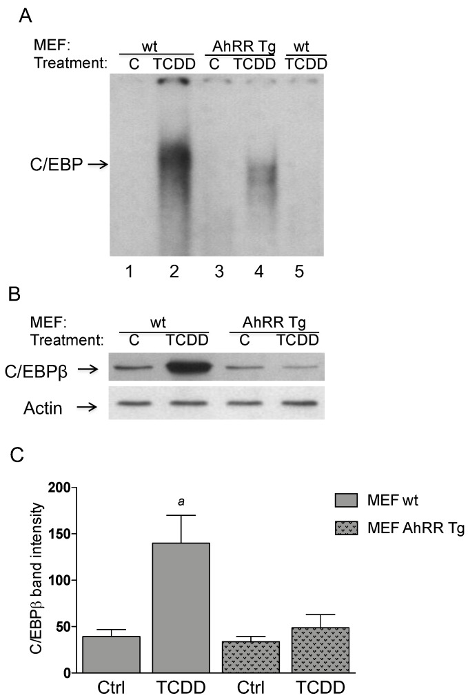
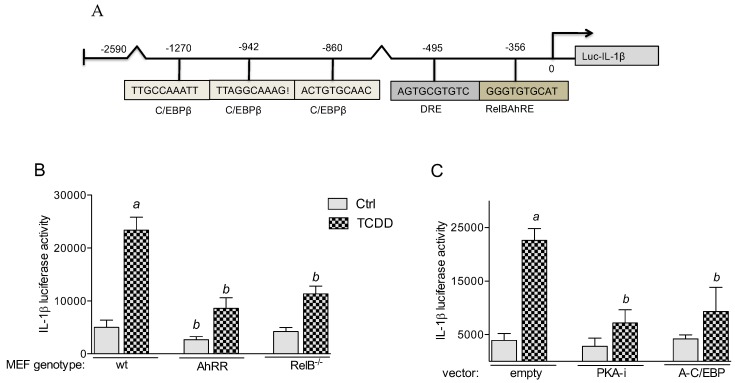
The aryl hydrocarbon receptor (AhR) is known for mediating the toxicity of environmental pollutants such as dioxins and numerous dioxin-like compounds, and is associated with the promotion of various malignancies, including lymphoma. The aryl hydrocarbon receptor repressor (AhRR), a ligand-independent, transcriptionally inactive AhR-like protein is known to repress AhR signaling through its ability to compete with the AhR for dimerization with the AhR nuclear translocator (ARNT). While AhRR effectively blocks AhR signaling, several aspects of the mechanism of AhRR's functions are poorly understood, including suppression of inflammatory responses and its putative role as a tumor suppressor. In a transgenic mouse that overexpresses AhRR (AhRR Tg) we discovered that these mice suppress 2,3,7,8-tetrachlorodibenzo-p-dioxin (TCDD)- and inflammation-induced tumor growth after subcutaneous challenge of EL4 lymphoma cells. Using mouse embryonic fibroblasts (MEF) we found that AhRR overexpression suppresses the AhR-mediated anti-apoptotic response. The AhRR-mediated inhibition of apoptotic resistance was associated with a suppressed expression of interleukin (IL)-1β and cyclooxygenase (COX)-2, which was dependent on activation of protein kinase A (PKA) and the CAAT-enhancer-binding protein beta (C/EBPβ). These results provide mechanistic insights into the role of the AhRR to suppress inflammation and highlight the AhRR as a potential therapeutic target to suppress tumor growth.

Keywords: AhR; AhRR; C/EBPβ; TCDD; carcinogenicity; cyclooxygenase 2; inflammation; interleukin 1; lymphoma.


Conflict of interest statement

The authors declare no conflict of interest.

Figures

Figure 1
Figure 1
Suppressed tumor growth of EL4 lymphoma cells in AhRR Tg mice. Tumor volume of 10-week-old AhRR Tg mice and littermate WT mice (n = 6 for each group) following subcutaneous injection with EL4 lymphoma cells is shown. (A) After 24 h mice were treated with corn oil (Ctrl, black lines) or i.p. injected with 10 μg/kg TCDD (blue lines). (B) WT mice were treated with NS398 or Anakinra (green lines) 30 min. before and every 4 days after injection of TCDD. Tumor volume was measured over a period of 18 days. Means of tumor volume are shown. a significantly higher than AhRR Tg TCDD. b significantly lower than WT Ctrl. c significantly higher than mice treated with NS398 or Anakinra. (p < 0.01).
Figure 2
Figure 2
Resistance of AhRR Tg mice to LPS. (A) Suppressed growth of LPS-induced tumor volume of EL4 lymphoma cells in AhRR Tg mice. WT and AhRR Tg mice were treated i.p. with a single injection of LPS (2.5 mg/kg bw; red lines). Tumor volume was measured over a period of 18 days. Means of tumor volume are shown. a significantly higher than LPS-treated AhRR Tg and WT Ctrl mice (p < 0.01). (B) 6-week-old male AhRR Tg mice and littermate WT mice (n = 8 for each group) were i.p. injected with 25 mg/kg of LPS (red lines) or with the vehicle control PBS (black lines). Lethality was observed over 120 h after LPS challenge. (C) AhRR suppresses LPS-induced expression of IL-1β and COX-2 in BMM. BMM derived from WT and AhRR Tg mice were treated with 100 ng/mL LPS for 24 h and mRNA was analyzed by qPCR. b significantly lower than LPS-treated BMM WT (p < 0.005).
Figure 3
Figure 3
AhRR suppresses TCDD-induced expression of COX-2 and IL-1β in MEF. (A) MEF from wt and AhRR Tg mice were treated with 1 nM TCDD for 24 h and mRNA expression of COX-2 and IL-1β was analyzed by qPCR. a significantly higher than Ctrl; b significantly lower than wt, p < 0.05. (B) TCDD-induced expression of COX-2 and IL-1β is PKA and C/EBP dependent. MEF wt were transfected with an empty vector as control and dominant negative expression vectors PKA-i or A-C/EBP. After 16 h transfection MEF wt were treated with 1 nM TCDD for 24 h and mRNA expression was analyzed by qPCR. a significantly higher than Ctrl; b significantly lower than wt, p < 0.05. (C) Repressed protein level of IL-1β and COX-2 in AhRR Tg MEF determined by Western blot. MEF derived from wt or AhRR Tg mice were treated for 24 h with 1 nM TCDD or vehicle (0.1% DMSO). (D) densitometric evaluation of band intensities of the western blot bands of MEF wt (open bars) and MEF AhRR Tg (shaded bars) is presented. Results of three separate experiments are shown as mean values ± S.D. a significantly different from control cells (p < 0.05).
Figure 4
Figure 4
AhRR represses the levels and activity of C/EBP. (A) Repressed DNA binding activity to a C/EBP consensus element in AhRR Tg MEF compared to wt MEF. MEF from wt (lanes 1 and 2) and AhRR Tg mice (lanes 3 and 4) were treated with 1 nM TCDD (lanes 2 and 4). After 4 h nuclear proteins were extracted. For specificity a 200-fold molar excess of unlabeled probe was added as competitor (lane 5). (B) Repressed protein level of C/EBPβ in AhRR Tg MEF. MEF from wt and AhRR Tg mice were treated with 1 nM TCDD for 4h. Nuclear proteins were extracted and protein level of C/EBPβ was determined by western blot. (C) densitometric evaluation of band intensities of the western blot bands of MEF wt (open bars) and MEF AhRR Tg (shaded bars) is presented. Results of three separate experiments are shown as mean values ± S.D. a significantly different from control cells (p < 0.05).
Figure 5
Figure 5
TCDD-induced IL-1β promoter activity is RelB and PKA-C/EBPβ-dependent. (A) Potential binding sites of the full-length promoter construct of the mouse IL-1β gene containing 2590 bp upstream of the transcriptional start site (indicated by an arrow) cloned into a luciferase (luc) reporter vector. Positions of one putative DRE consensus, one non-consensus DRE (RelBAhRE) and three recognition sites for C/EBPβ are presented. (B) MEF from wt, AhRR Tg, and RelB−/− mice were transfected with a luciferase reporter construct containing 2590 bp (IL-1β) of the mouse gene promoter region. (C) MEF wt were cotransfected with an empty, PKA inhibitor (PKA-i) and C/EBP dominant negative expression plasmid. 24 h after transfection, MEF were treated with 1 nM TCDD for 6 h. Relative luciferase activity units are given as mean values of triplicates as a result of three independent experiments. a significantly different from control cells (p < 0.05); b significantly lower than MEF wt or cells co-transfected with empty vector (p < 0.05).
Figure 6
Figure 6
TCDD stimulates the recruitment of AhR and RelB to the RelBAhRE region of the mouse IL-1β promoter. (A) MEF derived from wt and AhRR Tg mice were treated with 1 nM TCDD for 90 min. ChIP assays were performed using AhR- and RelB-specific antibodies followed by PCR analysis with primer pairs covering the specified RelBAhRE region of mouse IL-1β promoter. (B) Association of AhRR with ARNT and RelB. Co-IP of AhRR, ARNT, and RelB with RelB and AhRR antibody. MEF from wt and AhRR Tg mice were treated with 1 nM TCDD (T) or DMSO (C) for 90 min and cell lysates were used for co-IP. Western blot analysis was performed to detect specific association of AhRR with ARNT and RelB. Rabbit lgG was used as negative control.
Figure 7
Figure 7
TCDD-mediated apoptotic resistance in MEF. (A) MEF derived from wt, AhRR Tg, and RelB−/− mice were treated with 1 nM TCDD for 24 h. Apoptosis was induced by UV light and the number of apoptotic cells was counted after 4 h. (B) MEF wt were transfected with dominant negative expression vectors PKA-i, A-CREB and A-C/EBP for 16 h followed by treatment with 1 nM TCDD for 24 h. Apoptosis was induced by UV light and the number of apoptotic cells was counted after 4 h. Values are averages of duplicates from three independent experiments. a Significantly lower than control p < 0.05.
Figure 8
Figure 8
A schematic diagram illustrates our hypothesis how AhRR may interact with the canonical and non-canonical AhR signaling pathways as described recently [11]. AhRR may suppress inflammatory responses and tumor promotion via interaction with RelB in complex with AhR and ARNT. Double arrows depict the interaction of AhRR with ARNT and RelB with AhR.

References

    1. Van den Berg M., Denison M.S., Birnbaum L.S., Devito M.J., Fiedler H., Falandysz J., Rose M., Schrenk D., Safe S., Tohyama C., et al. Polybrominated dibenzo-p-dioxins, dibenzofurans, and biphenyls: Inclusion in the toxicity equivalency factor concept for dioxin-like compounds. Toxicol. Sci. 2013;133:197–208. doi: 10.1093/toxsci/kft070. - DOI - PMC - PubMed
    1. Pitot H.C., Goldsworthy T., Campbell H.A., Poland A. Quantitative evaluation of the promotion by 2,3,7,8-tetrachlorodibenzo-p-dioxin of hepatocarcinogenesis from diethylnitrosamine. Cancer Res. 1980;40:3616–3620. - PubMed
    1. Castro D.J., Löhr C.V., Fischer K.A., Pereira C.B., Williams D.E. Lymphoma and lung cancer in offspring born to pregnant mice dosed with dibenzo[a,l]pyrene: The importance of in utero vs. lactational exposure. Toxicol. Appl. Pharmacol. 2008;233:454–468. doi: 10.1016/j.taap.2008.09.009. - DOI - PMC - PubMed
    1. Vogel C.F., Li W., Sciullo E., Newman J., Hammock B., Reader J.R., Tuscano J., Matsumura F. Pathogenesis of aryl hydrocarbon receptor-mediated development of lymphoma is associated with increased cyclooxygenase-2 expression. Am. J. Pathol. 2007;171:1538–1548. doi: 10.2353/ajpath.2007.070406. - DOI - PMC - PubMed
    1. Landgren O., Shim Y.K., Michalek J., Costello R., Burton D., Ketchum N., Calvo K.R., Caporaso N., Raveche E., Middleton D., et al. Agent orange exposure and monoclonal gammopathy of undetermined significance: An operation ranch hand veteran cohort study. JAMA Oncol. 2015;1:1061–1068. - PMC - PubMed