. 2019 Apr 29;11(5):399.
doi: 10.3390/v11050399.

Adeno-Associated Virus VP1u Exhibits Protease Activity

Affiliations

Adeno-Associated Virus VP1u Exhibits Protease Activity

Justin J Kurian et al. Viruses. .

Abstract

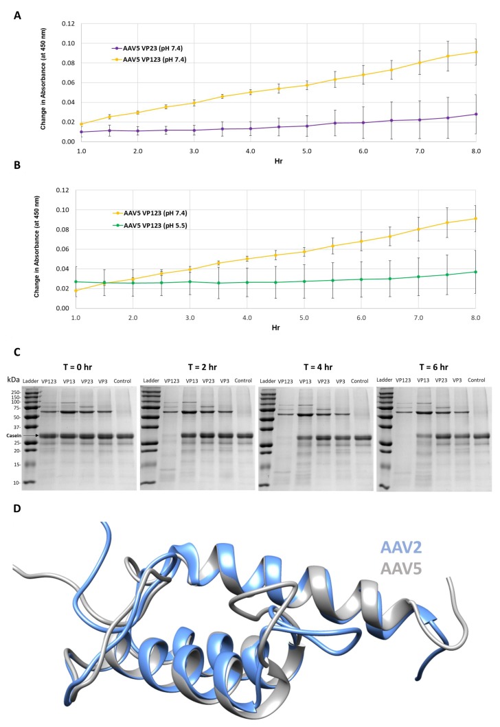
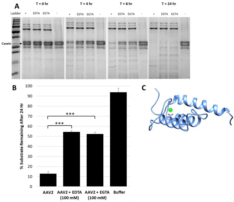
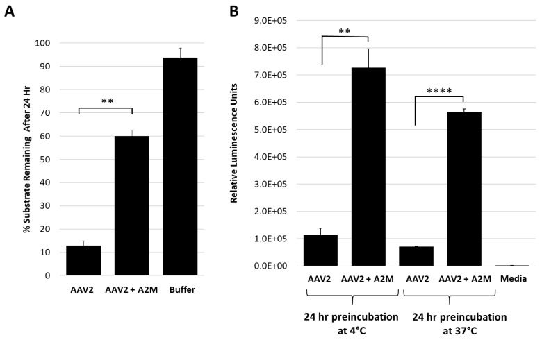
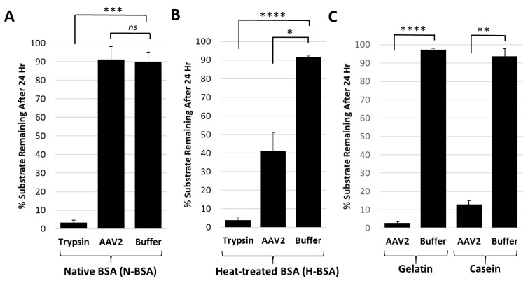
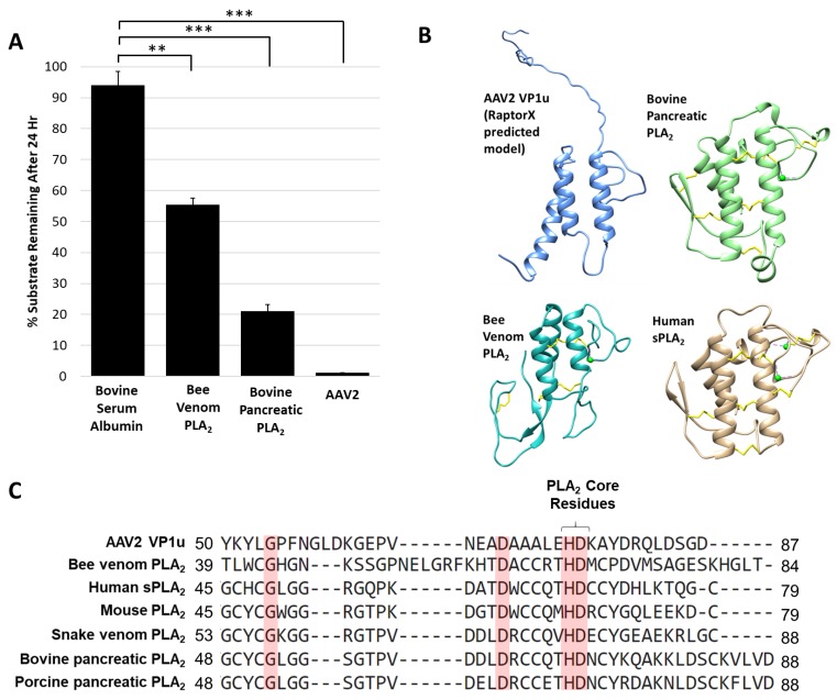
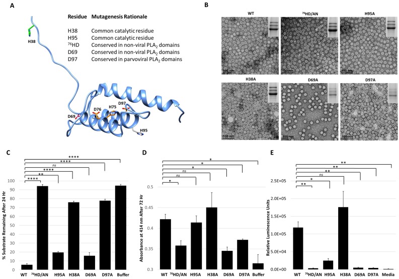
Adeno-associated viruses (AAVs) are being developed for gene delivery applications, with more than 100 ongoing clinical trials aimed at the treatment of monogenic diseases. In this study, the unique N-terminus of AAV capsid viral protein 1 (VP1u), containing a canonical group XIII PLA2 enzyme domain, was observed to also exhibit proteolytic activity. This protease activity can target casein and gelatin, two standard substrates used for testing protease function but does not self-cleave in the context of the capsid or target globular proteins, for example, bovine serum albumin (BSA). However, heated BSA is susceptible to VP1u-mediated cleavage, suggesting that disordered proteins are substrates for this protease function. The protease activity is partially inhibited by divalent cation chelators ethylenediaminetetraacetic acid (EDTA) and ethylene-bis(oxyethylenenitrilo)tetraacetic acid (EGTA), and human alpha-2-macroglobulin (A2M), a non-specific protease inhibitor. Interestingly, both the bovine pancreatic (group VIIA) and bee venom (group III) PLA2 enzymes also exhibit protease function against casein. This indicates that PLA2 groups, including VP1u, have a protease function. Amino acid substitution of the PLA2 catalytic motif (76HD/AN) in the AAV2 VP1u resulted in attenuation of protease activity, suggesting that the protease and PLA2 active sites are related. However, the amino acid substitution of histidine H38, which is not involved in PLA2 function, to alanine, also affects protease activity, suggesting that the active site/mechanism of the PLA2 and protease function are not identical.

Keywords: AAV; Adeno-associated virus; PLA2; phospholipase-A2; protease.


Conflict of interest statement

M.A.M. is a SAB member for Voyager Therapeutics, Inc. and AGTC, has a sponsored research agreement with AGTC, Voyager Therapeutics and Intima Biosciences, Inc. and is a consultant for Intima Biosciences, Inc. M.A.M. is a co-founder of StrideBio, Inc. This is a biopharmaceutical company with interest in developing AAV vectors for gene delivery application.

Figures

Figure 1
Figure 1
Protease activity is VP1 and neutral pH dependent. (A) Colorimetric readout (y-axis) over time (hr, x-axis) for a casein substrate degradation by AAV5 capsids assembled from VP1, VP2 and VP3 or only VP2 and VP3. (B) Same as in (A) but for AAV5 VP1, VP2 and VP3 capsids at pH 7.4 and 5.5. (C) SDS-PAGE of AAV2 VLPs, containing the VPs shown, incubated with a casein substrate at 37 °C for 2, 4 and 6 h. Observations confirm the need to have VP1 and physiological pH for activity. (D) Superposition of AAV2 (blue) and AAV5 (gray) VP1u models. N-terminal domains of models with predicted disorder not shown.
Figure 2
Figure 2
Metal chelators EDTA and EGTA reduce protease activity. (A) SDS-PAGE showing casein proteolysis by AAV2 in presence of 4 mM EDTA or EGTA. “+” indicates control with no inhibitor and “-” indicates control with no AAV2, that is, casein alone. The lane with AAV2 only (“+”) shows fastest decrease in casein with time. (B) Quantification of casein proteolysis by AAV2 in presence of either 100 mM EDTA or 100 mM EGTA. The y-axis indicates the amount of remaining protein after 24 h. (C) Predicted RaptorX structure model of AAV2 VP1u. PLA2 catalytic residues H75 and D76 are in orange colored sticks, the predicted site of bound calcium is denoted by a green sphere next to D76. A predicted disordered portion of AAV2 VP1u N-terminal residues is not shown. Statistical significance as indicated by asterisk annotations (*) are described in Material and Methods section.
Figure 3
Figure 3
Alpha-2-macroglobulin inhibits protease function and correlates with increased cellular transduction. (A) AAV2 protease function inhibition in presence of A2M. The y-axis shows the amount of casein substrate remaining after 24 h at the conditions indicated (x-axis). (B) Luciferase gene expression (y-axis in relative luminescence units) for AAV2 with or without A2M (x-axis) prior to cell infection. Statistical significance as indicated by asterisk annotations (*) are described in Material and Methods section.
Figure 4
Figure 4
The AAV2 VP1u protease preferentially degrades disordered proteins. (A) Quantification of trypsin or AAV2 protease activity against native BSA (N-BSA). The y-axis shows the percentage of substrate remaining after 24 h under the conditions indicated in the x-axis. (B) Quantification of trypsin or AAV2 activity against heated BSA (H-BSA). Axes are as defined in (A). (C) Quantification of AAV2 activity against casein and gelatin. Axes are as defined in (A). Data for casein was shown in Figure 2. Statistical significance as indicated by asterisk annotations (*) are described in Material and Methods section.
Figure 5
Figure 5
Protease activity is a general property of PLA2 enzymes: (A) Quantification of casein substrate degraded by bee venom PLA2, bovine pancreatic PLA2 and AAV2. The y- and x-axes are as described in Figure 4. (B) Predicted structure of AAV2 VP1u compared to bee venom PLA2 (PDB ID: 1POC), bovine pancreatic PLA2 (PDB ID: 1UNE) and human sPLA2 (PDB ID: 1KQU). (C) Partial amino acid sequence alignment of PLA2 enzymes (conserved residues are highlighted in red). Statistical significance as indicated by asterisk annotations (*) are described in Material and Methods section.
Figure 6
Figure 6
Functional phenotypes of AAV2 VP1u variants. (A) Location of amino acid substitutions in the AAV2 VP1u listed and shown in stick representation on a RaptorX model. Residues colored in orange represent those with PLA2 and protease defects when substituted, pink for defect to PLA2 only, green for defect to protease only and gray for no defect to either PLA2 or protease function. (B) Negative-stain EMs of wt AAV2 and variant capsids, confirming the capsid assembly of the viruses. SDS-PAGE gel insert shows expression of VP1, VP2 and VP3; (C) Quantification of the proteolysis of a casein substrate by wt AAV2 and the variants listed (x-axis) after 24 h. The y-axis is as described in Figure 2. (D) PLA2 activity of wt AAV2 and variants. The y-axis depicts the level of lipid modification after 72 h at pH 8.0 and x-axis lists the samples tested. (E) Luciferase reporter activity expression (RLU) for wt AAV2 and variants. The y-axis depicts the relative luminescence units and x-axis lists the samples tested. Statistical significance of asterisk annotations (*) are described in Material and Methods section.

References

    1. Gao G., Vandenberghe L.H., Alvira M.R., Lu Y., Calcedo R., Zhou X., Wilson J.M. Clades of Adeno-Associated Viruses Are Widely Disseminated in Human Tissues. J. Virol. 2004;78:6381–6388. doi: 10.1128/JVI.78.12.6381-6388.2004. - DOI - PMC - PubMed
    1. Van Vliet K., Mohiuddin Y., McClung S., Blouin V., Rolling F., Moullier P., Agbandje-McKenna M., Snyder R.O. Adeno-Associated Virus Capsid Serotype Identification: Analytical Methods Development and Application. J. Virol. Methods. 2009;159:167–177. doi: 10.1016/j.jviromet.2009.03.020. - DOI - PubMed
    1. Ylä-Herttuala S. Endgame: Glybera Finally Recommended for Approval as the First Gene Therapy Drug in the European Union. Mol. Ther. 2012;20:1831–1832. doi: 10.1038/mt.2012.194. - DOI - PMC - PubMed
    1. Smalley E. First AAV Gene Therapy Poised for Landmark Approval. Nat. Biotechnol. 2017;35:998–999. doi: 10.1038/nbt1117-998. - DOI - PubMed
    1. Snijder J., van de Waterbeemd M., Damoc E., Denisov E., Grinfeld D., Bennett A., Agbandje-McKenna M., Makarov A., Heck A.J.R. Defining the Stoichiometry and Cargo Load of Viral and Bacterial Nanoparticles by Orbitrap Mass Spectrometry. J. Am. Chem. Soc. 2014;136:7295–7299. doi: 10.1021/ja502616y. - DOI - PMC - PubMed

Publication types

MeSH terms